

Biomaterial design strategies to address obstacles in craniomaxillofacial bone repair
- PMID: 34540206
- PMCID: PMC8443006
- DOI: 10.1039/d1ra02557k
Biomaterial design strategies to address obstacles in craniomaxillofacial bone repair
Abstract
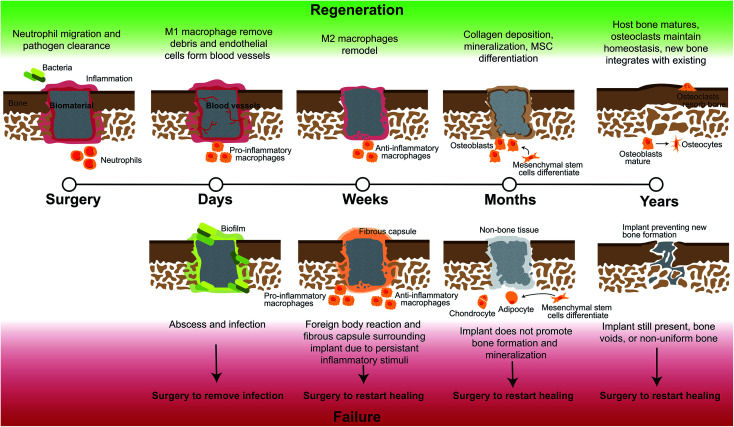
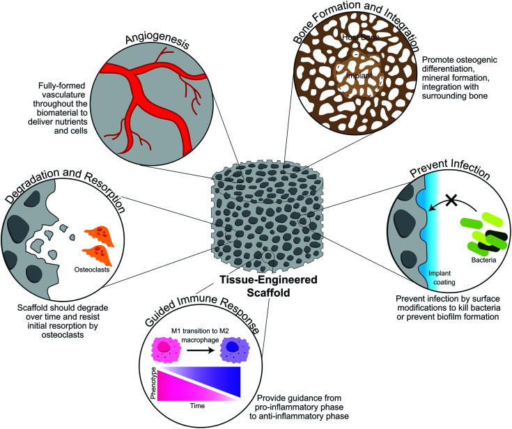
Biomaterial design to repair craniomaxillofacial defects has largely focused on promoting bone regeneration, while there are many additional factors that influence this process. The bone microenvironment is complex, with various mechanical property differences between cortical and cancellous bone, a unique porous architecture, and multiple cell types that must maintain homeostasis. This complex environment includes a vascular architecture to deliver cells and nutrients, osteoblasts which form new bone, osteoclasts which resorb excess bone, and upon injury, inflammatory cells and bacteria which can lead to failure to repair. To create biomaterials able to regenerate these large missing portions of bone on par with autograft materials, design of these materials must include methods to overcome multiple obstacles to effective, efficient bone regeneration. These obstacles include infection and biofilm formation on the biomaterial surface, fibrous tissue formation resulting from ill-fitting implants or persistent inflammation, non-bone tissue formation such as cartilage from improper biomaterial signals to cells, and voids in bone infill or lengthy implant degradation times. Novel biomaterial designs may provide approaches to effectively induce osteogenesis and new bone formation, include design motifs that facilitate surgical handling, intraoperative modification and promote conformal fitting within complex defect geometries, induce a pro-healing immune response, and prevent bacterial infection. In this review, we discuss the bone injury microenvironment and methods of biomaterial design to overcome these obstacles, which if unaddressed, may result in failure of the implant to regenerate host bone.
Conflict of interest statement
Conflicts of interest There are no conflicts of interest to declare.
Figures


References
-
- Bankoff A. D. P.. Biomechanical Characteristics of the Bone. in Human Musculoskeletal Biomechanics, ed. T. Goswami, InTech, 2012
Grants and funding
LinkOut - more resources
Full Text Sources