. 2021 Sep 9;8(17):2100717.
doi: 10.1002/admi.202100717. Epub 2021 Aug 18.

Dosage-dependent antimicrobial activity of DNA-histone microwebs against Staphylococcus aureus

Affiliations

Dosage-dependent antimicrobial activity of DNA-histone microwebs against Staphylococcus aureus

Ting Yang et al. Adv Mater Interfaces. .

Abstract

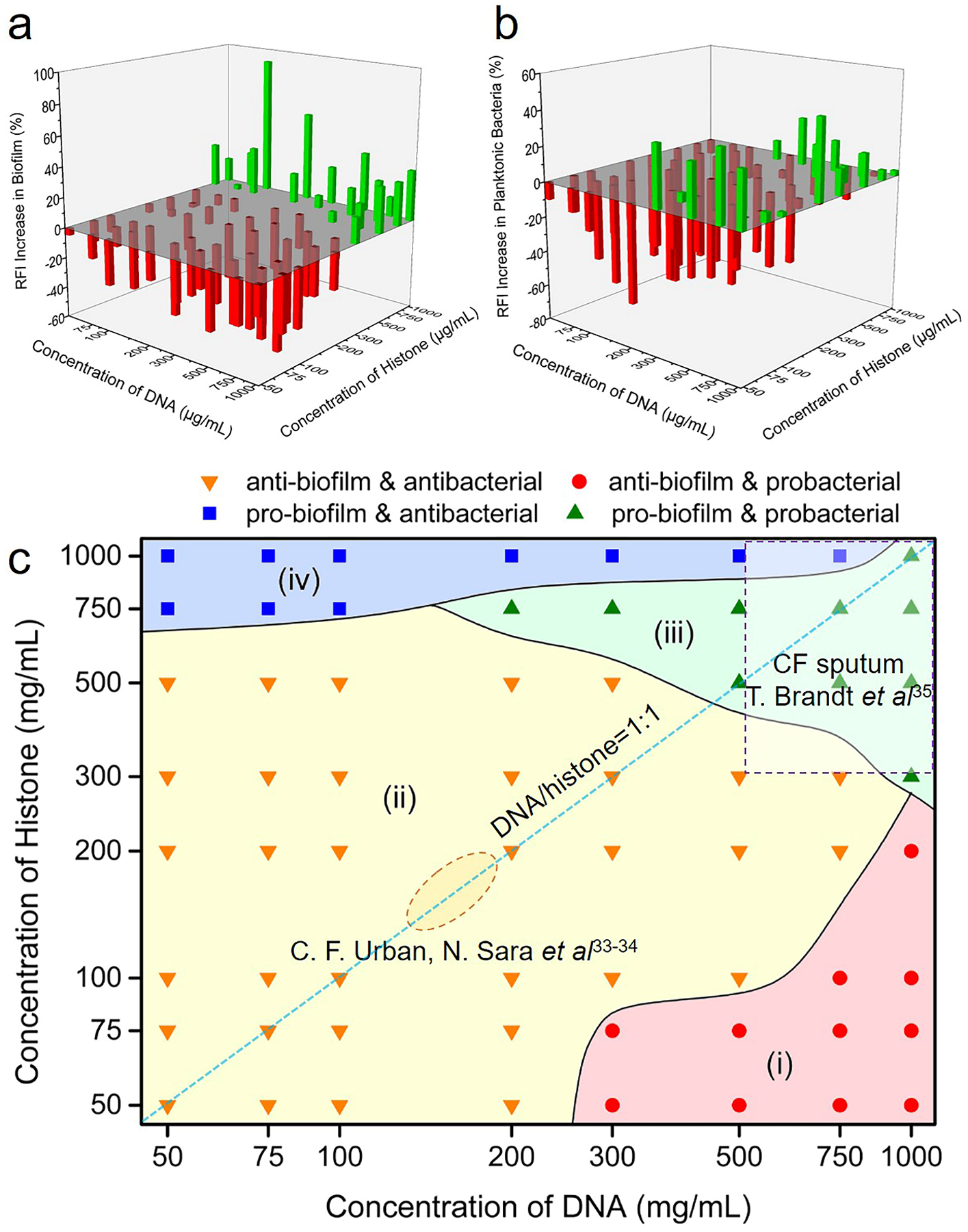
Neutrophil extracellular traps (NETs) is an antimicrobial cobweb-structured material produced by immune cells for clearance of pathogens in the body, but paradoxically associated with biofilm formation and exacerbated lung infections. To provide a better materials perspective on the pleiotropic roles played by NETs at diverse compositions/concentrations, a NETs-like material (called 'microwebs', abbreviated as μwebs) is synthesized for decoding the antimicrobial activity of NETs against Staphylococcus aureus in infection-relevant conditions. We show that μwebs composed of low-to-intermediate concentrations of DNA-histone complexes successfully trap and inhibit S. aureus growth and biofilm formation. However, with growing concentrations and histone proportions, the resulting microwebs appear gel-like structures accompanied by reduced antimicrobial activity that can even promote formation of S. aureus biofilms. Our simplified model of NETs provides a materials-based evidence on NETs-relevant pathology in the development of biofilms.

Keywords: Biofilm; Biomimetics; Neutrophil Extracellular Traps; S. aureus; microwebs.


Conflict of interest statement

Conflict of Interest The authors declare no competing financial interest.

Figures

Figure 1.
Figure 1.
Preparation and characterization of DHMs and microweb (μweb) suspensions. (a) Schematic showing the preparation of a DHM from dehydrated DNA-rich droplets. (b) A fluorescent image of the DHM membrane, showing DNA (green) resembles into a mesh in the DHMs overall structure. (c) SEM observation of mesh-like DHMs with submicron pores. (d) Preparation of μweb suspensions. (e) Fluorescence microscopy image of surface-bound μwebs. (f) Scanning electron microscopy image of surface-bound μwebs.
Figure 2.
Figure 2.
DNA-histone mesostructures (DHMs) promote adhesion of S. aureus in nutrient poor HBSS medium. (a,b) SEM images showing the adhesion of S. aureus on a) non-coated and b) DHM-coated substrates. t =1.5 hour. (c,d) CLSM 3D reconstructions showing the attached S. aureus aggregate into small clusters. Red: propidium iodide stained DHM and dead S. aureus; Green: live S. aureus. t =1.5 hour. (e) Statistics of the relative fluorescence intensity of S. aureus attached to non-coated, DNA-coated, histone-coated, or DHM-coated microplates. The statistics were performed by ANOVA with post hoc Tukey test: ns, not significant; *p < 0.05, **p < 0.01 and ***p < 0.001, N=6 for each condition.
Figure 3.
Figure 3.
Evaluation of the bacterial killing potency of DNA, histone and DNA-histone μweb suspensions against S. aureus. a) Enumeration of colony forming units of S. aureus (seeding density: 106 CFU mL−1) after culture in nutrient-poor HBSS media, DNA-histone μwebs suspensions (200 μg mL−1 DNA + 200 μg mL−1 histone), DNA solution (200 μg mL−1) and histone solution (200 μg mL−1). ANOVA followed by Tukey’s test was used for statistical analysis: ns, not significant; *p < 0.05, **p < 0.01 and ***p < 0.001, N=6 for each condition. b-d) Growth curves of S. aureus in nutrient-rich TSBg medium containing (b) eDNA, (c) histone, or (d) μwebs at physiologically relevant concentrations. Seeding density: 107 CFU mL−1.
Figure 4.
Figure 4.
Aggregated clumps of μwebs promote formation of S. aureus biofilm in nutrient rich TSBg medium. (a-c) SEM images showing that 400 μg mL−1 μweb promotes formation of S. aureus biofilm. Time of bacterial incubation, (a) t=6hr, (b) t=12hr, (c) t=24hr. The white arrows indicate the EPS or bacteria lysed by μweb. (d-f) SEM images showing formation of S. aureus biofilm without addition of μwebs in the culture medium. (d) t=6hr, (e) t=12hr, (f) t=24hr.
Figure 5.
Figure 5.
Concentration-dependent antimicrobial/biofilm promoting effects of μwebs on S. aureus. S. aureus were cultured with μwebs for 24 hours to evaluate the effect of μwebs with different compositions on the formation of S. aureus biofilm. The RFIs of planktonic bacteria and biofilm were measured separately. a) Changes in the RFIs of S. aureus biofilm in the presence of μwebs with different compositions. b) Changes in the RFIs of the planktonic S. aureus in the presence of μwebs with different compositions. c) A state diagram showing the effect of μweb compositions on the mode of bacteria growth in TSBg. Four different regions are defined in the diagram: (i) red, inhibits the formation of biofilm and slightly promotes growth of planktonic bacteria. (ii) yellow, inhibits both growth of biofilm and planktonic bacteria. (iii) green, promotes the growth of planktonic bacteria and formation of biofilm. (iv) blue, inhibits the growth of planktonic bacteria but prompts biofilm formation. The solid lines dividing boundaries between different zones are served as a guide for the eye.
Figure 6.
Figure 6.
A schematic illustration of the S. aureus responses to μwebs with varying compositions.

References

    1. Miller SI, Tsolis RM, Current Opinion in Microbiology. 2017, 35, v; - PubMed
    2. Brady RA, O’May GA, Leid JG, Prior ML, Costerton JW, Shirtliff ME, Infection and immunity. 2011, 79, 1797; - PMC - PubMed
    3. Thurlow LR, Hanke ML, Fritz T, Angle A, Aldrich A, Williams SH, Engebretsen IL, Bayles KW, Horswill AR, Kielian T, Journal of Immunology. 2011, 186, 6585. - PMC - PubMed
    1. Sugimoto S, Sato F, Miyakawa R, Chiba A, Onodera S, Hori S, Mizunoe Y, Sci Rep. 2018, 8, 2254. - PMC - PubMed
    1. Galdiero E, Lombardi L, Falanga A, Libralato G, Guida M, Carotenuto R, Pharmaceutics. 2019, 11, 322. - PMC - PubMed
    1. Schilcher K, Andreoni F, Dengler Haunreiter V, Seidl K, Hasse B, Zinkernagel AS, Antimicrob Agents Chemother. 2016, 60, 5957. - PMC - PubMed
    1. Sultan AR, Hoppenbrouwers T, Lemmens-den Toom NA, Snijders SV, van Neck JW, Verbon A, de Maat MPM, van Wamel WJB, Infection and Immunity. 2019, 87, e00605. - PMC - PubMed

LinkOut - more resources