

Long-term outcomes of focal laser photocoagulation for the treatment of polypoidal choroidal vasculopathy
- PMID: 34540617
- PMCID: PMC8403861
- DOI: 10.18240/ijo.2021.09.16
Long-term outcomes of focal laser photocoagulation for the treatment of polypoidal choroidal vasculopathy
Abstract
Aim: To evaluate the long-term effect and safety of focal laser photocoagulation treatment in eyes with polypoidal choroidal vasculopathy (PCV).
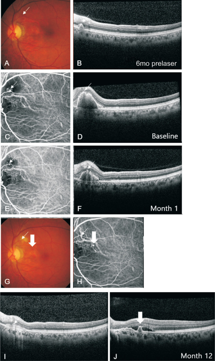
Methods: Medical records of 13 eyes of 13 patients with PCV were followed-up for more than 2y after focal laser photocoagulation treatment. The patients were diagnosed with PCV using indocyanine green angiography, and eyes with other comorbid ocular diseases were excluded. The measurement outcomes of the study were the post-treatment regression and recurrence of polyps, complications, and changes in visual acuities. Paired t-test was performed to compare visual outcome before and after the treatment.
Results: The mean age of the 13 patients was 70.2±5.5y, and the follow-up period was 72.3±31.0 (range, 25-118)mo. Three eyes had juxtafoveal polyps and 10 eyes had extrafoveal polyps. Of the 13 eyes, 9 eyes (69.2%) had regression of polyps 1.7±1.2 (range, 0.9-4)mo after focal laser photocoagulation. Five eyes (55.6%) showed recurrence of polyps during the follow-up periods, and the recurrence period was 12.8±18.9 (range, 1.9-48)mo. Mild subretinal hemorrhage occurred in two eyes (15.4%) 27 and 72d after laser treatment, respectively. There were no statistically significant differences in visual acuities at baseline; 1, 2, 3y post-treatment (all P>0.05); and last follow-up (0.63±0.5, 0.73±0.70, 0.67±0.57, 0.75±0.7, and 0.95±0.8 logMAR, respectively).
Conclusion: Focal laser photocoagulation is beneficial for early regression of polyps in eyes with PCV and does not result in significant submacular hemorrhage during the long-term follow-up. Furthermore, it can be primarily considered in eyes with PCV with extrafoveal or juxtafoveal polyps to regress risky polyps as well as to maintain visual acuity without serious hemorrhagic complications.
Keywords: focal laser photocoagulation; long-term efficacy; polypoidal choroidal vasculopathy.
International Journal of Ophthalmology Press.
Figures


References
-
- Cheung CMG, Lai TYY, Ruamviboonsuk P, Chen SJ, Chen Y, Freund KB, Gomi F, Koh AH, Lee WK, Wong TY. Polypoidal choroidal vasculopathy: definition, pathogenesis, diagnosis, and management. Ophthalmology. 2018;125(5):708–724. - PubMed
-
- Eriş E, Perente İ, Vural E, Yaşa D, Ozkaya A. Assessment of focal laser photocoagulations' early effect on polypoidal choroidal vasculopathy with optical coherence tomography angiography. Lasers Med Sci. 2018;33(8):1833–1835. - PubMed
-
- Wong CW, Cheung CM, Mathur R, Li X, Chan CM, Yeo I, Wong E, Lee SY, Wong D, Wong TY. Three-year results of polypoidal choroidal vasculopathy treated with photodynamic therapy: retrospective study and systematic review. Retina. 2015;35(8):1577–1593. - PubMed
LinkOut - more resources
Full Text Sources