Review
. 2021 Aug 12:9:704939.
doi: 10.3389/fcell.2021.704939. eCollection 2021.

Born to Run? Diverse Modes of Epithelial Migration

Affiliations
Review

Born to Run? Diverse Modes of Epithelial Migration

Pengfei Lu et al. Front Cell Dev Biol. .

Abstract

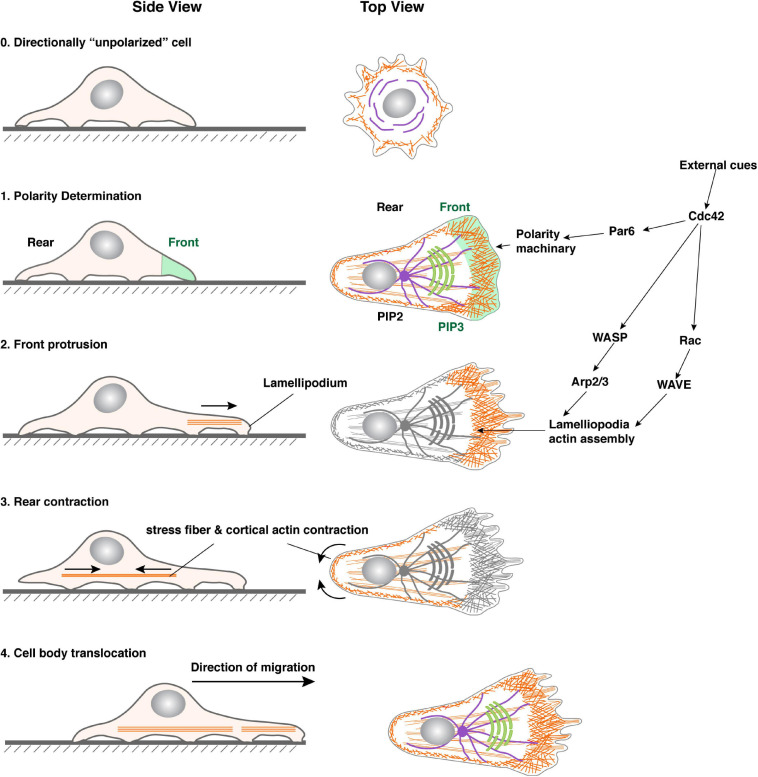
Bundled with various kinds of adhesion molecules and anchored to the basement membrane, the epithelium has historically been considered as an immotile tissue and, to migrate, it first needs to undergo epithelial-mesenchymal transition (EMT). Since its initial description more than half a century ago, the EMT process has fascinated generations of developmental biologists and, more recently, cancer biologists as it is believed to be essential for not only embryonic development, organ formation, but cancer metastasis. However, recent progress shows that epithelium is much more motile than previously realized. Here, we examine the emerging themes in epithelial collective migration and how this has impacted our understanding of EMT.

Keywords: EMT; apicobasal polarity; cell polarity; collective migration; epithelial polarity; extracellular matrix; mechanosensing; mesenchymal-epithelial transition.


Conflict of interest statement

The authors declare that the research was conducted in the absence of any commercial or financial relationships that could be construed as a potential conflict of interest.

Figures

FIGURE 1
FIGURE 1
Transitions between epithelial, mesenchymal, and intermediate cell states. Typical vertebrate epithelium structure with apicobasal polarity. Tight junctions separate the apical domain from the basolateral domain. Upon EMT, cells lose apicobasal polarity, reorganize their cytoskeleton, and become motile. Increasing evidence shows that cells undergo collective migration rather than individual migration after EMT. Both epithelial and mesenchymal states are plastic and mesenchymal cells can return to the epithelial state in both development and cancer situations through a process called MET. It is believed that cells, especially in cancer situations, can enter a “partial EMT” state where they express mixed epithelial and mesenchymal gene signatures. Microfilaments are indicated in red, microtubules in green. The position of the nucleus (light blue oval) is also shown.
FIGURE 2
FIGURE 2
Steps in single-cell migration. Diagram with both side and top views of a cell before and during migration to illustrate signaling events leading to polarity determination, microfilament dynamics, and morphological changes of the cell. For illustration purposes, a cell is theoretically “unpolarized” before being stimulated by an external cue (0). Upon stimulation, the area with the highest Cdc42, Par6, and downstream signaling activities becomes the front, which is marked by an increase in PIP3 levels in the membrane and focal adhesions assembly (1). As a result, lamellipodia formation starts in the front, leading to membrane forward protrusion, where new focal adhesions can be made (2). Concordantly, the rear of the cell undergoes a contraction process (arrowheads) involving actomyosin activities along the cell periphery and in the stress fibers (3). The cell retracts when focal adhesions in the rear break apart due to the contraction force and, as a result, the cell body translocates forward. The net results of the above steps are a forward movement of the cell.
FIGURE 3
FIGURE 3
Representative models of collective migration. Front-rear polarity in most of these models, including the Xenopus neural crest, Drosophila border cells, zebrafish lateral line, and mouse mammary gland, is triggered by a chemoattractant indicated, except for the wound healing model (using human skin as an example), where the external cue is thought to be mechanical forces. Leader cells are colored in green. Blue bars indicate the boundary of the apical and basolateral domains, which in vertebrates is where tight junctions locate, and in invertebrates is the subapical complex. They denote the presence of apicobasal polarity and the epithelial state. Note that neural crest cells do not have the blue bars because they are thought to have undergone a complete EMT and have lost the apicobasal polarity. Leader cells are colored in green.
FIGURE 4
FIGURE 4
A common polarity machinery regulates distinct cell behavior. (A) Yeast cell division, anterior-posterior determination during one-cell stage embryo development of C. elegans, and neuroblasts differentiation can all be viewed as different forms of cell fate determination. They all involve the asymmetric distribution of cell fate determinants (purple) upon polarity determination by regulatory machinery (green). (B) Mature immotile epithelium whose most salient feature is apicobasal polarity, as indicated by cell morphology and the presence of tight junctions (blue bars). (C) A migrating single cell, whose front is colored in green. Note that a neuron can be viewed as a specialized migrating cell, with its growth cone being the cell front, capable of moving and sensing the environment. (D) A migrating epithelium with a leader cell (green) and a follower cell, the latter of which still has typical apicobasal polarity. Pink arrows indicate polarity directions.
FIGURE 5
FIGURE 5
Roles of follower cells during collective migration. (A) Periphery follower cells in some models, including neural crest, form actin “cable” whose contraction could propel the forward movement of the collective. Note that actin cable could be a misnomer as it might not represent what the actin-polymer looks like in 3D. (B) Follower cells in some models have been shown to form cryptic lamellipodia, which can generate force and promote collective migration. However, as shown in the side view, an essential question is how the follower solves the potential conflict of having two polarities, namely apicobasal and front-rear polarities simultaneously in the same cell when they are mutually exclusive in all other models examined.

References

    1. Affolter M., Weijer C. J. (2005). Signaling to cytoskeletal dynamics during chemotaxis. Dev. Cell 9 19–34. 10.1016/j.devcel.2005.06.003 - DOI - PubMed
    1. Aiello N. M., Brabletz T., Kang Y., Nieto M. A., Weinberg R. A., Stanger B. Z. (2017). Upholding a role for EMT in pancreatic cancer metastasis. Nature 547 E7–E8. - PMC - PubMed
    1. Andrew D. J., Ewald A. J. (2009). Morphogenesis of epithelial tubes: insights into tube formation, maintenance and elaboration. Dev. Biol. 341 34–55. 10.1016/j.ydbio.2009.09.024 - DOI - PMC - PubMed
    1. Aoki K., Kondo Y., Naoki H., Hiratsuka T., Itoh R. E., Matsuda M. (2017). Propagating wave of ERK activation orients collective cell migration. Dev. Cell 43 305–317.e5. - PubMed
    1. Barriga E. H., Franze K., Charras G., Mayor R. (2018). Tissue stiffening coordinates morphogenesis by triggering collective cell migration in vivo. Nature 554 523–527. 10.1038/nature25742 - DOI - PMC - PubMed

LinkOut - more resources