. 2017 Jul 20;7(14):e2394.
doi: 10.21769/BioProtoc.2394.

Analyzing the Properties of Murine Intestinal Mucins by Electrophoresis and Histology

Affiliations

Analyzing the Properties of Murine Intestinal Mucins by Electrophoresis and Histology

Ran Wang et al. Bio Protoc. .

Abstract

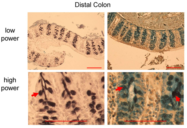
Specialized secretory cells known as goblet cells in the intestine and respiratory epithelium are responsible for the secretion of mucins. Mucins are large heavily glycosylated proteins and typically have a molecular mass higher than 106 Da. These large proteins are densely substituted with short glycan chains, which have many important functional roles including determining the hydration and viscoelastic properties of the mucus gel that lines and protects the intestinal epithelium. In this protocol, we comprehensively describe the method for extraction of murine mucus and its analysis by agarose gel electrophoresis. Additionally we describe the use of High Iron Diamine-Alcian Blue, Periodic Acid Schiff's-Alcian Blue and immune-staining methods to identify and differentiate between the different states of glycosylation on these mucin glycoproteins, in particular with a focus on sulphation and sialylation.

Keywords: Glycosylation; High Iron Diamine; Mucin; Secreted mucin; Sialylation; Stored mucin; Sulphation.


Figures

Figure 1.
Figure 1.. Electrophoresis gel tank (right) and electrophoresis power supply (left) for gel electrophoresis analyses of mucins
Figure 2.
Figure 2.. Vacuum Blotting Unit with vacuum blotter chamber (front) and vacuum pump (back)
Figure 3.
Figure 3.. Outlines of the procedures described
Figure 4.
Figure 4.. Scanned nitrocellulose membrane stained with PAS and anti-Muc2 antibody.
Total mucus from the colon was extracted from 4 individual C57BL/6 mice and analysed using 1% agarose gel electrophoresis and transferred to nitrocellulose membrane. Nitrocellulose membrane was stained with PAS (A) or anti-Muc2 antibody (B, H300, from Santa Cruz) and developed using infrared-labelled secondary antibody. Membrane was scanned using the Odyssey® CLx Imager.
Figure 5.
Figure 5.. Mouse intestine dissection and ‘Swiss Roll’ technique for histology.
Mouse intestine is dissected (A) followed by opening longitudinally using spring scissor (B) and removing feces using a P200 tip (C). Intestine is rolled around a 16 gauge needle with the help of tweezers (D, E). Swiss roll is secured on polystyrene cube with two needles pin in opposite direction (make sure needle is not placed in tissue) (F) before putting into specimen jar with 10% buffer formalin for fixation (G). Fixed Swiss roll is transferred into histology cassette (H) for paraffin embedding (I).
Figure 6.
Figure 6.. Pictures of PAS-AB stain in small intestine, proximal colon and distal colon of C57BL/6 mice.
Neutral glycoproteins are stained in magenta (red arrow, depicting the glycocalyx), mucins that are both acidic and neutrally charged are stained in purple (blue plus magenta stain, green arrow) and acidic glycoproteins are stained in blue (black arrow). These slides were counterstained with haematoxylin (as described in step B3f). Scale bars = 200 µm (low power), 100 µm (high power).
Figure 7.
Figure 7.. HID-AB staining in distal colon of C57BL/6 mice.
Black stain indicates sulfomucins dominated area (left panel, red arrow), whereas goblet cells with sialomucins (blue) and mixed sulfo-sialomucins (black and blue colors in the right panel, red arrows). Scale bars = 200 µm (low power), 100 µm (high power).
Figure 8.
Figure 8.. Quantification of positive staining by using NIS-Elements software.
A. Original picture showing Muc2 positive staining in dark brown color. B. Positive staining was defined by RGB pixel picking tool in NIS-Elements software. For example, to quantify goblet cell volume in a longitudinally sectioned crypt, the percent of positive staining per crypt is calculated as the area covered by positive staining divided by the area of the crypt. Three to five individual crypts are analysed for each sample before taking the average value.

References

    1. Arike L., Holmen-Larsson J. and Hansson G. C.(2017). Intestinal Muc2 mucin O-glycosylation is affected by microbiota and regulated by differential expression of glycosyltranferases. Glycobiology 27(4): 318-328. - PMC - PubMed
    1. Hasnain S. Z., Dawson P. A., Lourie R., Hutson P., Tong H., Grencis R. K., McGuckin M. A. and Thornton D. J.(2017). Immune-driven alterations in mucin sulphation is an important mediator of Trichuris muris helminth expulsion . PLoS Pathog 13(2): e1006218. - PMC - PubMed
    1. Reilly R. W. and Kirsner J. B.(1965). Runt intestinal disease. Lab Invest 14: 102-107. - PubMed
    1. Sheehan J. K. and Thornton D. J.(2000). Heterogeneity and size distribution of gel-forming mucins. Methods Mol Biol 125: 87-96. - PubMed
    1. Spicer S. S.(1965). Diamine methods for differentialing mucosubstances histochemically. J Histochem Cytochem 13: 211-234. - PubMed

LinkOut - more resources