. 2021 Sep 9:11:146-150.
doi: 10.1016/j.artd.2021.07.004. eCollection 2021 Oct.

Removing Well-Fixed, Collared and Noncollared Tapered Hip Stems Without an Extended Trochanteric Osteotomy Using a Novel Stem Removal System

Affiliations

Removing Well-Fixed, Collared and Noncollared Tapered Hip Stems Without an Extended Trochanteric Osteotomy Using a Novel Stem Removal System

Kevin M Sagers et al. Arthroplast Today. .

Abstract

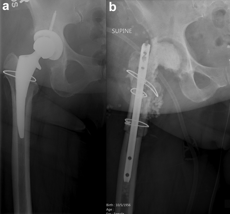
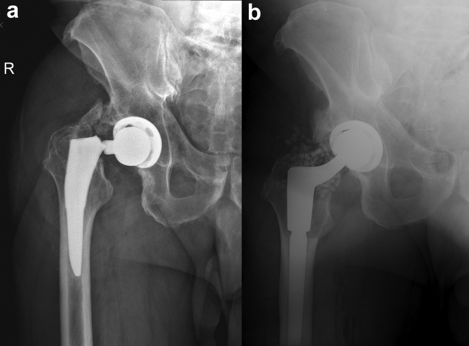
Improvements in materials, components, and surgical techniques in cementless total hip arthroplasty are resulting in improved femoral stem fixation through bony ongrowth or ingrowth. While improved femoral stem fixation is one reason for the current excellent total hip survivorship, indications for stem removal such as infection, implant fracture, or osteolysis remain. A commonly used technique for fully ingrown femoral stems is an extended trochanteric osteotomy which can result in comminuted fractures of the proximal femur during stem removal requiring additional fixation. Therefore, a novel hip stem removal was developed to facilitate removal of these well-ingrown stems without the need for an extended trochanteric osteotomy. This study describes the removal system and surgical technique and presents a case series of successfully removed ingrown stems.

Keywords: Cementless; Explantation; Femoral stem removal; Resection arthroplasty; Revision hip arthroplasty.


Figures

Figure 1
Figure 1
(a) Preoperative radiograph of a patient with a well-fixed collared stem who underwent explant for infection. (b) Postoperative radiograph after explantation with ETO. ETO resulted in a comminuted fracture of the proximal femur. An antibiotic-coated intramedullary nail was used as a spacer.
Figure 2
Figure 2
(a) The starting lateral osteotome in the Watson Extraction System. (b) The custom osteotome is designed to follow the geometry and taper of the stem. (c) A Sawbone model demonstrating the passage of the starting lateral osteotome.
Figure 3
Figure 3
(a) The second lateral osteotome. (b) The additional length allows freeing of the stem from any ongrowth at the hydroxyapatite-coated portion. (c) A Sawbone model demonstrating the passage of the second osteotome.
Figure 4
Figure 4
(a) A commonly used cementless triple tapered collared stem (Actis; DePuy Synthes). (b) The medial osteotome is passed along the medial ingrowth portion of the stem once the collar is removed. (c) A Sawbone model demonstrating the passage of the medial osteotome.
Figure 5
Figure 5
(a) Preoperative radiograph of a patient that was indicated for explant of a well-fixed collared stem due to infection. (b) Postoperative radiographs after successful removal of the stem without ETO or fracture using the Watson Extraction System. (c and d) Clinical photographs of the removed stem demonstrate minimal bone loss associated with stem removal.
Figure 6
Figure 6
(a) Preoperative radiograph of a patient indicated for revision of the femoral stem due to progressive lucent lines and pain. (b) Postoperative radiographs after successful removal of the stem without ETO or fracture using the Watson Extraction System and stem revision. (c) Clinical photograph of stem after removal.
Figure 7
Figure 7
(a) Preoperative radiograph of a patient with fractured neck of the previously implanted cementless stem. (b) Postoperative radiograph after successful removal of stem without ETO or fracture and stem revision.
Figure 8
Figure 8
(a) Preoperative radiograph of a catastrophic polyethylene liner wear or polyethylene liner dislocation with destruction of acetabular component and screw with superior migration of the femoral component. The neck of the femoral component appeared to have worn on the acetabular rim creating a notch concerning for risk of fracture and necessitating stem removal in addition to acetabular revision. (b) Postoperative radiograph after successful removal of the stem without an ETO and insertion of a monolithic revision stem and addition of a trochanteric hook plate with cable fixation due to osteolysis of the greater trochanter. (c) Clinical photograph of the stem after removal with minimal bone loss.

References

    1. Kurtz S., Ong K., Lau E., Mowat F., Halpern M. Projections of primary and revision hip and knee arthroplasty in the United States from 2005 to 2030. J Bone Joint Surg Am. 2007;89(4):780. - PubMed
    1. Brown J.M., Mistry J.B., Cherian J.J. Femoral component revision of total hip arthroplasty. Orthopedics (Online) 2016;39(6):e1129. - PubMed
    1. Aribindi R., Paprosky W., Nourbash P., Kronick J., Barba M. Extended proximal femoral osteotomy. Instr Course Lect. 1999;48:19. - PubMed
    1. Miner T.M., Momberger N.G., Chong D., Paprosky W.L. The extended trochanteric osteotomy in revision hip arthroplasty: a critical review of 166 cases at mean 3-year, 9-month follow-up. J Arthroplasty. 2001;16(8 Suppl 1):188. - PubMed
    1. Huffman G.R., Ries M.D. Combined vertical and horizontal cable fixation of an extended trochanteric osteotomy site. J Bone Joint Surg Am. 2003;85(2):273. - PubMed

LinkOut - more resources