. 2021 Jul 5:8:296-308.
doi: 10.1016/j.bioactmat.2021.06.030. eCollection 2022 Feb.

In vivo printing of growth factor-eluting adhesive scaffolds improves wound healing

Affiliations

In vivo printing of growth factor-eluting adhesive scaffolds improves wound healing

Kristo Nuutila et al. Bioact Mater. .

Abstract

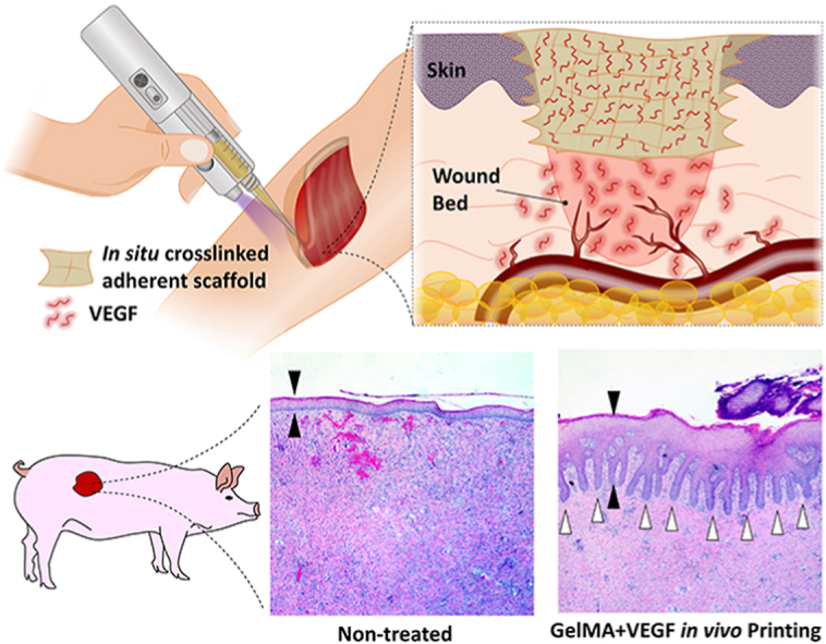
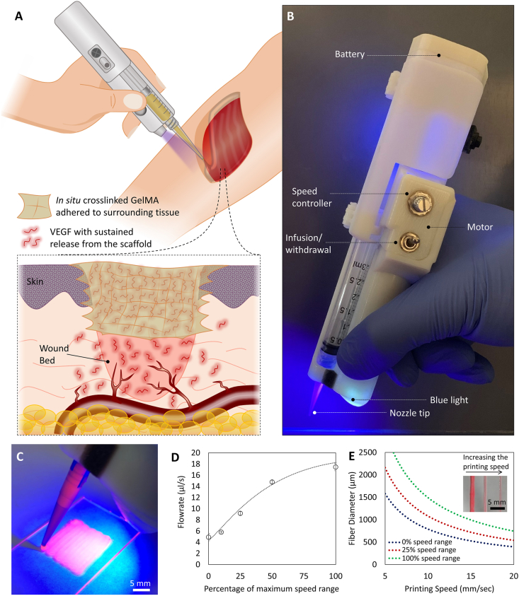
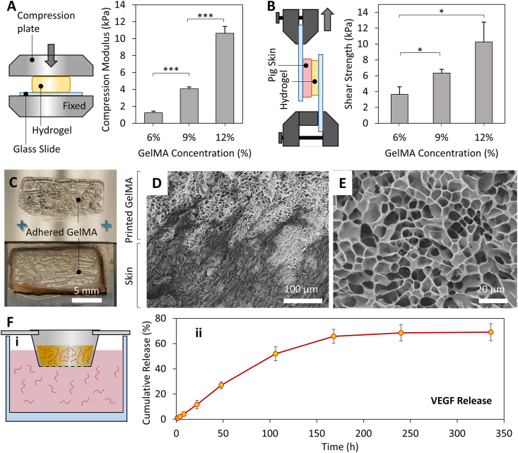
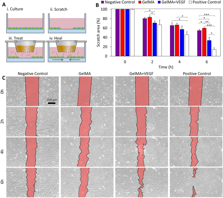
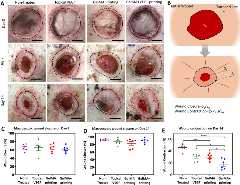
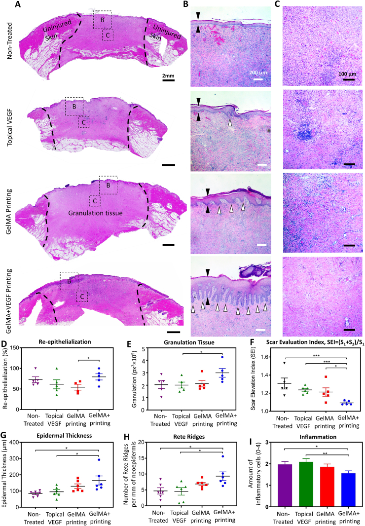
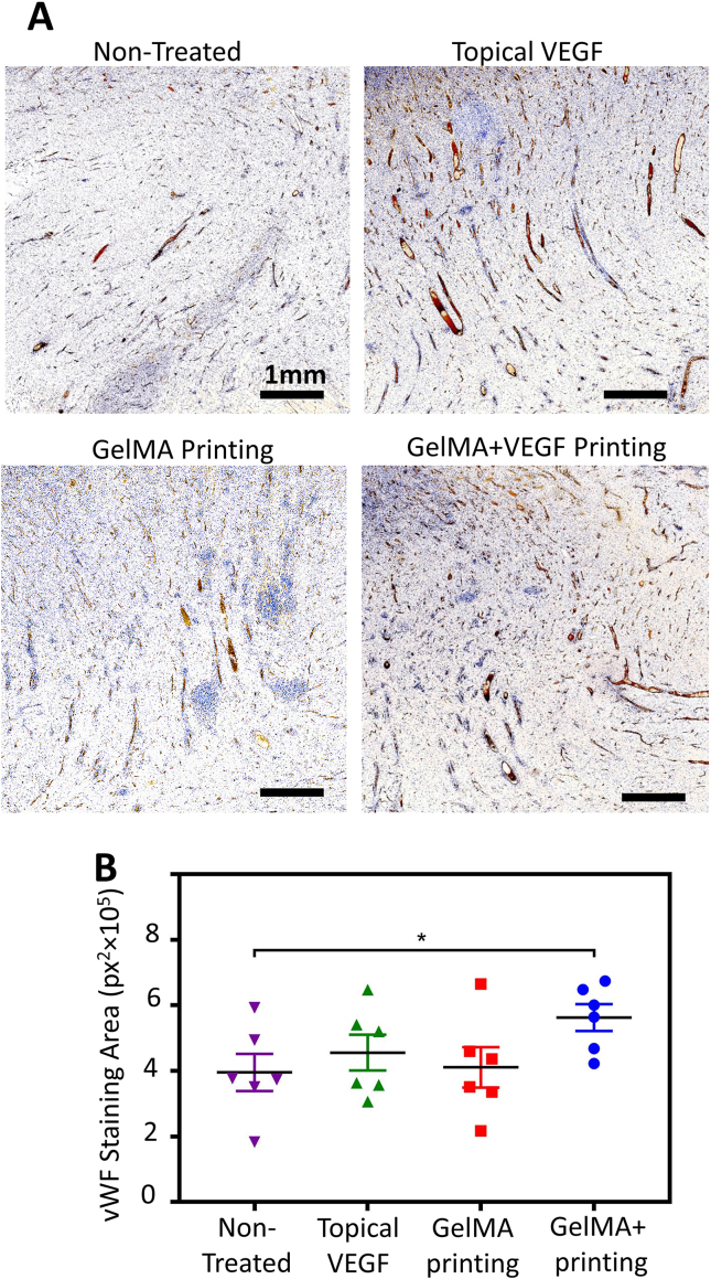
Acute and chronic wounds affect millions of people around the world, imposing a growing financial burden on patients and hospitals. Despite the application of current wound management strategies, the physiological healing process is disrupted in many cases, resulting in impaired wound healing. Therefore, more efficient and easy-to-use treatment modalities are needed. In this study, we demonstrate the benefit of in vivo printed, growth factor-eluting adhesive scaffolds for the treatment of full-thickness wounds in a porcine model. A custom-made handheld printer is implemented to finely print gelatin-methacryloyl (GelMA) hydrogel containing vascular endothelial growth factor (VEGF) into the wounds. In vitro and in vivo results show that the in situ GelMA crosslinking induces a strong scaffold adhesion and enables printing on curved surfaces of wet tissues, without the need for any sutures. The scaffold is further shown to offer a sustained release of VEGF, enhancing the migration of endothelial cells in vitro. Histological analyses demonstrate that the administration of the VEGF-eluting GelMA scaffolds that remain adherent to the wound bed significantly improves the quality of healing in porcine wounds. The introduced in vivo printing strategy for wound healing applications is translational and convenient to use in any place, such as an operating room, and does not require expensive bioprinters or imaging modalities.

Keywords: Adhesive scaffolds; GelMA; Handheld bioprinter; In vivo printing; VEGF; Wound healing.


Conflict of interest statement

The authors declare that they have no known competing financial interests or personal relationships that could have appeared to influence the work reported in this paper.

Figures

Image 1
Graphical abstract
Fig. 1
Fig. 1
In vivo printing of growth factor-eluting hydrogel for wound healing applications. (A) Schematic representation of the in vivo printing strategy. GelMA precursor supplemented with VEGF is extruded using a handheld printer and photo-crosslinked in situ. This approach enables the treatment of a wound with irregular shapes and on curved surfaces. In situ crosslinking offers the adhesion of the scaffold to the tissue and eliminates the requirement of fixation modalities. The released VEGF promotes angiogenesis in the wound bed and consequently enhances the quality and rate of wound healing. (B) The portable handheld printer used in this study. The device is a partially automated system allowing the adjustment of deposition rate and in situ photo-crosslinking with an integrated UV light. (C) Printing of hydrogel scaffolds for precise deposition of the bioink using the handheld printer. (D) The deposition rate of the device can be adjusted in a continuous manner, in the range of 4 μL/s to 18 μL/s (n = 4 was used for each point). (E) The diameter of the deposited filaments can be adjusted based on the printing speed and the applied flow rate. Inset shows the hydrogel filaments printed with different sizes. Here, a 22-gauge tapered nozzle was used.
Fig. 2
Fig. 2
Characterization of the printing bioink for wound healing application. (A) Evaluating the elastic modulus of GelMA hydrogel with different concentrations through compression tests. While a softer hydrogel is preferred for enhanced cell spreading and migration, 9% GelMA was selected as the bioink formulation in this study due to the limited printability of material having lower concentrations. (B) The assessment of the in situ crosslinked GelMA adhesion strength to skin using a modified shear test. The setup used for shear tests is schematically shown. (C) A representative sample from the shear tests demonstrating that the failure occurred from the bulk GelMA rather than adhesion interface with the skin. (D) SEM cross-section micrograph of the GelMA/pig skin interface demonstrating a proper binding between two microstructures. GelMA is the top brighter region in the image while the pig skin is the darker bottom area. (E) A magnified SEM micrograph showing the internal porous structure of GelMA hydrogel. (F) Release assessment studies demonstrating the capability of the hydrogel network to offer a sustained release of VEGF molecules. The setup used in this study is shown in (i), while the VEGF release profile is shown in (ii). n = 6 in mechanical properties evaluation and n = 4 for release experiments were utilized.
Fig. 3
Fig. 3
The effect of VEGF released from the GelMA network on the functionality of HUVECs. (A) A scratch assay was designed to examine the effectiveness of the release strategy on the migration of endothelial cells. Following the formation of a confluent monolayer of HUVECs, a scratch was made and treated with different conditions placed into cell culture media. (B) Quantitative assessment of the scratch assay results. Four different groups were compared to understand the effect of GelMA releasing VEGF on the activity of endothelial cells. The results confirm the beneficial effect of strategy for enhancing cellular activity. n = 3 was used at each time-point. (C) Representative images of the scratch area over time.
Fig. 4
Fig. 4
Macroscopic wound closure. (A) Representative pictures of the wounds administrated with different treatments on day 7 and day 14 post surgery. (B) Schematic illustration of the method for quantification of wound closure and contraction. Quantitative evaluation of macroscopic wound closure on (C) day 7 and (D) day 14 post surgery. (E) Wound contraction relative to the original wound size. While no statistically significant difference was observed in macroscopic wound closure, the wounds treated by in vivo printing of GelMA releasing VEGF had contracted significantly less than the other groups. In addition, the blank GelMA treated wounds exhibit significantly less contraction than the non-treated wounds. n = 6 used for all quantifications.
Fig. 5
Fig. 5
Microscopic evaluation of wound healing quality. (A–C) Representative histology pictures of the wound area on day 14 post surgery. The whole wound area is shown in (A), while the epidermis and wound bed are shown in (B) and (C), respectively. Different groups are the wounds treated with (i) in vivo printed GelMA containing VEGF (GelMA + VEGF printing), (ii) in vivo printed blank GelMA (GelMA printing), (iii) BPS containing VEGF administrated topically (topical VEGF), and (iv) wounds left untreated (non-treated). A thicker epithelial layer, more rete ridges, and lower amount of infiltrative cells in the wound bed demonstrates a significantly higher quality of healing in the wounds treated GelMA + VEGF printing. (D–I) Quantitate analysis of wound healing quality. (D) Wound re-epithelialization, calculated based on the area of new epithelium over total wound area. Highest amount of re-epithelialization was observed in the wounds treated with GelMA + VEGF. (E) Amount of granulation tissue. The GelMA + VEGF treated wounds had the largest area of granulation tissue. (F) Scar elevation index (SEI), calculated based on the total area of the healed skin over the area of normal skin below the buildup hypertrophic scarring. The wounds treated with GelMA + VEGF demonstrated significantly less scarring compared to the other groups. (G) Epidermal thickness. The thickness of epidermis was increased in the wounds treated with the GelMA + VEGF. (H) Rete ridges. The number of rete ridges in the GelMA + VEGF group was significantly higher compared to the non-treated and VEGF treated wounds. (I) Inflammation. The degree of inflammation was studied by analyzing the H&E stained wound sections for infiltration of inflammatory cells to the wound bed (Representative figures are shown in (C)). The inflammation was present in all wounds while wounds treated with GelMA + VEGF contained the smallest number of inflammatory cells. n = 6 was considered for quantifications.
Fig. 6
Fig. 6
Immunohistochemical analysis of angiogenesis in the wound bed on day 14 post surgery. The wound bed was examined for the expression of von Willebrand Factor (vWF). (A) Representative images of wound bed angiogenesis in non-treated wounds compared to those treated with BPS containing VEGF administrated topically (topical VEGF), in vivo printed blank GelMA (GelMA printing), and in vivo printed GelMA containing VEGF (GelMA + VEGF printing). The brown color indicates the presence of vWF. (B) Quantitative results of wound bed angiogenesis through measurement of vWF signal in different wounds (n = 6).

References

    1. J.E. Calonje, T. Brenn, A.J. Lazar, S.D. Billings, McKee's pathology of the skin, vol. 2 Volume Set E-Book, Elsevier Health Sciences2018.
    1. Barnum L., Samandari M., Schmidt T.A., Tamayol A. Microneedle arrays for the treatment of chronic wounds. Expet Opin. Drug Deliv. 2020;17(12):1767–1780. - PMC - PubMed
    1. Gurtner G.C., Werner S., Barrandon Y., Longaker M.T. Wound repair and regeneration. Nature. 2008;453(7193):314–321. - PubMed
    1. Derakhshandeh H., Aghabaglou F., McCarthy A., Mostafavi A., Wiseman C., Bonick Z., Ghanavati I., Harris S., Kreikemeier-Bower C., Moosavi Basri S.M., Rosenbohm J., Yang R., Mostafalu P., Orgill D., Tamayol A. A wirelessly controlled smart bandage with 3D-printed miniaturized needle arrays. Adv. Funct. Mater. 2020;30(13):1905544. - PMC - PubMed
    1. Tomasek J.J., Gabbiani G., Hinz B., Chaponnier C., Brown R.A. Myofibroblasts and mechano-regulation of connective tissue remodelling. Nat. Rev. Mol. Cell Biol. 2002;3(5):349–363. - PubMed

LinkOut - more resources