. 2021 Jun 16:8:381-395.
doi: 10.1016/j.bioactmat.2021.05.053. eCollection 2022 Feb.

Anti-infection mechanism of a novel dental implant made of titanium-copper (TiCu) alloy and its mechanism associated with oral microbiology

Affiliations

Anti-infection mechanism of a novel dental implant made of titanium-copper (TiCu) alloy and its mechanism associated with oral microbiology

Hui Liu et al. Bioact Mater. .

Abstract

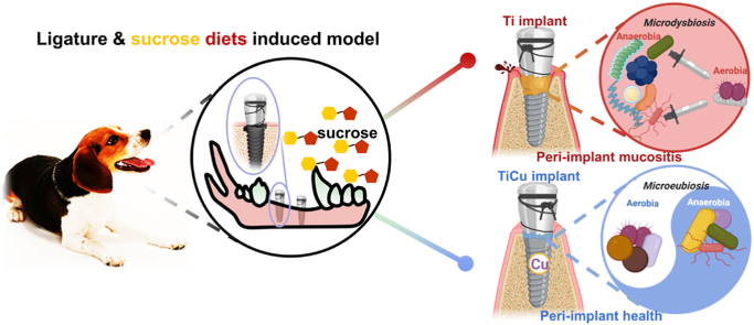
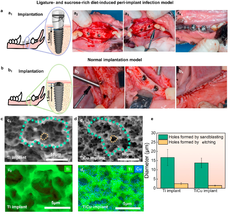
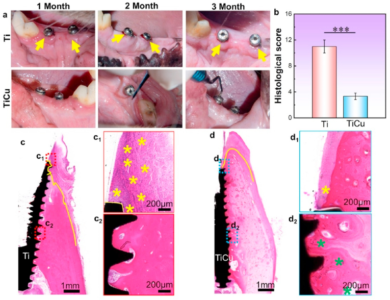
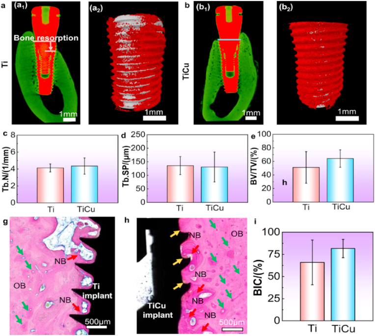
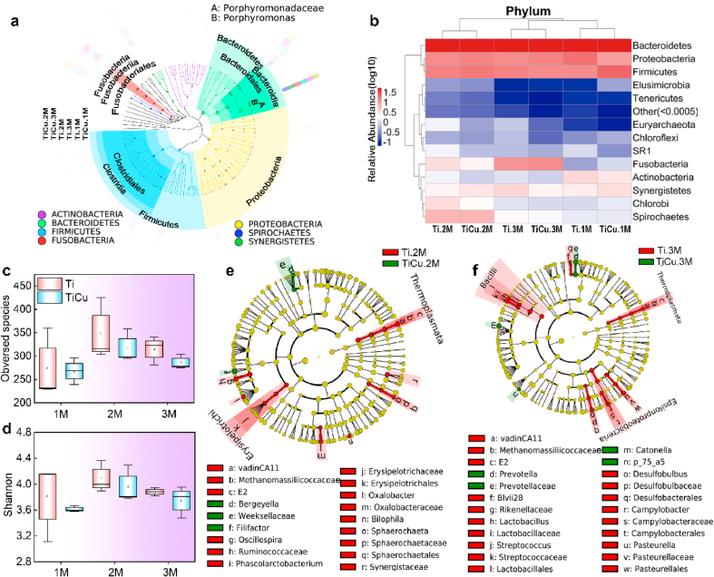
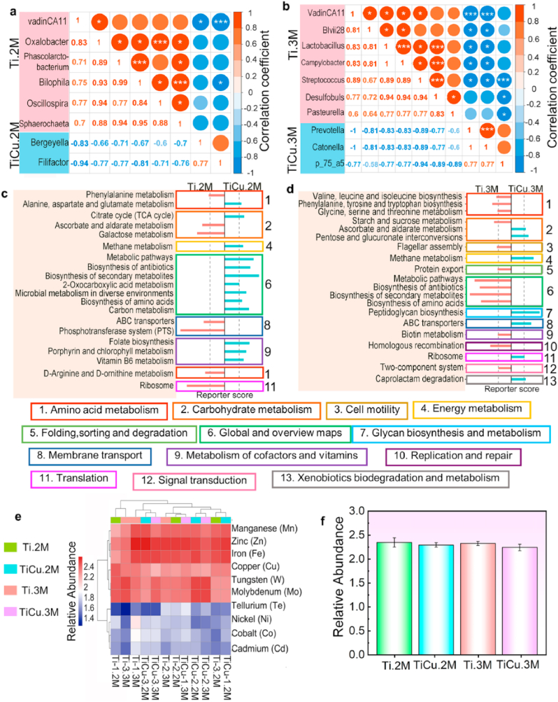
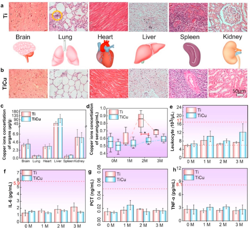
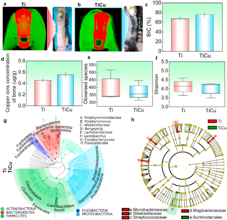
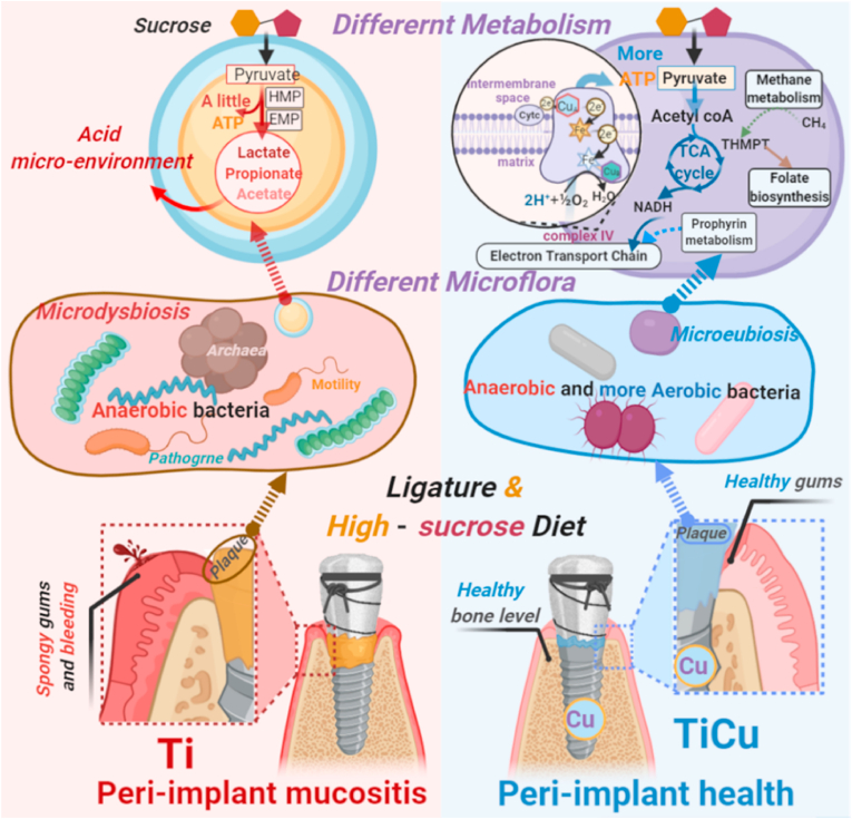
This work was focused on study of anti-infection ability and its underlying mechanism of a novel dental implant made of titanium-copper (TiCu) alloy. In general, most studies on antibacterial implants have used a single pathogen to test their anti-infection ability using infectious animal models. However, dental implant-associated infections are polymicrobial diseases. We innovatively combine the classic ligature model in dogs with sucrose-rich diets to induce oral infections via the canine native oral bacteria. The anti-infection ability, biocompatibility and underlying mechanism of TiCu implant were systematically investigated in comparison with pure Ti implant via general inspection, hematology, imageology (micro-CT), microbiology (16S rDNA and metagenome), histology, and Cu ion detections. Compared with Ti implant, TiCu implant demonstrated remarkable anti-infection potentials with excellent biocompatibility. Additionally, the underlying anti-infection mechanism of TiCu implant was considered to involve maintaining the oral microbiota homeostasis. It was found that the carbohydrates in the plaques formed on the surface of TiCu implant were metabolized through the tricarboxylic acid cycle (TCA) cycles, which prevented the formation of an acidic microenvironment and inhibited the accumulation of acidogens and pathogens, thereby maintaining the microflora balance between aerobic and anaerobic bacteria.

Keywords: Anti-infection; Biosafety; Oral microbiology; Titanium-copper alloy implants.


Conflict of interest statement

The authors declare that they have no known competing financial interests or personal relationships that could have appeared to influence the work reported in this paper.

Figures

Image 1
Graphical abstract
Fig. 1
Fig. 1
Implantation surgical procedures and surface characters of implants. (a) For the animals in ligature and sucrose-rich diet-induced peri-implant infection model, four same implants were inserted into the dog mandible (a1 and a2, two implants/side), the healing abutments were applied with ligatures (a3), and then the flaps sutured through the abutments(a4), these animals were then fed on sucrose-rich diets. (b) In normal implantation model, four same implants were also inserted into the mandible (b1 and b2, two implants/side), cover screws were used after implantations(b3), the flaps were sutured over the screws and implants(b4), and these animals were fed normal diets. (c1) and (d1) Surface morphologies of Ti and TiCu implants, green rings indicate the holes formed by sandblasting treatment and the yellow rings show the holes formed by acid etching treatment. (c2) and (d2) EDS surface scanning images of Ti and TiCu implants, respectively. (e) Diameters of holes on the implant surfaces. (n = 3 per group) [Data are mean ± s.d.].
Fig. 2
Fig. 2
Anti-inflammatory analysis of Ti and TiCu implants in the infection model. (a) Visual inspections of gingival tissues surrounding Ti or TiCu implants, yellow arrows showing swelling and bleeding of the gums. (b) Statistical analysis of histological scores, ***P < 0.001. (c) and (d) H&E staining analysis of mandibles tissues around Ti or TiCu implants at 3 months. (c1, c2, d1, and d2) Local images of tissues surrounding the Ti or TiCu implants, yellow stars showing the inflammatory cells, and green stars showing the osteoblasts (n = 3 per group) [Data are mean ± s.d. and ***p < 0.001 compared with the pure Ti implant].
Fig. 3
Fig. 3
Osseointegration analysis of Ti and TiCu implants in the infection model. (a1) and (b1) micro-CT images in the buccal-glossal direction, green color reflecting bone density. (a2) and (b2) enlarged images of Ti and TiCu groups, grey representing the implant, and red representing the bone tissues. Quantitative analysis of (c) trabecular number, (d) trabecular separation, (e) bone volume/tissue volume. (g) and (h)histological observations of the bone tissues around Ti and TiCu implants. OB: original bone; NB: newly formed bone marked by red arrows; yellow arrows show the hydroxyapatite layers, and green arrows show the Haversian system. (i) Bone implant contact rate (n = 3 per group) [Data are mean ± s.d. and *p < 0.05 compared with the pure Ti implant].
Fig. 4
Fig. 4
Dental plaque microbiome compositions in the infection model. (a) Species composition tree, colorful circles showing the richest phyla in all samples and colorful peripheral rings representing the relative abundance of taxonomic microflora of Ti and TiCu groups from 1 M to 3 M. (b) Heatmap analysis at the phylum level, “Others” representing the sum of species whose abundance was less than 0.5% in all samples. Alpha diversity analyses of (c) Observed species and (d) Shannon indexes. (e) and (f) LEfSe analysis of microbiome profiles up to the genus level at 2 M and 3 M, respectively (n = 3 per group) [Data are mean ± s.d.].
Fig. 5
Fig. 5
Metagenomics analysis in the infection model. (a) and (b) correlation analysis of different genera at 2 M and 3 M, red circles showing positive correlations, and blue circles showing negative relationships. (c) and (f) Different metabolisms at 2 M and 3 M, pink and blue bars representing metabolism from the plaque on the Ti (scores < −1.65) and TiCu implants (scores > 1.65). (e) Heatmap analysis of metal resistance genes (top 10). (f) Relative abundance of Cu resistance gene (n = 3 per group) [Data are mean ± s.d.].
Fig. 6
Fig. 6
Biosafety analysis in the infection model. (a) and (b) Histopathological analysis of major organs in animals with the Ti and TiCu implants, respectively. (c) Copper content in the major organs. (d) Copper content in the serum at each month. (e) Leukocytes. (f–h) Quantitative analysis of cytokines, including interleukin-6, tumor necrosis factor-α, and procalcitonin (n = 3 per group) [Data are mean ± s.d. and *p < 0.05 compared with the pure Ti implant].
Fig. 7
Fig. 7
Analysis in the normal implantation model. (a) and (b) Visual study and micro-CT images in animals with Ti and TiCu implants. (c) Quantitative analysis of BIC ratio. (d) Copper content in peri-implant bones. (e) Observed species analysis. (f) Shannon analysis. (g) Species composition tree of saliva samples. (h) LEfSe analysis of saliva microbiome profiles up to the genus level (n = 3 per group) [Data are mean ± s.d.].
Fig. 8
Fig. 8
Schematic representation for anti-infective mechanism of TiCu implant in the ligature and sucrose-rich diet-induced model. The microflora in animals with Ti implant metabolized the sucrose to acidify the environment and disturb the plaque microflora, and this microdysbiosis resulted in the peri-implant mucositis to occur. While the microflora on the TiCu implant sustained a metabolic balance, avoiding the acid plaque and maintaining the microflora balance of aerobic and anaerobic bacteria. TiCu implant preserved healthy peri-implant tissues. This diagram was created with BioRender.com.

References

    1. Jayachandran S., Walmsley A.D., Hill K. Challenges in dental implant provision and its management in general dental practice. J. Dent. 2020;99:103414. doi: 10.1016/j.jdent.2020.103414. - DOI - PubMed
    1. Andersen O.Z., Offermanns V., Sillassen M., Almtoft K.P., Andersen I.H., Sørensen S., Jeppesen C.S., Kraft D.C.E., Bøttiger J., Rasse M., Kloss F., Foss M. Accelerated bone ingrowth by local delivery of strontium from surface functionalized titanium implants. Biomaterials. 2013;24:5883–5890. doi: 10.1016/j.biomaterials.2013.04.031. - DOI - PubMed
    1. Lee C.T., Huang Y.W., Zhu L., Weltman R. Prevalences of peri-implantitis and peri-implant mucositis: systematic review and meta-analysis. J. Dent. 2017;62:1–12. doi: 10.1016/j.jdent.2017.04.011. - DOI - PubMed
    1. Subbiahdoss G., Kuijer R., Grijpma D.W., van der Mei H.C., Busscher H.J. Microbial biofilm growth vs. tissue integration: “The race for the surface” experimentally studied. Acta Biomater. 2009;5:1399–1404. doi: 10.1016/j.actbio.2008.12.011. - DOI - PubMed
    1. Burghardt I., Lüthen F., Prinz C., Kreikemeyer B., Zietz C., Neumann H.-G., Rychly J. A dual function of copper in designing regenerative implants. Biomaterials. 2015;44:36–44. doi: 10.1016/j.biomaterials.2014.12.022. - DOI - PubMed

LinkOut - more resources