. 2021 Mar;113(2):e24184.
doi: 10.1002/pep2.24184. Epub 2020 Jul 28.

Fluorinated peptide biomaterials

Affiliations

Fluorinated peptide biomaterials

Janna N Sloand et al. Pept Sci (Hoboken). 2021 Mar.

Abstract

Fluorinated compounds, while rarely used by nature, are emerging as fundamental ingredients in biomedical research, with applications in drug discovery, metabolomics, biospectroscopy, and, as the focus of this review, peptide/protein engineering. Leveraging the fluorous effect to direct peptide assembly has evolved an entirely new class of organofluorine building blocks from which unique and bioactive materials can be constructed. Here, we discuss three distinct peptide fluorination strategies used to design and induce peptide assembly into nano-, micro-, and macrosupramolecular states that potentiate high-ordered organization into material scaffolds. These fluorine-tailored peptide assemblies employ the unique fluorous environment to boost biofunctionality for a broad range of applications, from drug delivery to antibacterial coatings. This review provides foundational tactics for peptide fluorination and discusses the utility of these fluorous-directed hierarchical structures as material platforms in diverse biomedical applications.

Keywords: biomaterials; fluorine biochemistry; peptides; supramolecular assembly.


Conflict of interest statement

CONFLICT OF INTEREST The authors declare they have no conflict of interest.

Figures

FIGURE 1
FIGURE 1
De novo peptides (blue) incorporating fluorinated substitutes (green) access diverse supramolecular morphologies, including ribbons, fibrils, tubes and particles (shown in order from left to right). The unique assembly phenomena that underly these higher ordered structural states are driven by fluorine-fluorine interactions that occur both intra- and inter-molecularly
FIGURE 2
FIGURE 2
Left, fluorinated amino acids can be readily incorporated into non-natural sequences via solid-phase techniques. Examples of commonly employed fluorine-containing residues are shown. Middle, fluoroalkyl appendages ligated to the peptide terminus can drive unique assembly phenomena of natural sequences. Right, backbone fluorination of cyclic peptides can improve cyclisation efficiency, allow for tailoring of molecular conformations, and enhance self-assembly via the fluorous effect
FIGURE 3
FIGURE 3
Co-assembly of dodecanoic acid and peptide amphiphiles (PA), which contain F5-Phe (Z), residues in different positions adjacent to the assembly domain (Z1-Z3), produce divergent supramolecular structures dependent on fluorine placement and microenvironment. A-C, Cryo-TEM images of mixtures of PA Z1, PA Z2, and PA Z3 with dodecanoic acid [DA] in a molar ratio of 1:0.4, and E-G, conventional TEM images of mixtures in a 1:1 M ratio without staining (schematic illustrations of the morphologies are shown in the insets). DLS data of the vesicles formed by PA Z1 and dodecanoic acid in 1:1 M ratio are shown in the bottom inset in E. D,H, SAXS profiles of the DA-PA aqueous solutions in molar ratios of 1:0.4, D and 1:1, H. The profiles plot scattered intensity vs the scattering vector q (log-log plot); scattering intensities are offset vertically for clarity, and the fitting curves for the scattering data are shown in black in D. Reprinted with permission from J. Am. Chem. Soc. 2017, 139, 7823. Copyright © 2017 American Chemical Society
FIGURE 4
FIGURE 4
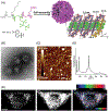
Fluoropeptoid assembly yields bioactive crystalline nanoflowers. A, Peptoid structures and the schematic representation showing the self-assembly of these amphiphilic peptoids into crystalline nanoflower-like particles composed of bilayer-like packing of peptoids, in which the peptoid backbone to backbone distance is 4.6 Å along the x-direction and is 1.6 nm along the y-direction; peptoids were highlighted with elements in various colors (nitrogen, blue; oxygen, red; carbon, gray; fluorine, green). B, TEM image of FPC, followed by negative staining. Scale bar: 100 nm. C, AFM image of FPC. D, X-ray diffraction data of FPC. E, Cell incubated for 1 hour with FPC (100 nM). Scale bars: 10 μm. Reprinted with permission from Small 2018, 14, 1803544. Copyright © 2018 Wiley-VCH
FIGURE 5
FIGURE 5
A, Linear structure of the fluorinated 21-residue peptide LX2, with 15L-leucines (L) and 6L-2, 2, 2-trifluoroethylglycine (F); B, Axial wheel projection illustrating the amphiphilic fluorous/hydrophobic faces of LX2 α-helix; C, Schematic representation of a tetrameric arrangement of LX2, showing in green the fluorinated faces of the fourhelix bundle, and in red the hydrophobic contours. Reprinted with permission from PLoS ONE, 2016, 11(11) Copyright © 2016 PLOS ONE
FIGURE 6
FIGURE 6
Fluorination enhances mucus penetration of polypeptide/siRNA polyplexes. A, Apparent permeability (Papp) of polypeptide/Cy3-siRNA polyplexes across the air-interface culture of Calu-3 cells. B, Representative trajectories of polyplexes in cystic fibrosis (CF) mucus. C, Mean squared displacement (MSD) of polyplexes as a function of the time scale (τ). D, Distribution of the logarithmic MSD of an individual polyplex at τ = 1 second. E, Fluorescence emission spectra of Cy3-siRNA, Cy5-polypeptide, and Cy5-polypeptide/Cy3-siRNA polyplexes after incubation with 5% CF mucus for 0 or 4 hours. F, Fluorescence intensity of the aggregates between mucin (0.3% or 0.5%) and polypeptide/Cy3-siRNA polyplexes following 4-hours incubation. G, Distribution of PG1/Cy3-siRNA, P3F16/Cy3-siRNA, and P7F7/Cy3-siRNA polyplexes in lung epithelial tissues following intratracheal administration (scale bar = 75 μm). Reprinted with permission from Nano Lett. 2020, 20, 1738. Copyright © 2020 American Chemical Society
FIGURE 7
FIGURE 7
A, 19F-MRI signals of C7-Glu2 and C7-Glu3 (2 mM) upon titration of CaCl2. B, Signal reduction is observed for C7-Glu2 and C7-Glu3 in response to decreasing Ca2+ concentrations. Each image is 1.8 × 1.8 cm2. In the figure C7-Glu2 and C7-Glu3 are labeled as C7E2 and C7E3, respectively. Adapted with permission from ACS Appl. Mater. Interfaces 2017, 9, 46, 39890. Copyright © 2017 American Chemical Society
FIGURE 8
FIGURE 8
Ultrasound-guided cytosolic protein delivery from fluoropeptide nanoemulsions (NE). A, Doppler ultrasound imaging (5 MHz) of NE vaporization and inertial cavitation of microbubbles as the acoustic pressure is increased. No signal is observed at pressures below the particle's inertial threshold (~0.9 MPa). Cavitation above this pressure threshold is observed as transient Doppler twinkling (1.0 MPa image shown as stacked twinkling events collected over a 40 seconds interval). B, Mean intracellular fluorescence (in relative fluorescence units; RFU) of A549 cells following delivery of labeled phalloidin from NEs at varying US intensity and duty cycle (DC). C, Live-cell image showing delivery of phalloidin (green) from NEs, spatially resolved to a circular area of the A549 cell monolayer subjected to ultrasound. Cell nuclei are stained blue, scale bar = 200 μm. D, Live A549 cells stained with the endosomal marker transferrin (red) following NE mediated delivery of labeled phalloidin. Scale bar = 80 μm. E, Immunofluorescent micrographs of squamous epidermal tumor sections 5 days after administration of antibody (IgG)-loaded NE without (−US) and with (+US; 2 W/cm2) application of the ultrasound trigger (scale bar = 1 mm). ROI magnification (white dashed square) demonstrating intracellular delivery of antibody following US activation of NEIgG particles (scale bar = 10 μm). F, Relative intracellular fluorescence (in relative fluorescence units) of squamous tumor cells isolated from InvtTA × tetORas mice following treatment with NEIgG particles ± the ultrasound trigger. B-D, Adapted with permission from Angew. Chem. Int. Ed. 2017, 56, 11404. Copyright © 2017 Wiley-VCH. A, E-F, Adapted with permission from ACS Nano 2020, 14, 4, 4061. Copyright © 2020 American Chemical Society
FIGURE 9
FIGURE 9
Antibacterial capabilities and biocompatibility of the enhanced resin composite restoratives. A, Bacterial growth inhibition kinetics evaluated by turbidity analysis following direct contact of S mutans bacteria with the Fmoc-F5-Phe-incorporated restoratives for 1 hour. B, Bacterial viability evaluation following direct contact analysis with either the control composite restorative (Filtek resin) or Fmoc-F5-Phe nanoassembly-incorporated resin composite restorative using the Live/Dead backlight bacterial viability kit. C,D, 3-(4,5-Dimethylthiazolyl-2)-2,5-diphenyltetrazolium bromide (MTT) cell viability analysis. The cytotoxicity of the Fmoc-F5-Phe-incorporated restoratives toward (C) 3 T3 fibroblast and D, HeLa cells was evaluated by the MTT assay at 2 w/w %, as well as control restoratives treated in the same manner that were not incorporated with nanoassemblies, E,F, Mammalian cell viability utilizing a fluorescent Live/Dead staining assay containing fluorescein diacetate (live cells) and propidium iodide (dead cells). E, 3T3 fibroblasts and F, HeLa cells. The scale bar is 500 μm. Reprinted with permission from ACS Appl. Mater. Interfaces 2019, 11, 24, 21 334. Copyright © 2019 American Chemical Society
FIGURE 10
FIGURE 10
Ion transport of fluorinated sugar amino acid derived α,γ-cyclic hexapeptides (2.5 μM) across model membranes (EYPCLUVs⊃HPTS) determined for varying cations, A, and anions, B, in the extravesicular buffer. Reprinted with permission from Org. Lett. 2017, 19, 21, 5948. Copyright © 2017 American Chemical Society

References

    1. Tao K, Makam P, Aizen R, Gazit E, Science 2017, 358(6365), eaam9756. - PMC - PubMed
    1. Nguyen V, Zhu R, Jenkins K, Yang R, Nat. Commun 2016, 7(1), 1. - PMC - PubMed
    1. Lee JS, Yoon I, Kim J, Ihee H, Kim B, Park CB, Angew. Chem. Int. Ed 2011, 50(5), 1164. - PubMed
    1. Makam P, Gazit E, Chem. Soc. Rev 2018, 47(10), 3406. - PMC - PubMed
    1. Lee J-H, Heo K, Schulz-Schönhagen K, Lee JH, Desai MS, Jin H-E, Lee S-W, ACS Nano. 2018, 12(8), 8138. - PubMed

LinkOut - more resources