Cerebellar Activation Deficits in Schizophrenia During an Eyeblink Conditioning Task
- PMID: 34541537
- PMCID: PMC8443466
- DOI: 10.1093/schizbullopen/sgab040
Cerebellar Activation Deficits in Schizophrenia During an Eyeblink Conditioning Task
Abstract
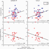
The cognitive dysmetria theory of psychotic disorders posits that cerebellar circuit abnormalities give rise to difficulties coordinating motor and cognitive functions. However, brain activation during cerebellar-mediated tasks is understudied in schizophrenia. Accordingly, this study examined whether individuals with schizophrenia have diminished neural activation compared to controls in key regions of the delay eyeblink conditioning (dEBC) cerebellar circuit (eg, lobule VI) and cerebellar regions associated with cognition (eg, Crus I). Participants with schizophrenia-spectrum disorders (n = 31) and healthy controls (n = 43) underwent dEBC during functional magnetic resonance imaging (fMRI). Images were normalized using the Spatially Unbiased Infratentorial Template (SUIT) of the cerebellum and brainstem. Activation contrasts of interest were "early" and "late" stages of paired tone and air puff trials minus unpaired trials. Preliminary whole brain analyses were conducted, followed by cerebellar-specific SUIT and region of interest (ROI) analyses of lobule VI and Crus I. Correlation analyses were conducted between cerebellar activation, neuropsychological test scores, and psychotic symptom scores. In controls, the largest clusters of cerebellar activation peaked in lobule VI during early dEBC and Crus I during late dEBC. The schizophrenia group showed robust cortical activation to unpaired trials but no significant conditioning-related cerebellar activation. Crus I ROI activation during late dEBC was greater in the control than schizophrenia group. Greater Crus I activation correlated with higher working memory scores in the full sample and lower positive psychotic symptom severity in schizophrenia. Findings indicate functional cerebellar abnormalities in schizophrenia which relate to psychotic symptoms, lending direct support to the cognitive dysmetria framework.
Keywords: associative learning; cerebellum; functional magnetic resonance imaging; psychosis.
© The Author(s) 2021. Published by Oxford University Press on behalf of the University of Maryland's school of medicine, Maryland Psychiatric Research Center.
Figures


References
-
- Andreasen NC, Paradiso S, O’Leary DS. “Cognitive dysmetria” as an integrative theory of schizophrenia: a dysfunction in cortical-subcortical-cerebellar circuitry? Schizophr Bull. 1998;24:203–218. - PubMed
-
- Chua SE, Cheung C, Cheung V, et al. . Cerebral grey, white matter and csf in never-medicated, first-episode schizophrenia. Schizophr Res. 2007;89:12–21. - PubMed
-
- Kühn S, Romanowski A, Schubert F, Gallinat J. Reduction of cerebellar grey matter in Crus I and II in schizophrenia. Brain Struct Funct. 2012;217:523–529. - PubMed
-
- McDonald C, Bullmore E, Sham P, et al. . Regional volume deviations of brain structure in schizophrenia and psychotic bipolar disorder: computational morphometry study. Br J Psychiatry 2005;186:369–377. - PubMed
Grants and funding
LinkOut - more resources
Full Text Sources

