

Diffusion of Protein Molecules through Microporous Nanofibrous Polyacrylonitrile Membranes
- PMID: 34541542
- PMCID: PMC8445001
- DOI: 10.1021/acsapm.0c01394
Diffusion of Protein Molecules through Microporous Nanofibrous Polyacrylonitrile Membranes
Abstract
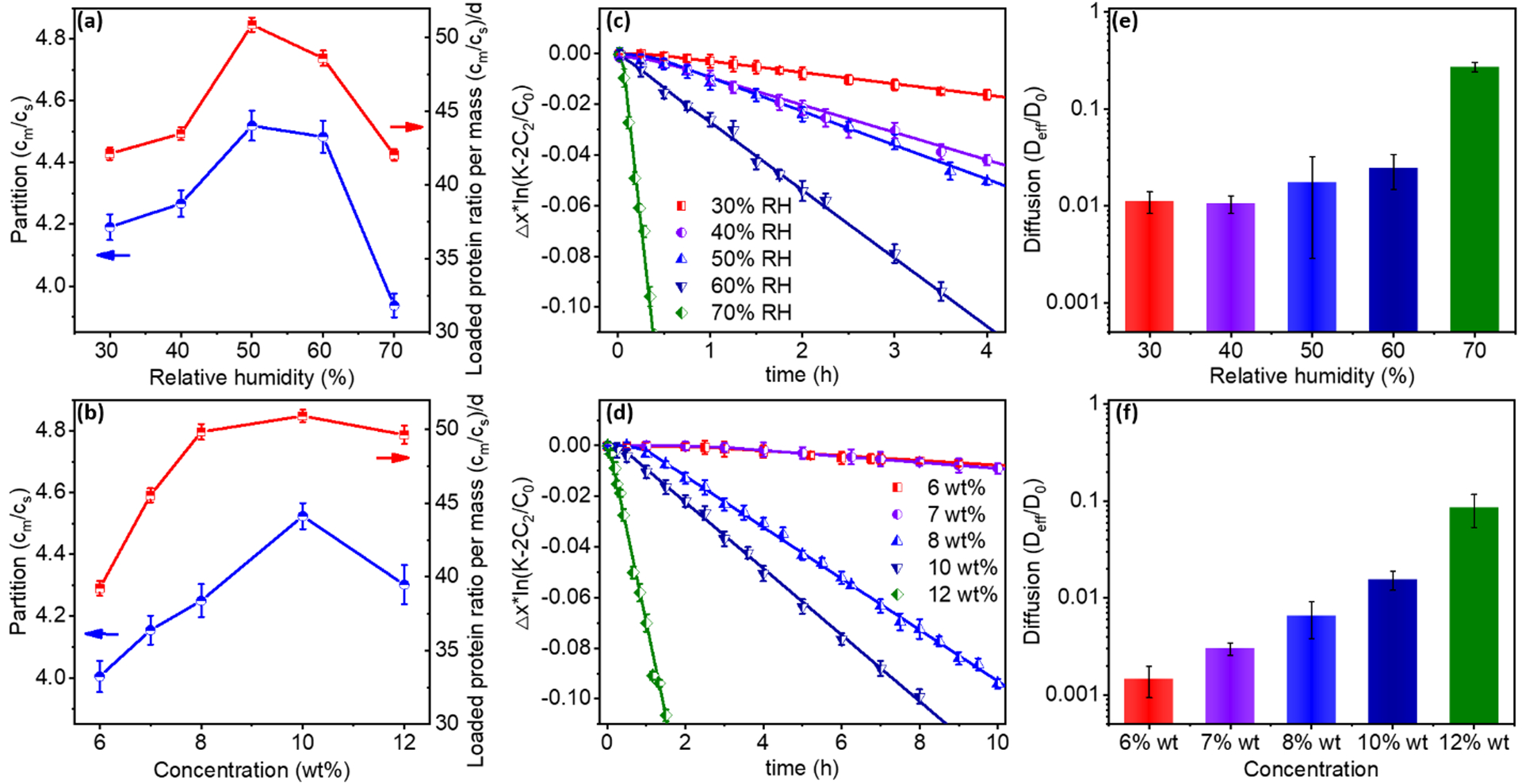
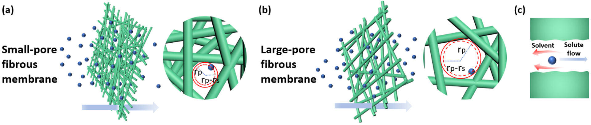
Porous nanofibrous membranes have ultrahigh specific surface areas and could be broadly employed in protein purification, enzyme immobilization, and biosensors with enhanced selectivity, sensitivity, and efficiency. However, large biomolecules, such as proteins, have hindered diffusion behavior in the micro-porous media, significantly reducing the benefits provided by the nanofibrous membranes. The study of protein diffusion in polyacrylonitrile (PAN) nanofibrous membranes produced under varied humidity and polymer concentration of electrospinning revealed that heterogeneous structures of the nanofibrous membranes possess much smaller effective pore sizes than the measured pore sizes, which significantly affects the diffusion of large molecules through the system though sizes of proteins and pH conditions also have great impacts. Only when the measured membrane pore size is at least 1000 times higher than the protein size, the diffusion behavior of the protein is predictable in the system. The results provide insights into the design and applications of proper nanofibrous materials for improved applications in protein purification and immobilizations.
Keywords: Micro-porous structure; Nanofibrous membrane; Protein diffusion; Protein-polymer interaction; diffusion coefficient; effective pore size; partition coefficient.
Figures


References
-
- Sundarrajan S; Tan KL; Lim SH; Ramakrishna S, Electrospun nanofibers for air filtration applications. Procedia Eng. 2014, 75, 159–163.
-
- Ge J; Zong D; Jin Q; Yu J; Ding B, Biomimetic and superwettable nanofibrous skins for highly efficient separation of oil‐in‐water emulsions. Adv. Funct. Mater 2018, 28 (10), 1705051.
-
- Zhu J; Sun G, Facile fabrication of hydrophilic nanofibrous membranes with an immobilized metal–chelate affinity complex for selective protein separation. ACS Appl. Mater. Interfaces 2014, 6 (2), 925–932. - PubMed
-
- Tang P; Zhang M; Ji B; Yong T; Sun G, Hierarchical Nucleophilic Nanofibrous Membranes for Fast, Durable, and Bare‐Eye Visible Detoxification of Carcinogenic Alkylating Toxicants. Adv. Funct. Mater 2019, 29 (50), 1905990.
Grants and funding
LinkOut - more resources
Full Text Sources
Other Literature Sources
