. 2022 Feb;240(2):253-267.
doi: 10.1111/joa.13551. Epub 2021 Sep 19.

Phenotypic regionalization of the vertebral column in the thorny skate Amblyraja radiata: Stability and variation

Affiliations

Phenotypic regionalization of the vertebral column in the thorny skate Amblyraja radiata: Stability and variation

Fidji Berio et al. J Anat. 2022 Feb.

Abstract

Regionalization of the vertebral column occurred early during vertebrate evolution and has been extensively investigated in mammals. However, less data are available on vertebral regions of crown gnathostomes. This is particularly true for batoids (skates, sawfishes, guitarfishes, and rays) whose vertebral column has long been considered to be composed of the same two regions as in teleost fishes despite the presence of a synarcual. However, the numerous vertebral units in chondrichthyans may display a more complex regionalization pattern than previously assumed and the intraspecific variation of such pattern deserves a thorough investigation. In this study, we use micro-computed tomography (µCT) scans of vertebral columns of a growth series of thorny skates Amblyraja radiata to provide the first fine-scale morphological description of vertebral units in a batoids species. We further investigate axial regionalization using a replicable clustering analysis on presence/absence of vertebral elements to decipher the regionalization of the vertebral column of A. radiata. We identify four vertebral regions in this species. The two anteriormost regions, named synarcual and thoracic, may undergo strong developmental or functional constraints because they display stable patterns of shapes and numbers of vertebral units across all growth stages. The third region, named hemal transitional, is characterized by high inter-individual morphological variation and displays a transition between the monospondylous (one centrum per somite) to diplospondylous (two centra per somite) conditions. The posteriormost region, named caudal, is subdivided into three sub-regions with shapes changing gradually along the anteroposterior axis. These regionalized patterns are discussed in light of ecological habits of skates.

Keywords: Amblyraja radiata; axial regionalization; elasmobranchs; evolution; vertebral column.


Conflict of interest statement

The authors declare that they have no conflicts of interests.

Figures

FIGURE 1
FIGURE 1
Schematics summarizing the 13 morphological vertebral characteristics used to describe vertebral units in Amblyraja radiata; two consecutive vertebral units are represented: n (anterior) and n + 1 (posterior). bd, basidorsal; bv, basiventral; ce, centrum; df, dorsal foramen; hs, hemal spine; id, interdorsal; ns, neural spine; tp, transverse process; vf, ventral foramen. Dotted lines in (6) indicate the lateral complete or partial fusion of centrum and interdorsal
FIGURE 2
FIGURE 2
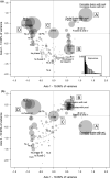
Correspondence analysis based on the presence/absence of vertebral elements in Amblyraja radiata. Data from 21 specimens for a total of n = 2935 vertebral units. Axes 1 and 2 (a) and axes 1 and 3 (b). The size of the circles is proportional to the relative n. ant, anterior centrum; C, centrum; df, dorsal foramen; hs, haemal spine; hs.a to hs.e, haemal spine of shape a to e; id, interdorsal; iv, interventral; ns, neural spine; ns.a to d, neural spine of shape a to d; PA, presence/absence; post, posterior centrum; tp, transverse process; tp.a to tp.c, transverse process of shape a to c; vf, ventral foramen
FIGURE 3
FIGURE 3
Partition Around Medoids (PAM) clustering for 21 vertebral columns of a growth series of Amblyraja radiata. For each specimen, a Dice similarity matrix based on presence/absence data is used by the PAM algorithm. The number of clusters is k = 5. The two anteriormost clusters are less variable in terms of shape and number of vertebral units than the posterior ones. The hemal transitional region displays inter‐individual high variability and the caudal region is divided into three sub‐regions [Colour figure can be viewed at wileyonlinelibrary.com]
FIGURE 4
FIGURE 4
Vertebral column of Amblyraja radiata (13.8 cm in DW) displaying the axial regionalization pattern. (a) Lateral view of the whole vertebral column. (b) Vertebral unit of the cervical region (synarcual). (c) Vertebral unit of the thoracic region. (d) Vertebral unit of the hemal transitional region. (e) Vertebral unit of the anterior caudal region. (f) Vertebral unit of the posterior caudal region. (a‐f) are camera lucida drawings. The tesserae are the round blocks covering B‐E. bd, basidorsal; bv, basiventral; ce, centrum; df, dorsal foramen; et, elongated tesserae; hf, haemal foramen; hs, hemal spine; id, interdorsal; ns, neural spine; s‐r, sub‐region; t, tessera; tb, tesseral bridge; tp, transverse process; vf, ventral foramen. Scale bars = 0.5 cm for a, 1 mm for b‐e, and 0.5 mm for f
FIGURE 5
FIGURE 5
Synarcual of Amblyraja radiata (13.8 cm in DW) showing the anterior complete fusion of vertebral units and their posterior partial separation. Camera lucida drawing. lsd, distal portion of lateral stay; pa, pectoral arch; pfvu, partial fusion of vertebral units; tfvu, total fusion of vertebral units. lsd and pa named after Claeson (2011). Scale bar = 0.5 cm
FIGURE 6
FIGURE 6
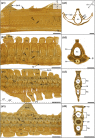
Micro‐CT images of vertebrae from the different regions in the vertebral column of Amblyraja radiata (16.8 cm in DW). (a) Cervical vertebrae (synarcual). (b) Thoracic vertebrae. (c) Hemal transitional vertebrae. (d) Caudal vertebrae, second subregion. (a1, b1, c1, and d1) Lateral views. (a2, b2, c2, and d2) Transverse sections located by double‐headed arrows in a1, b1, c1, and d1, respectively. A, anterior; bd, basidorsal; bv, basiventral; ce, centrum; dent., dermal denticle; D, dorsal; df, dorsal foramen; f, foramen on the posteriormost transverse process; ha, hemal arch; hc, hemal canal; hs, hemal spine; id, interdorsal; L, left; lsd, distal portion of lateral stay; na, neural arch; nc, neural canal; ns, neural spine; ns‐id, neural spine‐interdorsal complex; P, posterior; R, right; st, subtype; tb, tesseral bridge; tp, transverse process; tz, transitional zone; V, ventral; vf, ventral foramen. Scale bars = 100 µm [Colour figure can be viewed at wileyonlinelibrary.com]

References

    1. Arratia, G. , Schultze, H.‐P. & Casciotta, J. (2001) Vertebral column and associated elements in dipnoans and comparison with other fishes: development and homology. Journal of Morphology, 250(2), 101–172. - PubMed
    1. Aschliman, N.C. , Claeson, K.M. & McEachran, J.D. (2012) Phylogeny of batoidea. In: Carrier, J.C. , Musick, J.A. & Heithaus, M.R. (Eds.) Biology of sharks and their relatives, 2nd edition. Boca Raton: CRC Press, pp. 57–95.
    1. Berio, F. , Broyon, M. , Enault, S. , Pirot, N. , López‐Romero, F.A. & Debiais‐Thibaud, M. (2021) Diversity and evolution of mineralized skeletal tissues in chondrichthyans. Frontiers in Ecology and Evolution, 9, 223.
    1. Bird, N.C. & Mabee, P.M. (2003) Developmental morphology of the axial skeleton of the zebrafish, Danio rerio (Ostariophysi: Cyprinidae). Developmental Dynamics, 228(3), 337–357. - PubMed
    1. Boglione, C. , Pulcini, D. , Scardi, M. , Palamara, E. , Russo, T. & Cataudella, S. (2014) Skeletal anomaly monitoring in rainbow trout (Oncorhynchus mykiss, Walbaum 1792) reared under different conditions. PLoS One, 9(5), e96983. - PMC - PubMed

Publication types

LinkOut - more resources