Comparative Study
. 2021 Nov 1;78(11):1375-1382.
doi: 10.1001/jamaneurol.2021.3180.

Head-to-Head Comparison of 8 Plasma Amyloid-β 42/40 Assays in Alzheimer Disease

Affiliations
Comparative Study

Head-to-Head Comparison of 8 Plasma Amyloid-β 42/40 Assays in Alzheimer Disease

Shorena Janelidze et al. JAMA Neurol. .

Erratum in

  • Error in Figure.
    [No authors listed] [No authors listed] JAMA Neurol. 2023 Apr 1;80(4):422. doi: 10.1001/jamaneurol.2022.5184. JAMA Neurol. 2023. PMID: 36648932 Free PMC article. No abstract available.

Abstract

Importance: Blood-based tests for brain amyloid-β (Aβ) pathology are needed for widespread implementation of Alzheimer disease (AD) biomarkers in clinical care and to facilitate patient screening and monitoring of treatment responses in clinical trials.

Objective: To compare the performance of plasma Aβ42/40 measured using 8 different Aβ assays when detecting abnormal brain Aβ status in patients with early AD.

Design, setting, and participants: This study included 182 cognitively unimpaired participants and 104 patients with mild cognitive impairment from the BioFINDER cohort who were enrolled at 3 different hospitals in Sweden and underwent Aβ positron emission tomography (PET) imaging and cerebrospinal fluid (CSF) and plasma collection from 2010 to 2014. Plasma Aβ42/40 was measured using an immunoprecipitation-coupled mass spectrometry developed at Washington University (IP-MS-WashU), antibody-free liquid chromatography MS developed by Araclon (LC-MS-Arc), and immunoassays from Roche Diagnostics (IA-Elc); Euroimmun (IA-EI); and Amsterdam University Medical Center, ADx Neurosciences, and Quanterix (IA-N4PE). Plasma Aβ42/40 was also measured using an IP-MS-based method from Shimadzu in 200 participants (IP-MS-Shim) and an IP-MS-based method from the University of Gothenburg (IP-MS-UGOT) and another immunoassay from Quanterix (IA-Quan) among 227 participants. For validation, 122 participants (51 cognitively normal, 51 with mild cognitive impairment, and 20 with AD dementia) were included from the Alzheimer Disease Neuroimaging Initiative who underwent Aβ-PET and plasma Aβ assessments using IP-MS-WashU, IP-MS-Shim, IP-MS-UGOT, IA-Elc, IA-N4PE, and IA-Quan assays.

Main outcomes and measures: Discriminative accuracy of plasma Aβ42/40 quantified using 8 different assays for abnormal CSF Aβ42/40 and Aβ-PET status.

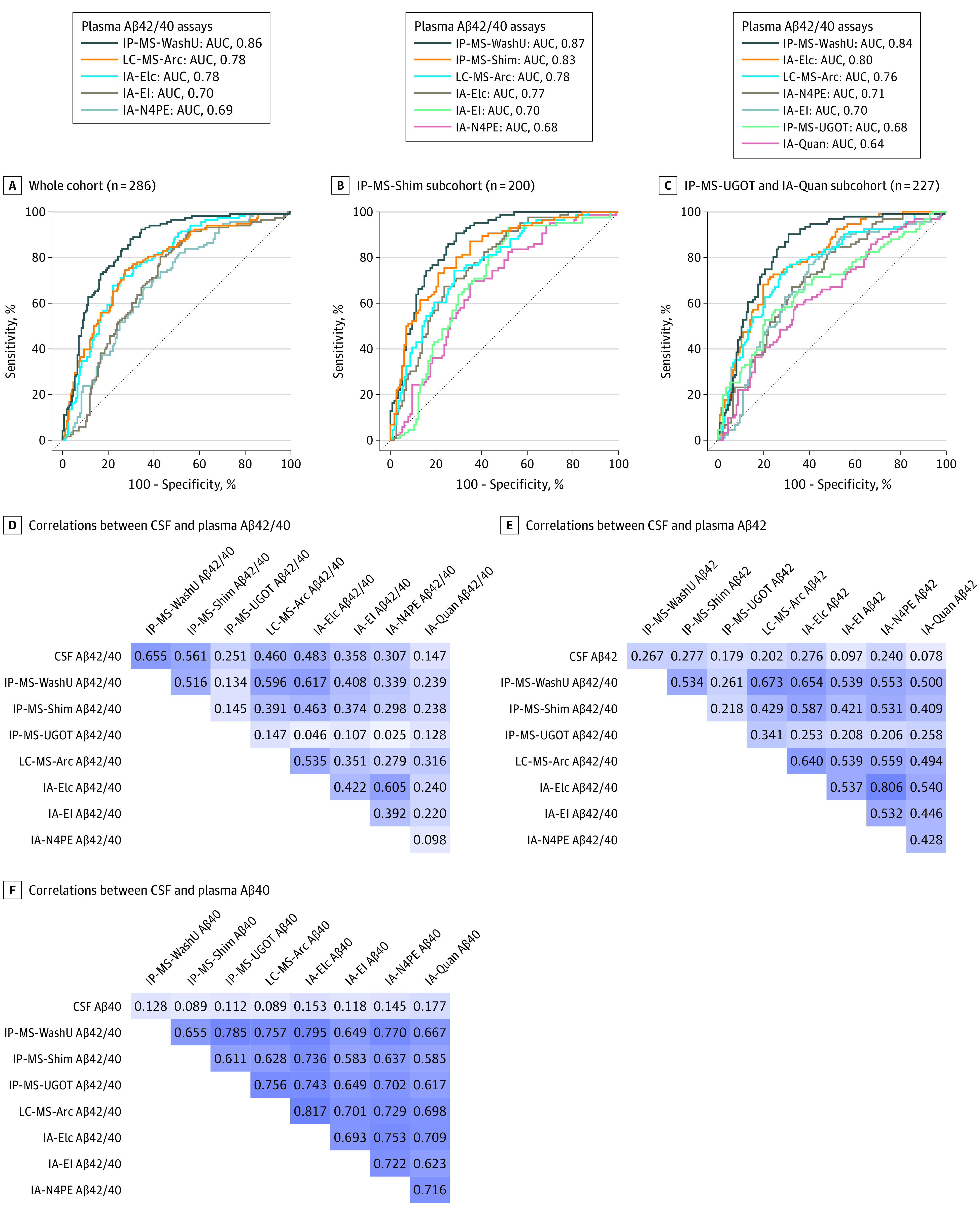
Results: A total of 408 participants were included in this study. In the BioFINDER cohort, the mean (SD) age was 71.6 (5.6) years and 49.3% of the cohort were women. When identifying participants with abnormal CSF Aβ42/40 in the whole cohort, plasma IP-MS-WashU Aβ42/40 showed significantly higher accuracy (area under the receiver operating characteristic curve [AUC], 0.86; 95% CI, 0.81-0.90) than LC-MS-Arc Aβ42/40, IA-Elc Aβ42/40, IA-EI Aβ42/40, and IA-N4PE Aβ42/40 (AUC range, 0.69-0.78; P < .05). Plasma IP-MS-WashU Aβ42/40 performed significantly better than IP-MS-UGOT Aβ42/40 and IA-Quan Aβ42/40 (AUC, 0.84 vs 0.68 and 0.64, respectively; P < .001), while there was no difference in the AUCs between IP-MS-WashU Aβ42/40 and IP-MS-Shim Aβ42/40 (0.87 vs 0.83; P = .16) in the 2 subcohorts where these biomarkers were available. The results were similar when using Aβ-PET as outcome. Plasma IPMS-WashU Aβ42/40 and IPMS-Shim Aβ42/40 showed highest coefficients for correlations with CSF Aβ42/40 (r range, 0.56-0.65). The BioFINDER results were replicated in the Alzheimer Disease Neuroimaging Initiative cohort (mean [SD] age, 72.4 [5.4] years; 43.4% women), where the IP-MS-WashU assay performed significantly better than the IP-MS-UGOT, IA-Elc, IA-N4PE, and IA-Quan assays but not the IP-MS-Shim assay.

Conclusions and relevance: The results from 2 independent cohorts indicate that certain MS-based methods performed better than most of the immunoassays for plasma Aβ42/40 when detecting brain Aβ pathology.


Conflict of interest statement

Conflict of Interest Disclosures: Dr Teunissen reports nonfinancial support from Quanterix during the conduct of the study; has a patent pending for glial fibrillary acidic protein as a biomarker; serves as an editorial board member of Alzheimer Research and Therapy, Medidact Neurologie, Springer, and Neurology: Neuroimmunology & Neuroinflammation; serves as an editor of Neuromethods Book Series, Springer; and reports grants from Roche Diagnostics, nonfinancial support from ADx Neurosciences and Eli Lilly, and compensation from ACImmune, Axon Neurosciences, Biogen, Brainstorm Cell Therapeutics, Celgene, Denali Therapeutics, EIP Pharma, Eisai, PeopleBio, Toyama Pharmaceutical Association, and Vivoryon Therapeutics outside the submitted work. Dr Zetterberg reports personal fees from Alector, AlzeCure Pharma, AZTherapies, Biogen, Biosplice Therapeutics, Cellectricon, Cognition Therapeutics, Denali Therapeutics, Eisai, Fujirebio Diagnostics, NervGen, Pinteon Therapeutics, Red Abbey Labs, Roche Diagnostics, Siemens Healthineers, and Wave Life Sciences outside the submitted work; and is a cofounder of Brain Biomarker Solutions in Gothenburg AB. Drs Allué and Sarasa report a patent pending for EP2020382352 and are employed at Araclon Biotech. Dr Eichenlaub is employed at Roche Diagnostics. Dr Bittner is employed at F. Hoffmann-La Roche, has a patent pending for blood-based biomarkers, and owns stock from F. Hoffmann-LaRoche. Mr Ovod has submitted the US provisional patent application “Plasma Based Methods for Detecting CNS Amyloid Deposition” as a coinventor and may receive royalty income based on technology (stable isotope labeling kinetics and blood plasma assay) licensed by Washington University to C2N Diagnostics. Dr Toba reports grants from Amedisys during the conduct of the study. Dr Nakamura reports grants from Amedisys during the conduct of the study. Dr Bateman reports consulting for Eisai and Janssen; personal fees from ACImmune, Amgen, C2N Diagnostics, and Pfizer; grants from AbbVie, Avid Radiopharmaceuticals, Biogen, Centene, Eisai, Eli Lilly, F. Hoffmann-LaRoche, Genentech, Janssen Pharmaceuticals, and United Neuroscience outside the submitted work; he is a member of the DIAN-TU Pharma Consortium (Eli Lilly and Company/Avid Radiopharmaceuticals, Hoffmann-La Roche/Genentech, Biogen, Eisai, Janssen, and United Neuroscience), the NFL Consortium (AbbVie, Biogen, Roche, BMS), and Tau SIK Consortium (AbbVie, Avid Radiopharmaceuticals, Biogen, Eli Lilly); has a patent for a plasma amyloid-beta test licensed to C2N Diagnostics; and cofounded C2N Diagnostics; in addition, Washington University and Dr. Bateman have equity ownership interest in C2N Diagnostics and receive royalty income based on blood plasma assay technology licensed by Washington University to C2N Diagnostics; Washington University and Dr Bateman have submitted the US provisional patent application “Plasma Based Methods for Detecting CNS Amyloid Deposition.” Dr Blennow reports personal fees from Abcam, Axon, Biogen, the Japanese Organization for Medical Device Development, Julius Clinical, Eli Lilly, MagQu, Novartis, Prothena, Roche Diagnostics, Shimadzu, and Siemens Healthineers and grants from the Swedish Research Council during the conduct of the study as well as research funding from Biogen, Eisai, and Roche Diagnostics and nonfinancial support from AC Immune and Eli Lilly outside the submitted work; and is a cofounder of Brain Biomarker Solutions in Gothenburg AB, which is a part of the GU Ventures Incubator Program. Dr Hansson reports research support paid to his institution from Avid Radiopharmaceuticals, Biogen, Eisai, Eli Lilly, General Electric Healthcare, Pfizer, and Roche Diagnostics and has received personal fees from AC Immune, Alzpath, Biogen, Cerveau, and Roche Diagnostics. No other disclosures were reported.

Figures

Figure.
Figure.. Receiver Operating Characteristic (ROC) Analysis for Abnormal Cerebrospinal Fluid (CSF) Amyloid-β42/40 (Aβ42/40) and Correlations Between CSF and Plasma Aβ
A, ROC curve analysis for differentiating participants with abnormal CSF Aβ42/40 from those with normal CSF Aβ42/40 (cutoff, 0.0597) in the whole cohort. B, ROC curve analysis in the subcohorts where IPMS-Shim Aβ42/40 was available. C, ROC curve analysis in the subcohorts where IPMS-UGOT and IA-Quan Aβ42/40 were available. D, Spearman correlations between plasma and CSF Aβ42/40 in a subcohort (n = 155) individuals where all plasma samples were analyzed using all 8 assays. E, Spearman correlations between plasma and CSF Aβ42 in a subcohort (n = 155) where all plasma samples were analyzed using all 8 assays. F, Spearman correlations between plasma and CSF Aβ40 in a subcohort (n = 155) where all plasma samples were analyzed using all 8 assays.

References

    1. Hansson O. Biomarkers for neurodegenerative diseases. Nat Med. 2021;27(6):954-963. doi:10.1038/s41591-021-01382-x - DOI - PubMed
    1. Olsson B, Lautner R, Andreasson U, et al. . CSF and blood biomarkers for the diagnosis of Alzheimer’s disease: a systematic review and meta-analysis. Lancet Neurol. 2016;15(7):673-684. doi:10.1016/S1474-4422(16)00070-3 - DOI - PubMed
    1. Janelidze S, Stomrud E, Palmqvist S, et al. . Plasma β-amyloid in Alzheimer’s disease and vascular disease. Sci Rep. 2016;6:26801. doi:10.1038/srep26801 - DOI - PMC - PubMed
    1. Nakamura A, Kaneko N, Villemagne VL, et al. . High performance plasma amyloid-β biomarkers for Alzheimer’s disease. Nature. 2018;554(7691):249-254. doi:10.1038/nature25456 - DOI - PubMed
    1. Ovod V, Ramsey KN, Mawuenyega KG, et al. . Amyloid β concentrations and stable isotope labeling kinetics of human plasma specific to central nervous system amyloidosis. Alzheimers Dement. 2017;13(8):841-849. doi:10.1016/j.jalz.2017.06.2266 - DOI - PMC - PubMed

Publication types