. 2022 Mar 2;225(5):912-923.
doi: 10.1093/infdis/jiab481.

Nasopharyngeal Codetection of Haemophilus influenzae and Streptococcus pneumoniae Shapes Respiratory Syncytial Virus Disease Outcomes in Children

Affiliations

Nasopharyngeal Codetection of Haemophilus influenzae and Streptococcus pneumoniae Shapes Respiratory Syncytial Virus Disease Outcomes in Children

Alejandro Diaz-Diaz et al. J Infect Dis. .

Abstract

Background: The role of nasopharyngeal bacteria in respiratory syncytial virus (RSV) disease has been underestimated. We measured the frequency and burden of respiratory bacteria in the upper respiratory tract of infants with RSV infection over 7 respiratory seasons, and their impact on clinical outcomes.

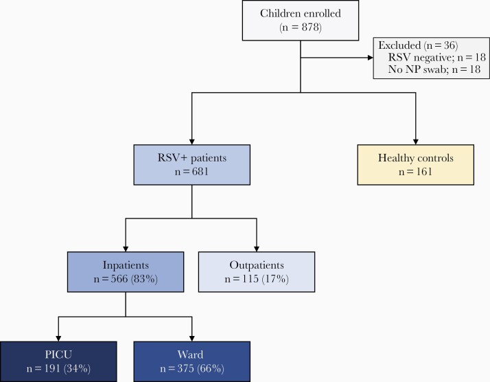
Methods: Children <2 years old with mild (outpatients, n=115) or severe (inpatients, n=566) RSV infection, and matched healthy controls (n=161) were enrolled. Nasopharyngeal samples were obtained for RSV, Streptococcus pneumoniae, Staphylococcus aureus, Moraxella catarrhalis, and Haemophilus influenzae detection and quantitation by PCR. Multivariable models were constructed to identify variables predictive of severe disease.

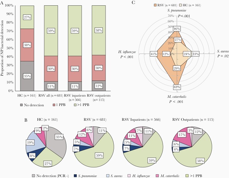
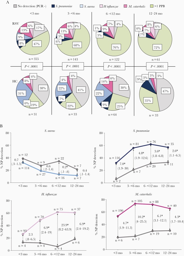
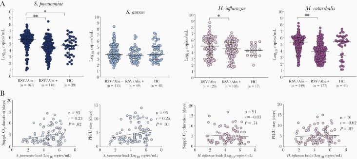
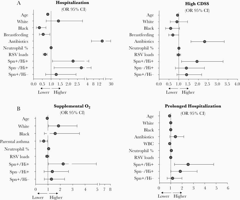
Results: S. pneumoniae, H. influenzae, and M. catarrhalis, but not S. aureus, were detected more frequently in RSV-infected children (84%) than healthy controls (46%; P<.001). Detection of S. pneumoniae and/or H. influenzae was associated with fever, more frequent antibiotic treatment, worse radiologic findings, and higher neutrophil counts (P<.01). In adjusted analyses, S. pneumoniae/H. influenzae codetection was independentlyassociated with greater odds of hospitalization, higher disease severity scores, need for supplemental oxygen, and longer hospitalization.

Conclusions: Nasopharyngeal codetection of S. pneumoniae and H. influenzae in infants with RSV infection is associated with increased disease severity.

Keywords: RSV; bacterial PCR; disease severity; infants; nasopharyngeal bacterial colonization.


Figures

Figure 1.
Figure 1.
Flowchart of study participants. The upper boxes in gray indicate the number of patients and controls enrolled and the reasons for exclusion. Children with RSV infection included in the study (blue boxes) were classified according to disease severity in RSV outpatients and RSV inpatients (ward and PICU). Yellow box indicates the number of healthy controls enrolled. Abbreviations: NP, nasopharyngeal; PICU, pediatric intensive care unit; RSV, respiratory syncytial virus.
Figure 2.
Figure 2.
Detection of potentially pathogenic bacteria (PPB) in the upper respiratory tract of young children with respiratory syncytial virus (RSV) infection and healthy age controls. A, Frequency of nasopharyngeal (NP) bacterial detection in study patients. The horizontal axis represents the study groups and the vertical axis represents the percentage of NP bacterial detection (Streptococcus pneumoniae, Staphylococcus aureus, Moraxella catarrhalis, Haemophilus influenzae): negative detection for any of the 4 bacteria (gray), detection of 1 of these 4 PPB (red), and detection of >1 PPB (green). B, Frequency of NP detection of specific bacteria. Pie charts for healthy controls and children with RSV infection (all, inpatients, and outpatients) showing the percentage detection of none of the 4 bacteria by PCR (gray), S. pneumoniae only (dark blue), S. aureus only (light blue), M. catarrhalis only (dark pink), H. influenzae only (light pink), and >1 PPB (green). C, Radar plot depicting the frequency of any detection of S. pneumoniae, S. aureus, M. catarrhalis, and H. influenzae in children with RSV infection (orange) compared to healthy controls (HC; light yellow). P values indicate the comparisons between RSV and HC for any detection (alone and in combination with other bacteria) of each of the 4 bacteria.
Figure 3.
Figure 3.
Nasopharyngeal (NP) bacterial detection stratified by age in children with respiratory syncytial virus (RSV) infection in relation to healthy controls. A, Pie charts depicting the proportion of nasopharyngeal bacterial detected in RSV patients (upper panels) and healthy controls (HC; lower panels) stratified by age: no detection of the 4 bacteria (PCR−; gray), single detection of Streptococcus pneumoniae (dark blue), Staphylococcus aureus (light blue), Moraxella catarrhalis (dark pink), Haemophilus influenzae (light pink), and >1 potentially pathogenic bacteria (PPB; green). Comparisons by χ2 for each age group. B, Odds of S. aureus, S. pneumoniae, H. influenzae, and M. catarrhalis detection in RSV patients (colored lines) in relation to healthy controls (gray lines) according to age. Thin arrows indicate the odds ratio (95% confidence interval) for each age group comparison. Asterisks indicate the time points that are significantly different. The numbers of RSV patients and controls per bacteria and age group are depicted in each plot. Analyses by χ2 test for trends.
Figure 4.
Figure 4.
Bacterial loads in children with respiratory syncytial virus (RSV) infection and healthy controls. A, Bacterial loads in healthy controls and children with RSV infection according to antibiotic use at study enrollment. The horizontal axis represents RSV-infected children not treated with antibiotics (RSV/Abx−), those treated (RSV/Abx+), and healthy controls (HC). The Y-axis represent bacterial loads in log10 copies/mL for each bacterium. Comparisons by Mann-Whitney U test between RSV/Abx− and RSV/Abx+, and between RSV/Abx− and HC. ∗P<.05; ∗∗P<.01. B, Spearman correlations between Streptococcus pneumoniae (blue) and Haemophilus influenzae (pink) loads and duration of supplemental oxygen and pediatric intensive care unit (PICU) stay among patients admitted to the PICU. The number of patients (n), correlation coefficient (r), and P value are included in each plot.
Figure 5.
Figure 5.
Predictors of disease severity in children with RSV infection. A, Adjusted need for hospitalization and high CDSS evaluated in the whole cohort of RSV patients. High CDSS was defined as a CDSS >3. B, Adjusted odds of oxygen use and prolonged hospital stay defined by a duration of hospitalization of ≥3 days. Reference group for race is other race. Reference group for bacterial classification is no detection of Streptococcus pneumoniae and Haemophilus influenzae (Spn−/Hi−). Data are adjusted ORs with point estimates and 95% CIs. Abbreviations: CDSS, clinical disease severity score; CI, confidence interval; Hi, H. influenzae; OR, odds ratio; RSV, respiratory syncytial virus; Spn, S. pneumoniae.

References

    1. Shi T, McAllister DA, O’Brien KL, et al. Global, regional, and national disease burden estimates of acute lower respiratory infections due to respiratory syncytial virus in young children in 2015: a systematic review and modelling study. Lancet 2017; 390:946–58. - PMC - PubMed
    1. Pneumonia Etiology Research for Child Health (PERCH) Study Group. Causes of severe pneumonia requiring hospital admission in children without HIV infection from Africa and Asia: the PERCH multi-country case-control study. Lancet 2019; 394:757–79. - PMC - PubMed
    1. García CG, Bhore R, Soriano-Fallas A, et al. Risk factors in children hospitalized with RSV bronchiolitis versus non-RSV bronchiolitis. Pediatrics 2010; 126:e1453–60. - PMC - PubMed
    1. Hall CB, Weinberg GA, Iwane MK, et al. The burden of respiratory syncytial virus infection in young children. N Engl J Med 2009; 360:588–98. - PMC - PubMed
    1. Mella C, Suarez-Arrabal MC, Lopez S, et al. Innate immune dysfunction is associated with enhanced disease severity in infants with severe respiratory syncytial virus bronchiolitis. J Infect Dis 2013; 207:564–73. - PMC - PubMed

Publication types