. 2021 Dec;600(7889):512-516.
doi: 10.1038/s41586-021-04005-0. Epub 2021 Sep 20.

High genetic barrier to SARS-CoV-2 polyclonal neutralizing antibody escape

Affiliations

High genetic barrier to SARS-CoV-2 polyclonal neutralizing antibody escape

Fabian Schmidt et al. Nature. 2021 Dec.

Abstract

The number and variability of the neutralizing epitopes targeted by polyclonal antibodies in individuals who are SARS-CoV-2 convalescent and vaccinated are key determinants of neutralization breadth and the genetic barrier to viral escape1-4. Using HIV-1 pseudotypes and plasma selection experiments with vesicular stomatitis virus/SARS-CoV-2 chimaeras5, here we show that multiple neutralizing epitopes, within and outside the receptor-binding domain, are variably targeted by human polyclonal antibodies. Antibody targets coincide with spike sequences that are enriched for diversity in natural SARS-CoV-2 populations. By combining plasma-selected spike substitutions, we generated synthetic 'polymutant' spike protein pseudotypes that resisted polyclonal antibody neutralization to a similar degree as circulating variants of concern. By aggregating variant of concern-associated and antibody-selected spike substitutions into a single polymutant spike protein, we show that 20 naturally occurring mutations in the SARS-CoV-2 spike protein are sufficient to generate pseudotypes with near-complete resistance to the polyclonal neutralizing antibodies generated by individuals who are convalescent or recipients who received an mRNA vaccine. However, plasma from individuals who had been infected and subsequently received mRNA vaccination neutralized pseudotypes bearing this highly resistant SARS-CoV-2 polymutant spike, or diverse sarbecovirus spike proteins. Thus, optimally elicited human polyclonal antibodies against SARS-CoV-2 should be resilient to substantial future SARS-CoV-2 variation and may confer protection against potential future sarbecovirus pandemics.


Figures

Extended Data Fig. 1.
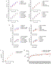
Extended Data Fig. 1.. Characterization of HIV-1 pseudotypes bearing the chimeric, mutant, and variant SARS-CoV-2 and sarbecovirus spike proteins
(a) Titration of pseudotyped viruses on 293T/ACE2cl.22 cells. Chimeric spike pseudotyped viruses in the upper left panel were built using the unaltered SARS-CoV-2Δ19 and SARS-CoVΔ19 spike protein constructs and a 3-plasmid HIV-1 pseudotyping system (see Methods). The other panels depict titration of pseudotypes derived using a furin cleavage site mutant SARS-CoV-2Δ19 spike protein (R683G) and a 2-plasmid HIV-1 pseudotyping system (see Methods). (b) The same pseudotyped viruses used in (a) were used to infect 3 different 293T/ACE2 clonal cell lines each expressing a different level of ACE2 (MFI = mean fluorescence intensity). (c) Titration of pseudotypes bearing an unaltered SARS-CoV-2Δ19 spike protein and a furin cleavage site mutant SARS-CoV-2Δ19 spike protein (R683G) generated using a 2-plasmid HIV-1 pseudotype system (see Methods). (d) Comparative neutralization potency (NT50 values) of high titer convalescent (RU27) plasmas against HIV-1 pseudotypes bearing R683G mutant (grey symbols) and unaltered (red symbols) SARS-CoV-2Δ19 spike proteins. For all panels, median ± range of two independent experiments is plotted.
Extended Data Fig. 2
Extended Data Fig. 2. The RU27 SARS-CoV-2 convalescent plasma panel contains neutralizing antibodies targeting RBD and non-RBD determinants
(a-c) Correlations of neutralizing potencies of the RU27 plasmas against psudotypes bearing the indicated pairs of spike proteins. Simple linear regression was used to calculate R2 and p-values, dashed lines indicate 95% confidence intervals for the regression line
Extended Data Fig. 3.
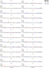
Extended Data Fig. 3.. Selection pressure on SARS-CoV-2 spike exerted by convalescent plasma
Frequencies of amino acid substitutions at each codon of the SARS-CoV-2 spike protein following the indicated number of passages (P2-P6) of two independent rVSV-SARS-CoV-2 populations (1D7 and 2E1), in each of the RU27 plasmas, determined by NGS sequencing.
Extended Data Fig. 4.
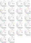
Extended Data Fig. 4.. Neutralization sensitivity of plasma-selected rVSV/SARS-CoV-2 mutants to RU1-20 plasmas.
Infection, relative to non-neutralized controls, by plaque purified rVSV/SARS-CoV-2 isolates in the presence of the indicated dilutions of the indicated plasmas from the RU27 panel. The same plasmas that were used to select the indicated mutants were used to determine neutralization potency against the respective plaque purified mutants (red) and parental (WT, grey) rVSV/SARS-CoV-2 1D7 or 2E1 viruses. Median ± range of two technical replicates is plotted.
Extended Data Fig. 5.
Extended Data Fig. 5.. Neutralization sensitivity of plasma-selected rVSV/SARS-CoV-2 mutants to RU 21-27 plasmas.
Infection, relative to non-neutralized controls, by plaque purified rVSV/SARS-CoV-2 isolates in the presence of the indicated dilutions of the indicated plasmas from the RU27 panel. The same plasmas that were used to select the indicated mutants were used to determine neutralization potency against the respective plaque purified mutants (red) and parental (WT, grey) rVSV/SARS-CoV-2 1D7 or 2E1 viruses. Median ± range of two technical replicates is plotted.
Extended Data Fig. 6.
Extended Data Fig. 6.. Neutralization sensitivity of rVSV/SARS-CoV-2 encoding the PMS1-1 spike.
(a) Design of the PMS1-1 polymutant spike protein with 13 plasma-selected spike mutations aggregated in a single spike. (b) Infection, relative to non-neutralized controls, by rVSV/SARS-CoV2PMS1-1 (red) and rVSV/SARS-CoV22E1 (grey) in the presence on the indicated dilutions of the plasmas from the RU27 panel. Median ± range of two technical replicates is plotted.
Extended Data Fig. 7.
Extended Data Fig. 7.. Synthetic polymutant and natural variant SARS-CoV-2 spike proteins
(a) Design of the PMS1-1 and PMSD4 polymutant spike proteins with 13 plasma-selected spike mutations aggregated in each spike. (b) Schematic representation of mutations in naturally occurring VOC/VOI SARS-CoV-2 spike proteins.
Extended Data Fig 8.
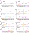
Extended Data Fig 8.. Neutralization potency of random convalescent and vaccine recipient plasmas against polymutant, VOC/VOI, and sarbecovirus HIV-1 pseudotypes
(a-c) Comparative neutralization potency (NT50 values) of random convalescent (Ran1-21) and vaccine recipient (Vac1-14) plasmas plasma against WT (grey symbols) and the indicated SARS-CoV-2 synthetic polymutant (a), natural variant (b) or sarbecovirus (c) (red symbol) HIV-1 pseudotypes. For all panels, median ± range of two independent experiments is plotted.
Extended Data Fig 9.
Extended Data Fig 9.. Neutralization potency of high titer convalescent plasma against PMS, VOC/VOI, and sarbecovirus HIV-1 pseudotypes
(a-c) Comparative neutralization potency (NT50 values) of high titer convalescent (RU27) plasma against WT (grey symbols) and indicated polymutant (a), SARS-CoV-2 natural variant (b) or sarbecovirus (c) (red symbol) HIV-1 pseudotypes. For all panels, median ± range of two independent experiments is plotted.
Extended Data Fig 10.
Extended Data Fig 10.. Neutralization potency of plasma from infected-then-vaccinated against VOC/VOI and diverse sarbecovirus HIV-1 pseudotypes
(a) Neutralization potency (NT50 values) of random convalescent plasmas (grey symbols) or ITV plasmas (red symbols) against SARS-CoV-2 prototype or variant or sarbecovirus HIV-1 pseudotypes. Median of two independent experiments is plotted. Dashed line indicated median NT50 for random convalescent plasmas against Wuhan-Hu-1 SARS-CoV-2. Numbers above each scatterplot indicate the median NT50 relative to the median NT50 for Wuhan-Hu-1 SARS-CoV-2. (b) Sequence diversity across sarbecovirus spike domains; SARS-CoV-2 and the indicated sarbecovirus spike sequences were aligned with Clustal and compared using Simplot; the percent identity relative to SARS-CoV2 was plotted within a rolling window of 100 amino acids.
Fig. 1
Fig. 1. Neutralizing antibodies in SARS-CoV-2 convalescent plasma targeting both RBD and non-RBD determinants
(a) Design of RBD-exchanged chimeric spike proteins. (b,c) Fifty percent neutralization titers (NT50) for 26 high titer convalescent plasmas (from the RU1-27 panel) against pseudotyped HIV-1 virions bearing the indicated spike proteins. Median of two independent experiments is plotted.
Fig. 2
Fig. 2. Selection of SARS-CoV-2 spike mutants by polyclonal antibodies
(a) Frequencies of amino acid substitutions at each codon of the SARS-CoV-2 spike protein in two independent rVSV-SARS-CoV-2 populations (1D7 and 2E1), determined by Illumina sequencing. Pooled results following selection with the RU27 plasma panel are displayed. (b) Locations of amino acid substitutions in 38 plaque purified rVSV/SARS-CoV-2 isolates obtained from rVSV/SARS-CoV-2 populations following passage in the RU27 plasmas. (c) Frequencies of naturally occurring amino acid substitutions (red circles) at each codon of the SARS-CoV-2 spike protein. Shaded gray bars in (a-c) indicate shared regions where variation is enriched. (d) Comparison of the averaged frequency of substitutions observed after passaging rVSV/SARS-CoV-2 with RU27 plasmas (center) and the frequency of sequence changes in natural populations (right), projected onto the SARS-CoV-2 spike structure (PDB 6VXX) with positions of the RBD and NTD domains indicated (left). The average frequency of substitutions in a 15 Å radius is represented using the color spectrum (scale = 0-20 center and 0-9 right). (e) Neutralization potency of RU27 plasmas against rVSV/SARS-CoV-2 encoding WT, individual selected mutants, or PMS1-1 spike proteins. Median of two independent determinations is plotted.
Fig. 3
Fig. 3. Neutralization resistance of polymutant SARS-CoV-2 spike proteins.
(a) Design of the PMS20 spike protein with 20 antibody-selected and VOC-associated mutations. (b) Replication of rVSV/SARS-CoV-2 chimeras encoding 2E1 (parental) or PMS20 spike proteins in 293T/ACE2cl.22 cells infected at a mutliplicity of 0.001 and 0.008 respectively. (c) Comparative neutralization potency of randomly selected convalescent (Ran 1-21) and vaccine recipient (Vac1-14) plasmas, against Wuhan-hu-1 and PMS20 (b) SARS-CoV-2 HIV-1 pseudotypes. For b, and c the median ± range of two independent determinations is plotted.
Fig 4.
Fig 4.. Neutralization breadth of polyclonal antibodies from infected-then-vaccinated individuals.
(a) Comparative neutralization potency (NT50 values) of random convalescent (Ran1-21) and infected-then-vaccinated (ITV1-14) plasmas against HIV-1 pseudotypes bearing SARS-CoV-2, PMS20, and RBD-exchanged chimeric spike proteins. (b) Fold difference in NT50, comparing neutralization of HIV-1 pseudotypes bearing SARS-CoV-2 and PMS20 spike proteins by Ran1-21, Vac1-15 and ITV1-14 plasmas (p-values calculated using 2-sided Mann Whitney test). (c) Neutralization curves for ITV plasmas and the indicated sarbecovirus HIV-1 pseudotypes. For (a) and (c) median ± range of two independent determinations is plotted.

Comment in

References

    1. Weisblum Y et al. Escape from neutralizing antibodies by SARS-CoV-2 spike protein variants. eLife 9, e61312, doi:10.7554/eLife.61312 (2020). - DOI - PMC - PubMed
    1. Muecksch F et al. Affinity maturation of SARS-CoV-2 neutralizing antibodies confers potency, breadth, and resilience to viral escape mutations. Immunity, doi:10.1016/j.immuni.2021.07.008 (2021). - DOI - PMC - PubMed
    1. Wang Z et al. Naturally enhanced neutralizing breadth against SARS-CoV-2 one year after infection. Nature 595, 426–431, doi:10.1038/s41586-021-03696-9 (2021). - DOI - PMC - PubMed
    1. Sauer MM et al. Structural basis for broad coronavirus neutralization. Nature structural & molecular biology, Online ahead of print, doi:10.1038/s41594-021-00596-4 (2021). - DOI - PubMed
    1. Schmidt F et al. Measuring SARS-CoV-2 neutralizing antibody activity using pseudotyped and chimeric viruses. The Journal of experimental medicine 217, e20201181, doi:10.1084/jem.20201181 (2020). - DOI - PMC - PubMed

Methods references

    1. Gaebler C et al. Combination of quadruplex qPCR and next-generation sequencing for qualitative and quantitative analysis of the HIV-1 latent reservoir. The Journal of experimental medicine 216, 2253–2264, doi:10.1084/jem.20190896 (2019). - DOI - PMC - PubMed
    1. Korber B et al. Tracking Changes in SARS-CoV-2 Spike: Evidence that D614G Increases Infectivity of the COVID-19 Virus. Cell 182, 812–827.e819, doi:10.1016/j.cell.2020.06.043 (2020). - DOI - PMC - PubMed
    1. Walls AC et al. Structure, Function, and Antigenicity of the SARS-CoV-2 Spike Glycoprotein. Cell 181, 281–292.e286, doi:10.1016/j.cell.2020.02.058 (2020). - DOI - PMC - PubMed
    1. Guy AJ, Irani V, Richards JS & Ramsland PA BioStructMap: a Python tool for integration of protein structure and sequence-based features. Bioinformatics (Oxford, England) 34, 3942–3944, doi:10.1093/bioinformatics/bty474 (2018). - DOI - PMC - PubMed
    1. Guy AJ et al. Proteome-wide mapping of immune features onto Plasmodium protein three-dimensional structures. Scientific reports 8, 4355, doi:10.1038/s41598-018-22592-3 (2018). - DOI - PMC - PubMed

Publication types

Supplementary concepts