. 2021 Oct 26;12(5):e0233521.
doi: 10.1128/mBio.02335-21. Epub 2021 Sep 21.

SARS-CoV-2 Nsp5 Demonstrates Two Distinct Mechanisms Targeting RIG-I and MAVS To Evade the Innate Immune Response

Affiliations

SARS-CoV-2 Nsp5 Demonstrates Two Distinct Mechanisms Targeting RIG-I and MAVS To Evade the Innate Immune Response

Yongzhen Liu et al. mBio. .

Abstract

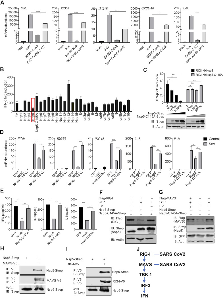
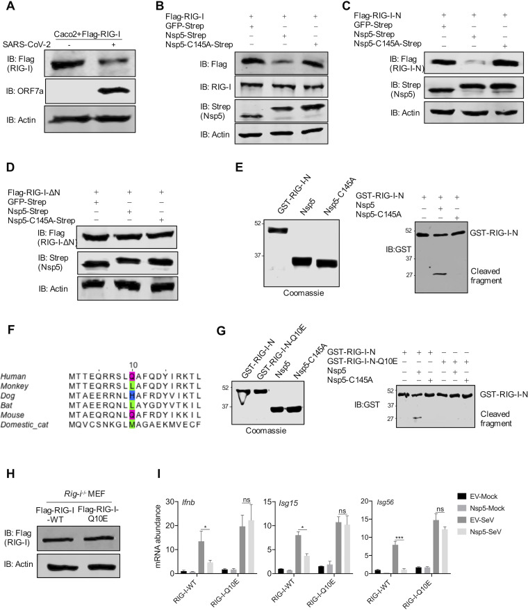
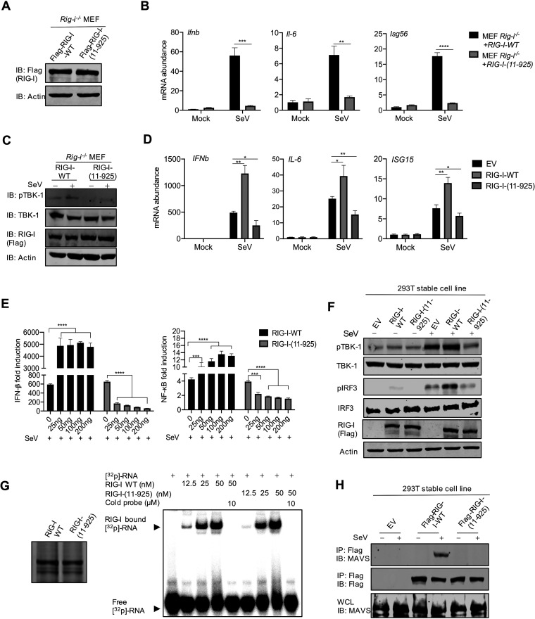
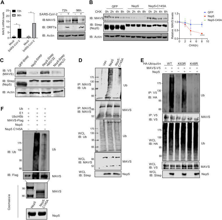
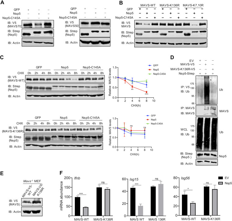
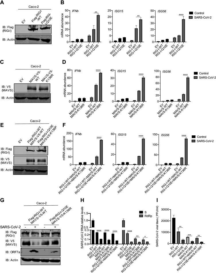
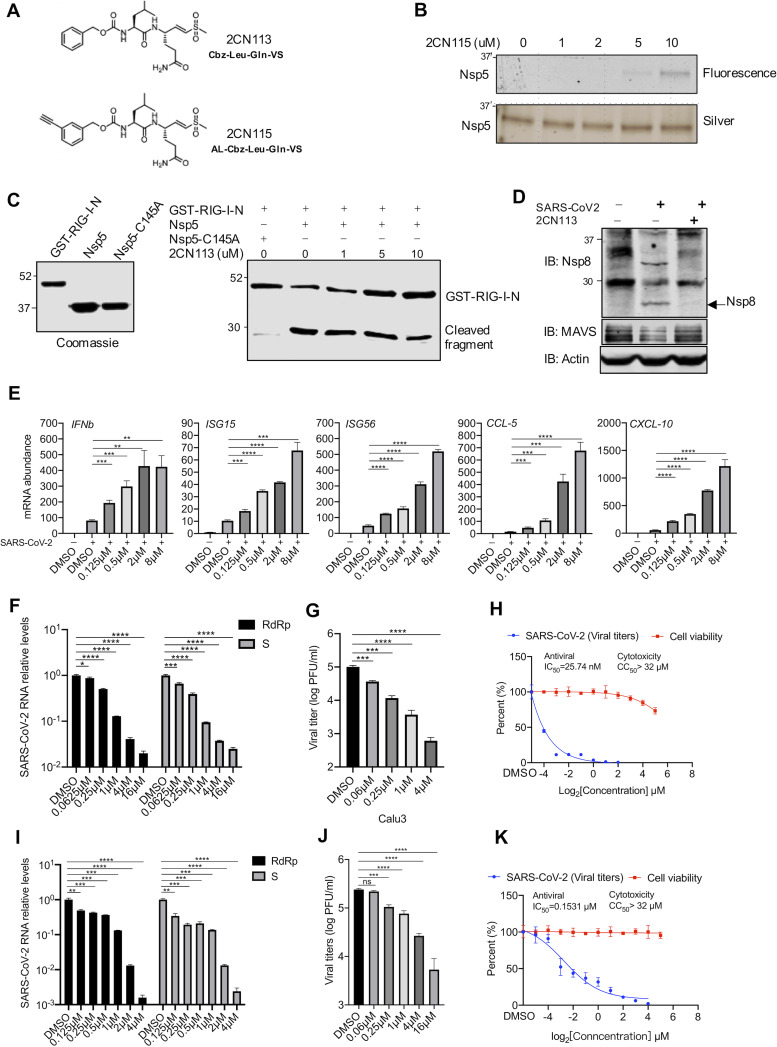
Newly emerged severe acute respiratory syndrome coronavirus 2 (SARS-CoV-2) caused a global pandemic with astonishing mortality and morbidity. The high replication and transmission of SARS-CoV-2 are remarkably distinct from those of previous closely related coronaviruses, and the underlying molecular mechanisms remain unclear. The innate immune defense is a physical barrier that restricts viral replication. We report here that the SARS-CoV-2 Nsp5 main protease targets RIG-I and mitochondrial antiviral signaling (MAVS) protein via two distinct mechanisms for inhibition. Specifically, Nsp5 cleaves off the 10 most-N-terminal amino acids from RIG-I and deprives it of the ability to activate MAVS, whereas Nsp5 promotes the ubiquitination and proteosome-mediated degradation of MAVS. As such, Nsp5 potently inhibits interferon (IFN) induction by double-stranded RNA (dsRNA) in an enzyme-dependent manner. A synthetic small-molecule inhibitor blunts the Nsp5-mediated destruction of cellular RIG-I and MAVS and processing of SARS-CoV-2 nonstructural proteins, thus restoring the innate immune response and impeding SARS-CoV-2 replication. This work offers new insight into the immune evasion strategy of SARS-CoV-2 and provides a potential antiviral agent to treat CoV disease 2019 (COVID-19) patients. IMPORTANCE The ongoing COVID-19 pandemic is caused by SARS-CoV-2, which is rapidly evolving with better transmissibility. Understanding the molecular basis of the SARS-CoV-2 interaction with host cells is of paramount significance, and development of antiviral agents provides new avenues to prevent and treat COVID-19 diseases. This study describes a molecular characterization of innate immune evasion mediated by the SARS-CoV-2 Nsp5 main protease and subsequent development of a small-molecule inhibitor.

Keywords: E3 ligase; MAVS; Nsp5; RIG-I; SARS-CoV-2; protease; small-molecule inhibitor.


Figures

FIG 1
FIG 1
SARS-CoV-2 Nsp5 targets RIG-I and MAVS to inhibit IFN induction. (A) Calu-3 cells were mock infected or infected with SARS-CoV-2 (MOI = 0.5) for 24 h and superinfected with Sendai virus (SeV; 100 hemagglutinating units [HAU]/ml) for 6 h. The expression of the indicated antiviral genes was analyzed by real-time PCR using total RNA. (B) A screen identified SARS-CoV-2 proteins that inhibit IFN induction by the ectopically expressed MAVS in 293T cells. (C) IFN induction in 293T cells stimulated by RIG-I-N was assessed by a reporter assay with wild-type Nsp5 or its enzyme-deficient Nsp5-C145A mutant. IB, immunoblotting. (D, E) Antiviral cytokine gene expression in HCT116 cells stably expressing Nsp5 or the Nsp5-C145A mutant in response to SeV infection (100 HAU/ml) was analyzed by real-time PCR (D) and ELISA (E). (F, G) Immunoblotting analysis of ectopically expressed RIG-I (F) and MAVS (G) with Nsp5 or the Nsp5-C145A mutant. (H, I) Interactions of SARS-CoV-2 Nsp5 with RIG-I (H) or MAVS (I) were analyzed by coimmunoprecipitation in transfected 293T cells. (J) Diagram of the point of inhibition by SARS-CoV-2 of the RIG-I-MAVS pathway. Data are means ± standard deviations (SD). Significance was calculated using Student's two-tailed, unpaired t test. *, P < 0.05; **, P < 0.01; ***, P < 0.001; ****, P < 0.0001; ns, nonsignificant. See also Fig. S1 in the supplemental material.
FIG 2
FIG 2
Nsp5 cleaves RIG-I at the Q10 residue. (A, B) Immunoblotting analysis of ectopically expressed RIG-I in Caco-2 cells infected with SARS-CoV-2 (A) and with the expression of Nsp5 or the Nsp5-C145A mutant using anti-Flag antibody or anti-RIG-I antibody (B). (C, D) Immunoblotting analysis of ectopically expressed RIG-I-N (C) and RIG-I-ΔN (D) in 293T cells with the expression of Nsp5 or the Nsp5-C145A mutant. (E, right) Numbers at the left of the gel are molecular weights in kDa. (E) Immunoblotting analysis of in vitro GST–RIG-I-N cleavage by Nsp5 or the Nsp5-C145A mutant with all proteins purified from E. coli and analyzed using Coomassie blue staining (left). (F) Alignment of the 20 N-terminal amino acids of RIG-I from human and five nonhuman mammalian species. The putative cleavage site, Q10, of human RIG-I and its equivalent residues are highlighted with colored boxes. (G) Immunoblotting analysis of GST–RIG-I-N and GST–RIG-I-N-Q10E after in vitro cleavage by Nsp5 or the Nsp5-C145A mutant with all proteins purified from E. coli and analyzed using Coomassie blue staining (left). (H) Immunoblotting analysis of whole-cell lysates of Rig-i−/− MEFs reconstituted with RIG-I-WT or RIG-I-Q10E. (I) Total RNA extracted from these cells without or with Nsp5 expression in response to Sendai virus infection (100 HAU/ml) was analyzed by RT-qPCR with primers specific for the indicated genes. Data are means ± SD. Significance was calculated using Student’s two-tailed, unpaired t test. *, P < 0.05; ***, P < 0.001; ns, nonsignificant. See also Fig. S2.
FIG 3
FIG 3
Loss of function and dominant negative effect of the cleaved RIG-I fragment [RIG-I-(11–925)]. (A) Immunoblotting analysis of WCLs of Rig-i−/− MEFs reconstituted with RIG-I-WT or RIG-I-Q10E. (B) Total RNA extracted from Rig-i−/− MEFs reconstituted with RIG-I-WT or RIG-I-11-925 infected with Sendai virus (100 HAU/ml) was analyzed by RT-qPCR with primers specific for the indicated genes. (C) Immunoblotting analysis of WCLs of Rig-i−/− MEFs reconstituted with RIG-I-WT or RIG-I-(11–925) and infected with Sendai virus (100 HAU/ml) with the indicated antibodies. (D) Total RNA extracted from 293T cells stably expressing RIG-I-WT or RIG-I-(11–925) infected with Sendai virus (100 HAU/ml) was analyzed by RT-qPCR with primers specific for the indicated genes. (E) IFN induction and NF-κB activation in 293T cells transfected with increasing amounts of plasmids containing RIG-I or RIG-I-(11–925) were analyzed by luciferase reporter assay. (F) Immunoblotting analysis of WCLs of 293T cells stably expressing RIG-I-WT or RIG-I-11-925 with the indicated antibodies. (G) Electrophoresis mobility shift assay of purified RIG-I or RIG-I-(11–925) incubated with 32P-labeled 5′-triphosphate 19-mer dsRNA. (H) Immunoblotting analysis of precipitated RIG-I (anti-Flag) and WCLs of 293T cells that stably express RIG-I-WT or RIG-I-(11–925) and that were infected with Sendai virus (100 HAU/ml) with the indicated antibodies. Data are means ± SD. Significance was calculated using Student’s two-tailed, unpaired t test. *, P < 0.05; **, P < 0.01; ***, P < 0.001; ****, P < 0.0001. See also Fig. S3.
FIG 4
FIG 4
Nsp5 promotes MAVS ubiquitination and degradation. (A) qRT-PCR analysis and immunoblotting analysis of Caco-2 cells infected with SARS-CoV-2 (MOI = 1) for 72 h and 96 h. (B) Immunoblotting analysis of WCLs of 293T cells transfected with plasmids containing Nsp5 or Nsp5C145A and treated with DMSO or 100 μg/ml cycloheximide (CHX) and with the indicated antibodies. MAVS was quantified by densitometry and plotted as shown on the right. Results represent one of three independent experiments. (C) Immunoblotting analysis of WCLs of 293T cells transfected with plasmids containing MAVS and Nsp5 and treated with the proteasome inhibitor MG132 (10 μM) or the lysosome inhibitor NH4Cl (10 mM). (D) Immunoblotting analysis of endogenous ubiquitination levels of MAVS and WCLs of 293T cells transfected with plasmids containing Nsp5 or Nsp5-C145A and treated with the indicated antibodies. Vec, vector; Ub, ubiquitin. (E) Immunoblotting analysis of endogenous ubiquitination levels of MAVS and WCLs of 293T cells transfected with plasmids containing Nsp5 or Nsp5-C145A and ubiquitin (WT) and treated with the K48R or K63R mutant and with the indicated antibodies. (F) In vitro ubiquitination assay of MAVS. Nsp5 or Nsp5-C145A and MAVS were incubated in a reaction mixture containing ubiquitin, E1, and E2(UbcH5b), and ubiquitination levels were determined by immunoblotting with anti-Ub antibody. Purified MAVS is shown at the bottom with SDS-PAGE and Coomassie blue staining. Data are means ± SD. Significance was calculated using Student's two-tailed, unpaired t test. *, P < 0.05.
FIG 5
FIG 5
Nsp5 targets the K136 residue of MAVS to promote ubiquitination and degradation. (A) Immunoblotting analysis of WCLs of 293T cells transfected with plasmids containing MAVS70 or MAVS50, together with those containing Nsp5 or Nsp5-C145A, and treated with the indicated antibodies. (B) Immunoblotting analysis of WCLs of 293T cells transfected with plasmids containing wild-type MAVS, MAVS-K136R, or MAVS-K7R/K10R and those containing Nsp5 or Nsp5-C145A and treated with the indicated antibodies. (C) Immunoblotting analysis of WCLs of 293T cells stably expressing MAVS-WT or MAVS-K136R transfected with plasmids containing Nsp5 or Nsp5C145A, treated with DMSO or cycloheximide (CHX, 100 μg/ml), and treated with the indicated antibodies. MAVS protein was quantified by densitometry and plotted as shown on the right. (D) Immunoblotting analysis of ubiquitination levels of MAVS and WCLs of 293T cells stably expressing MAVS-WT or MAVS-K136R transfected with plasmids containing Nsp5 and treated with the indicated antibodies. (E) Immunoblotting analysis of WCLs of Mavs−/− MEFs reconstituted with MAVS-WT or MAVS-K136R. (F) Total RNA extracted from these cells without or with Nsp5 expression was analyzed by RT-qPCR and treated with primers specific for the indicated genes. See also Fig. S4.
FIG 6
FIG 6
RIG-I-Q10E and MAVS-K136A resist destruction and restore innate immune activation during SARS-CoV-2 infection. (A, B) Immunoblotting analysis of WCLs of Caco-2 cells stably expressing RIG-I-WT or RIG-I-Q10E with the indicated antibodies (A). Total RNA extracted from Caco-2 cells stably expressing RIG-I-WT or RIG-I-Q10E infected with SARS-CoV-2 (MOI = 1) was analyzed by RT-qPCR with primers specific for the indicated genes (B). (C) Immunoblotting analysis of WCLs of Caco-2 cells stably expressing MAVS-WT or MAVS-K136R with the indicated antibodies. (D) Total RNA extracted from Caco-2 cells stably expressing MAVS-WT or MAVS-K136R infected with SARS-CoV-2 (MOI = 1) was analyzed by RT-qPCR with primers specific for the indicated genes. (E) Immunoblotting analysis of WCLs of Caco-2 cells stably expressing RIG-I-WT and MAVS-WT or RIG-I-Q10E and MAVS-K136R with the indicated antibodies. (F) Total RNA extracted from Caco-2 cells stably expressing RIG-I-WT and RIG-I-Q10E or MAVS-WT and MAVS-K136R infected with SARS-CoV-2 (MOI = 1) was analyzed by RT-qPCR with primers specific for the indicated genes. (G) Immunoblotting analysis of WCLs of Caco-2 cells stably expressing RIG-I-WT and RIG-I-Q10E or MAVS-WT and MAVS-K136R infected with SARS-CoV-2 for 72 h, with the indicated antibodies. (H, I) SARS-CoV-2 replication in Caco-2 cells stably expressing RIG-I-WT, RIG-I-Q10E, MAVS-WT, MAVS-K136R, RIG-I-WT, and MAVS-WT or RIG-I-Q10E and MAVS-K136R was analyzed by RT-qPCR for SARS-CoV-2 S and RdRp RNA (H) or determined by plaque assay (I) at 72 h postinfection. Data are means ± SD. Significance was calculated using Student’s two-tailed, unpaired t test. *, P < 0.05; **, P < 0.01; ***, P < 0.001; ****, P < 0.0001. See also Fig. S5.
FIG 7
FIG 7
A small-molecule inhibitor of Nsp5 restores innate immune activation and impedes SARS-CoV-2 replication. (A) Structures of 2CN113 and 2CN115. (B) Nsp5-Step-expressing 293T cells were treated with 2CN115 at the indicated doses for 2 h. Nsp5-Step was purified and subjected to binding analysis by in-gel fluorescence imaging and silver staining. (C) The effect of 2CN113 on in vitro GST–RIG-I-N cleavage by Nsp5 was analyzed with all proteins purified from E. coli by immunoblotting. (D) HeLa cells stably expressing hACE2 were treated with 2CN113 and infected with SARS-CoV-2 (MOI = 0.1). The effect of 2CN113 on SARS-CoV-2 Nsp8 processing was analyzed by immunoblotting with the indicated antibodies. (E) Calu-3 cells were treated with 2CN113 and infected with SARS-CoV-2 (MOI = 1). The mRNA abundance of antiviral genes was determined by real-time PCR at 72 h after SARS-CoV-2 infection. (F, G) Calu-3 cells were treated with 2CN113 and infected with SARS-CoV-2 (MOI = 0.1). The effect of 2CN113 on the SARS-CoV-2 RNA abundance (F) and viral titer (G) was determined at 72 h after SARS-CoV-2 infection by real-time PCR analysis of total RNA and plaque assay of the medium, respectively. S, SARS-CoV-2 S protein. (I, J) Vero cells stably expressing hACE2 were treated with 2CN113 and infected with SARS-CoV-2 (MOI = 0.001). The effect of 2CN113 on SARS-CoV-2 RNA abundance (I) and viral titer (J) was determined at 24 h after SARS-CoV-2 infection by real-time PCR analysis of total RNA and plaque assay of the medium, respectively. (H, K) The IC50 of 2CN113 on SARS-CoV-2 replication by plaque assay and the CC50 of 2CN113 on Calu-3 (H) and Vero (K) cells were determined and plotted. Data are means ± SD. Significance was calculated using Student’s two-tailed, unpaired t test. *, P < 0.05; **, P < 0.01; ***, P < 0.001; ****, P < 0.0001; ns, nonsignificant. See also Fig. S6.

References

    1. Hui DS, I Azhar E, Madani TA, Ntoumi F, Kock R, Dar O, Ippolito G, Mchugh TD, Memish ZA, Drosten C, Zumla A, Petersen E. 2020. The continuing 2019-nCoV epidemic threat of novel coronaviruses to global health—the latest 2019 novel coronavirus outbreak in Wuhan, China. Int J Infect Dis 91:264–266. doi:10.1016/j.ijid.2020.01.009. - DOI - PMC - PubMed
    1. Wu JT, Leung K, Leung GM. 2020. Nowcasting and forecasting the potential domestic and international spread of the 2019-nCoV outbreak originating in Wuhan, China: a modelling study. Lancet 395:689–697. doi:10.1016/S0140-6736(20)30260-9. - DOI - PMC - PubMed
    1. Polack FP, Thomas SJ, Kitchin N, Absalon J, Gurtman A, Lockhart S, Perez JL, Pérez Marc G, Moreira ED, Zerbini C, Bailey R, Swanson KA, Roychoudhury S, Koury K, Li P, Kalina WV, Cooper D, Frenck RW, Hammitt LL, Türeci Ö, Nell H, Schaefer A, Ünal S, Tresnan DB, Mather S, Dormitzer PR, Şahin U, Jansen KU, Gruber WC, C4591001 Clinical Trial Group . 2020. Safety and efficacy of the BNT162b2 mRNA Covid-19 vaccine. N Engl J Med 383:2603–2615. doi:10.1056/NEJMoa2034577. - DOI - PMC - PubMed
    1. Baden LR, El Sahly HM, Essink B, Kotloff K, Frey S, Novak R, Diemert D, Spector SA, Rouphael N, Creech CB, McGettigan J, Khetan S, Segall N, Solis J, Brosz A, Fierro C, Schwartz H, Neuzil K, Corey L, Gilbert P, Janes H, Follmann D, Marovich M, Mascola J, Polakowski L, Ledgerwood J, Graham BS, Bennett H, Pajon R, Knightly C, Leav B, Deng W, Zhou H, Han S, Ivarsson M, Miller J, Zaks T. 2021. Efficacy and safety of the mRNA-1273 SARS-CoV-2 vaccine. N Engl J Med 384:403–416. doi:10.1056/NEJMoa2035389. - DOI - PMC - PubMed
    1. Voysey M, Clemens SAC, Madhi SA, Weckx LY, Folegatti PM, Aley PK, Angus B, Baillie VL, Barnabas SL, Bhorat QE, Bibi S, Briner C, Cicconi P, Collins AM, Colin-Jones R, Cutland CL, Darton TC, Dheda K, Duncan CJA, Emary KRW, Ewer KJ, Fairlie L, Faust SN, Feng S, Ferreira DM, Finn A, Goodman AL, Green CM, Green CA, Heath PT, Hill C, Hill H, Hirsch I, Hodgson SHC, Izu A, Jackson S, Jenkin D, Joe CCD, Kerridge S, Koen A, Kwatra G, Lazarus R, Lawrie AM, Lelliott A, Libri V, Lillie PJ, Mallory R, Mendes AVA, Milan EP, Minassian AM, et al. . 2021. Safety and efficacy of the ChAdOx1 nCoV-19 vaccine (AZD1222) against SARS-CoV-2: an interim analysis of four randomised controlled trials in Brazil, South Africa, and the UK Lancet 397:99–111. doi:10.1016/S0140-6736(20)32661-1. - DOI - PMC - PubMed

Publication types

MeSH terms

LinkOut - more resources