. 2021 Sep 20;4(11):e202000703.
doi: 10.26508/lsa.202000703. Print 2021 Nov.

Choroid plexus APP regulates adult brain proliferation and animal behavior

Affiliations

Choroid plexus APP regulates adult brain proliferation and animal behavior

Karen Arnaud et al. Life Sci Alliance. .

Abstract

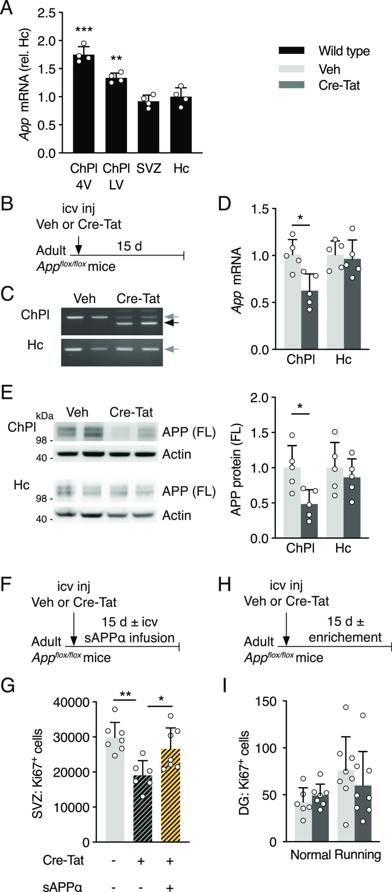
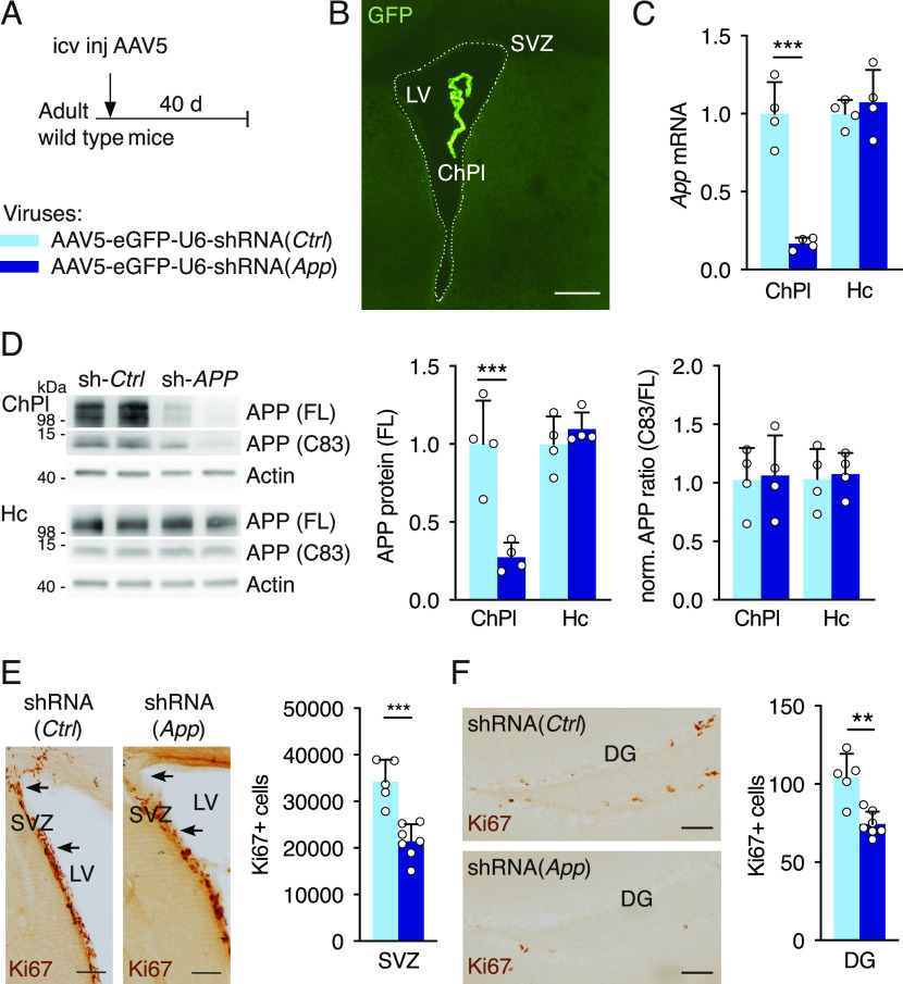
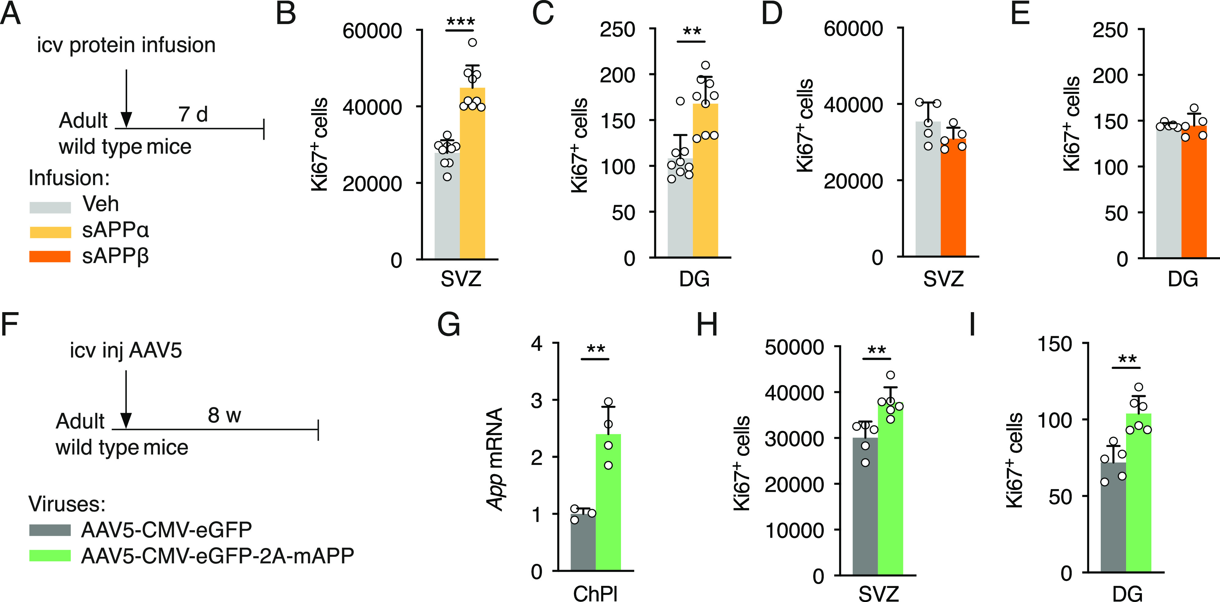
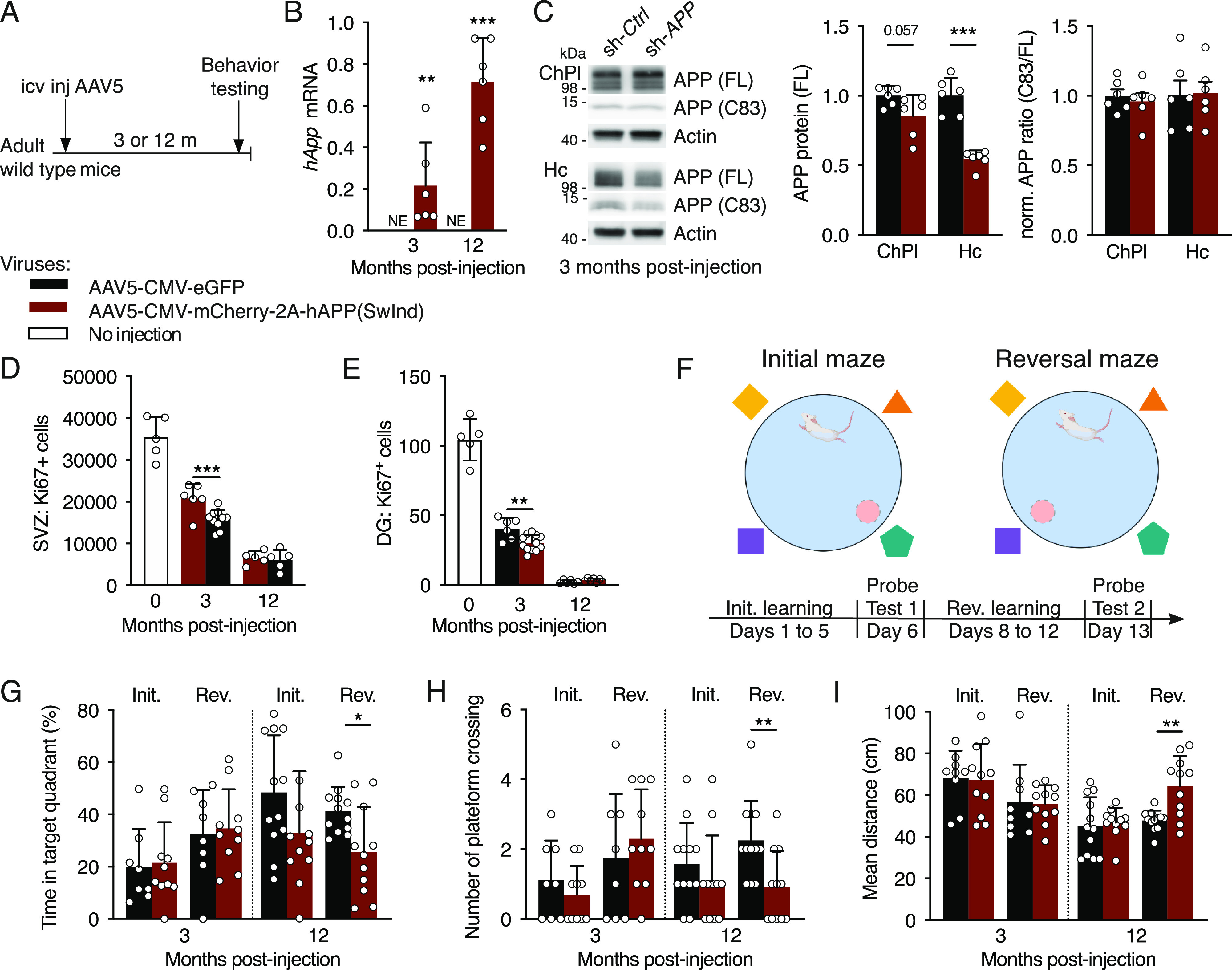
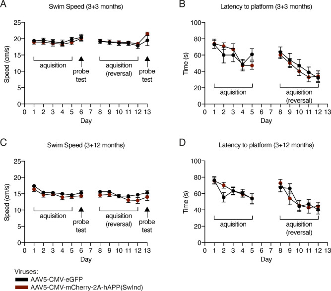
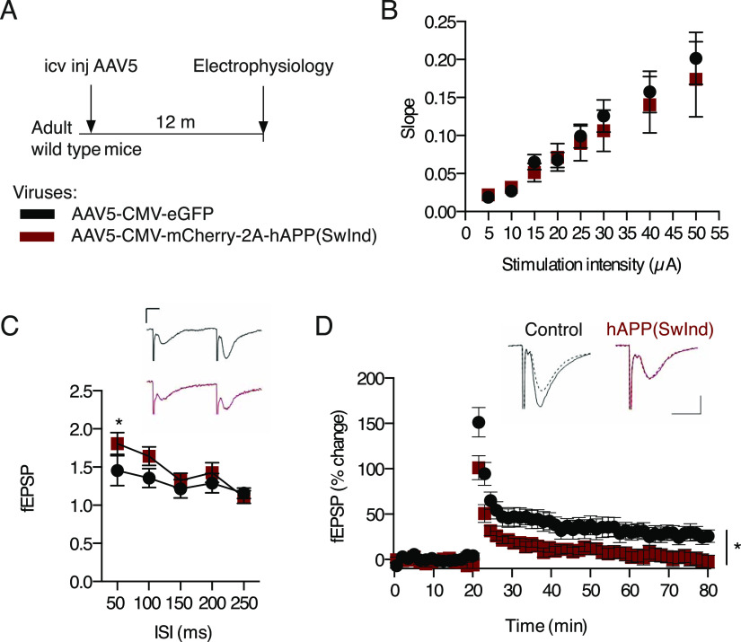
Elevated amyloid precursor protein (APP) expression in the choroid plexus suggests an important role for extracellular APP metabolites such as sAPPα in cerebrospinal fluid. Despite widespread App brain expression, we hypothesized that specifically targeting choroid plexus expression could alter animal physiology. Through various genetic and viral approaches in the adult mouse, we show that choroid plexus APP levels significantly impact proliferation in both subventricular zone and hippocampus dentate gyrus neurogenic niches. Given the role of Aβ peptides in Alzheimer disease pathogenesis, we also tested whether favoring the production of Aβ in choroid plexus could negatively affect niche functions. After AAV5-mediated long-term expression of human mutated APP specifically in the choroid plexus of adult wild-type mice, we observe reduced niche proliferation, reduced hippocampus APP expression, behavioral defects in reversal learning, and deficits in hippocampal long-term potentiation. Our findings highlight the unique role played by the choroid plexus in regulating brain function and suggest that targeting APP in choroid plexus may provide a means to improve hippocampus function and alleviate disease-related burdens.


Conflict of interest statement

A Prochiantz is a cofounder of BrainEver SAS; other authors report no biomedical financial interests or potential conflicts of interest.

Figures

Figure 1.
Figure 1.. Choroid plexus App loss-of-function in Appflox/flox mice decreases adult neural progenitor proliferation.
(A) Quantitative PCR analysis of App expression in ChPl from ventricles (LV and 4V), SVZ, and Hc in wild type mice. Values (n = 4 mice) are normalized to GAPDH and expression in Hc. (B) Schematic of App ChPl knock-down model involving single icv injection of Veh or Cre-Tat in adult Appflox/flox mice. (C) App specific recombination in LV choroid plexus after Cre-Tat icv injection. PCR of genomic DNA extracted from ChPl and Hc. Grey arrow indicates App wild type locus, black arrow indicates deleted App locus upon Cre recombination. (D) Quantitative PCR analysis of App expression normalized to GAPDH after Cre-Tat or Veh icv injection (n = 5 mice per group). (E) Western blot analysis of ChPl and Hc after Cre-Tat or Veh icv injection for quantification of APP protein levels normalized to actin (n = 5 mice per group). (F) Schematic of sAPPα rescue paradigm for evaluating SVZ cell proliferation. sAPPα was infused by 15-d osmotic mini-pump implanted immediately after Cre-Tat icv injection. (G) Analysis of SVZ cell proliferation by quantification of Ki67 positive cells after Veh or Cre-Tat icv injection followed by infusion of Veh or sAPPα (Veh/Veh n = 7 mice, Cre-Tat/Veh n = 6 mice, Cre-Tat/sAPPα n = 7 mice). (H) Schematic of App ChPl knock-down with environmental enrichment (running wheel) for evaluating Hc proliferation. (I) Analysis of DG cell proliferation by quantification of Ki67 positive cells after App recombination in mice in normal conditions or after physical exercise (Veh n = 7 mice, Cre-Tat n = 8 mice). *P < 0.05; **P < 0.01; ***P < 0.001; t test in (A, D, E, G); one-way ANOVA in (I); all values, mean ± SD. LV, lateral ventricle; 4V, fourth ventricle; ChPl, choroid plexus; Hc, hippocampus; SVZ, ventricular-subventricular zone; DG, dentate gyrus; icv, intracerebroventricular; Veh, vehicle.
Figure 2.
Figure 2.. Knock-down of choroid plexus App expression reduces cell proliferation in neurogenic niches.
(A) Schematic of App ChPl knock-down model involving a single icv injection of AAV5 expressing shRNA targeting the mouse App sequence in wild type mice. Control (Ctrl) virus expresses a scrambled shRNA devoid of targets in mouse. (B) An icv injection of AAV5 leads to specific choroid plexus expression. Note the absence of eGFP in brain parenchyma. Scale bar, 200 μm. (C) Quantitative PCR analysis of App expression in the ChPl and Hc after ChPl App knock-down (n = 4 mice per group). (D) Western blot analysis of ChPl and Hc after ChPl App knock-down for quantification of APP protein levels normalized to actin, and assessing normalized APP metabolite ratios (n = 4 mice per group). (E, F) Analysis of cell proliferation in SVZ (E) and DG (F) by quantification of Ki67 positive cells after ChPl App knock-down (shRNA-App n = 7 mice, shRNA-Ctrl n = 5 mice). Arrows in (E) highlight regional differences in SVZ cells. Scale bars, 100 μm. **P < 0.01; ***P < 0.001; t test; all values, mean ± SD. ChPl, choroid plexus; Hc, hippocampus; SVZ, ventricular-subventricular zone; DG, dentate gyrus; icv, intracerebroventricular.
Figure S1.
Figure S1.. Specificity of AAV5 expression after intracerebroventricular injection in adult mouse.
(A, B, C) Immunohistochemical analysis of GFP expression of different coronal sections (with different magnification) of ∼4-mo-old mice, 6 wk after intracerebroventricular injection of AAV5-eGFP-U6-shRNA(App) virus. GFP expression is only observed in choroid plexus (A, C). Scale bars, 200 μm. DG, dentate gyrus; D3V, dorsal third ventricle; LV, lateral ventricle.
Figure 3.
Figure 3.. sAPPα gain-of-function in the cerebral ventricles increases cell proliferation in neurogenic niches.
(A) Schematic of sAPP icv infusion by 7-d osmotic mini-pump in adult wild type mice. (B, C) Analysis of cell proliferation in SVZ (B) and dentate gyrus (DG) (C) by quantification of Ki67 positive cells after icv infusion of sAPPα or Veh (n = 9 mice per group). (D, E) Analysis of cell proliferation in SVZ (D) and DG (E) by quantification of Ki67 positive cells after icv infusion of sAPPβ or Veh (n = 5 mice per group). (F) Schematic of App ChPl gain-of-function model involving a single icv injection of AAV5 expressing mouse App or eGFP (control) in wild type mice. (G) Quantitative PCR analysis of App expression in ChPl after AAV5 icv injection (AAV5-mAPP n = 4 mice, AAV5-eGFP n = 3 mice). (H, I) Analysis of cell proliferation in SVZ (H) and DG (I) by quantification of Ki67 positive cells after icv injection (AAV5-mAPP n = 6 mice, AAV5-eGFP n = 5 mice). *P < 0.05; **P < 0.01; ***P < 0.001; t test; all values, mean ± SD. ChPl, choroid plexus; Hc, hippocampus; SVZ, ventricular-subventricular zone; DG, dentate gyrus; icv, intracerebroventricular; Veh, vehicle.
Figure 4.
Figure 4.. Choroid plexus hAPP(SwInd) expression decreases proliferation in neurogenic niches and impairs reversal learning.
(A) Schematic of mutant APP ChPl model involving a single icv injection of AAV5 expressing human mutated APP or eGFP (control) in wild type mice. (B) Quantitative PCR analysis of ChPl viral expression of hAPP(SwInd) in wild type mice at 3- and 12-mo post-injection (n = 6 mice per group). (C) Western blot analysis of ChPl and Hc after ChPl viral expression of hAPP(SwInd) in wild type mice at 3-mo post-injection for quantification of APP protein levels normalized to actin, and assessing normalized APP metabolite ratios (n = 6 mice per group). (D, E) Quantification of Ki67 positive cells in SVZ (D) and DG (E) after ChPl viral expression (AAV5-eGFP n = 6 mice, AAV5-hAPP(SwInd) n = 12 mice). (F) Reversal learning paradigm. (G, H, I) Behavioral responses after probe test 1 (Initial) and probe test 2 (Reversal) quantified by time (G), activity (H) and distance (I) (6 mo: n = 8–11 mice per group; 15 mo: n = 11–12 mice per group). *P < 0.05; **P < 0.01; ***P < 0.001; t test; all values: mean ± SD. SVZ, ventricular-subventricular zone; DG, dentate gyrus; NE, not expressed; icv, intracerebroventricular.
Figure S2.
Figure S2.. Morris water maze escape latency and speed measured during acquisition phases and probe tests.
(A, B, C, D) At 3-mo (A, B) and 12-mo (C, D) post viral injection, recordings were performed during the 5 d of initial and reversal learning phase to determine displacement speed (A, C) and mean escape latency (B, D). Displacement speed was also measured during probe tests (A, C). Escape latency cannot be measured during probe tests owing to the absence of a platform. Two-way repeated measures ANOVA; all values ± SEM.
Figure 5.
Figure 5.. Choroid plexus hAPP(SwInd) expression impairs hippocampal LTP.
(A) Schematic of mutant APP ChPl model involving a single icv injection of AAV5 expressing human mutated APP(SwInd) or eGFP (control) in wild type mice. (B) Comparison of stimulus-response (input/output) relationship in CA1 region between AAV5-hAPP(SwInd) (N = 5, n = 13, black squares) and AAV5-eGFP (N = 7, n = 10, red dots) injected mice. (C) Comparison of paired pulse facilitation between AAV5-hAPP(SwInd) (N = 6, n = 13) and AAV5-eGFP (N = 8, n = 13) injected mice. (D) LTP was induced by high frequency stimulation (HFS) at Schaffer collateral-CA1 synapses after 20 min baseline recording of slices from mice injected with AAV5-hAPP(SwInd) (N = 4, n = 9) or AAV5-eGFP (N = 5, n = 7). Representative traces showing responses before (dashed line) and 60 min after tetanus delivery (bold line). *P < 0.05; ANOVA; all values: mean ± SEM; N, number of animals; n, number of slices; icv, intracerebroventricular. Calibration bars: 10 ms, 0.2 mV.

References

    1. Arber C, Lovejoy C, Harris L, Willumsen N, Alatza A, Casey JM, Lines G, Kerins C, Mueller AK, Zetterberg H, et al. (2021) Familial Alzheimer’s disease mutations in PSEN1 lead to premature human stem cell neurogenesis. Cell Rep 34: 108615. 10.1016/j.celrep.2020.108615 - DOI - PMC - PubMed
    1. Balusu S, Brkic M, Libert C, Vandenbroucke RE (2016) The choroid plexus-cerebrospinal fluid interface in Alzheimer’s disease: More than just a barrier. Neural Regen Res 11: 534–537. 10.4103/1673-5374.180372 - DOI - PMC - PubMed
    1. Baratchi S, Evans J, Tate WP, Abraham WC, Connor B (2012) Secreted amyloid precursor proteins promote proliferation and glial differentiation of adult hippocampal neural progenitor cells. Hippocampus 22: 1517–1527. 10.1002/hipo.20988 - DOI - PubMed
    1. Baruch K, Deczkowska A, David E, Castellano JM, Miller O, Kertser A, Berkutzki T, Barnett-Itzhaki Z, Bezalel D, Wyss-Coray T, et al. (2014) Aging. Aging-induced type I interferon response at the choroid plexus negatively affects brain function. Science 346: 89–93. 10.1126/science.1252945 - DOI - PMC - PubMed
    1. Bjornsson CS, Apostolopoulou M, Tian Y, Temple S (2015) It takes a village: Constructing the neurogenic niche. Dev Cell 32: 435–446. 10.1016/j.devcel.2015.01.010 - DOI - PMC - PubMed

Publication types

Substances

LinkOut - more resources