. 2022 Jan;12(1):204-219.
doi: 10.1158/2159-8290.CD-21-0072. Epub 2021 Sep 20.

RTK-Dependent Inducible Degradation of Mutant PI3Kα Drives GDC-0077 (Inavolisib) Efficacy

Affiliations

RTK-Dependent Inducible Degradation of Mutant PI3Kα Drives GDC-0077 (Inavolisib) Efficacy

Kyung W Song et al. Cancer Discov. 2022 Jan.

Abstract

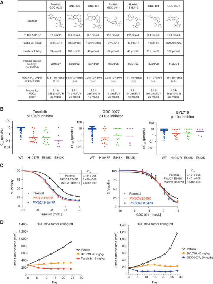
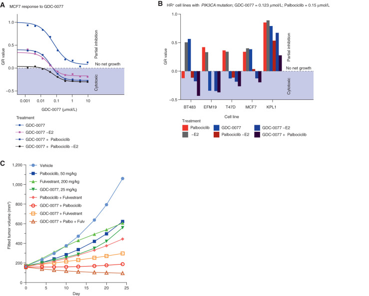
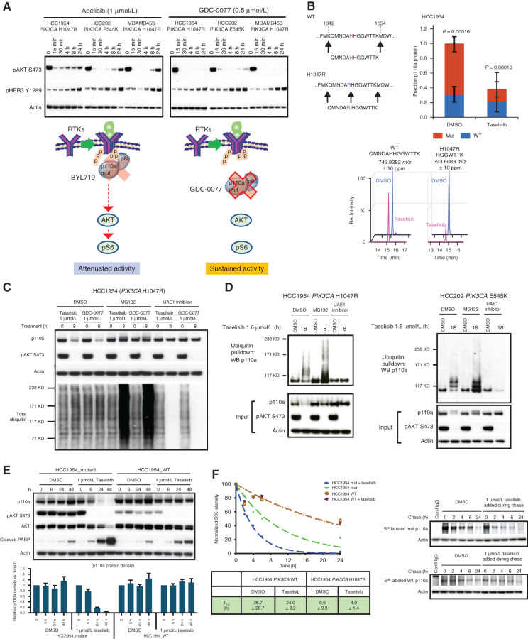
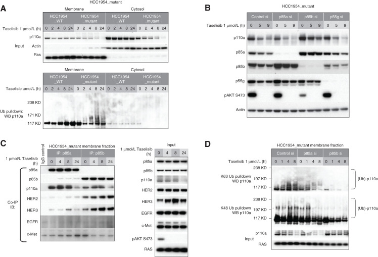
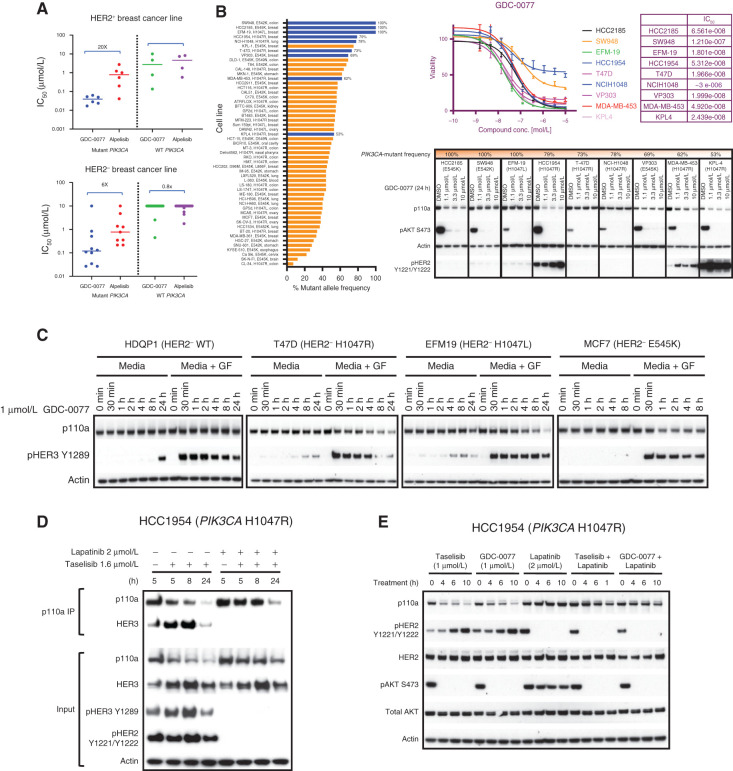
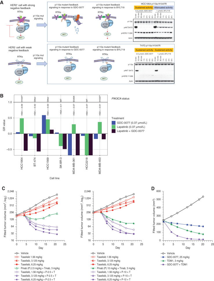
PIK3CA is one of the most frequently mutated oncogenes; the p110a protein it encodes plays a central role in tumor cell proliferation. Small-molecule inhibitors targeting the PI3K p110a catalytic subunit have entered clinical trials, with early-phase GDC-0077 studies showing antitumor activity and a manageable safety profile in patients with PIK3CA-mutant breast cancer. However, preclinical studies have shown that PI3K pathway inhibition releases negative feedback and activates receptor tyrosine kinase signaling, reengaging the pathway and attenuating drug activity. Here we discover that GDC-0077 and taselisib more potently inhibit mutant PI3K pathway signaling and cell viability through unique HER2-dependent mutant p110a degradation. Both are more effective than other PI3K inhibitors at maintaining prolonged pathway suppression. This study establishes a new strategy for identifying inhibitors that specifically target mutant tumors by selective degradation of the mutant oncoprotein and provide a strong rationale for pursuing PI3Kα degraders in patients with HER2-positive breast cancer. SIGNIFICANCE: The PI3K inhibitors GDC-0077 and taselisib have a unique mechanism of action; both inhibitors lead to degradation of mutant p110a protein. The inhibitors that have the ability to trigger specific degradation of mutant p110a without significant change in wild-type p110a protein may result in improved therapeutic index in PIK3CA-mutant tumors.See related commentary by Vanhaesebroeck et al., p. 20.This article is highlighted in the In This Issue feature, p. 1.


Figures

Figure 1. GDC-0032 and GDC-0077 have increased potency in PIK3CA-mutant cancer cells. A, Chemical structures and physicochemical properties of PI3K inhibitors. aInhibition of ATP hydrolysis by PI3K isoforms in a biochemical assay, with ADP production measured by ADP-Glo. bPlasma protein binding determined by equilibrium dialysis. cPermeability measured using Madin-Darby canine kidney (MDCK) epithelial cells; A, apical; B, basolateral. B→A/A→B used to estimate efflux potential. dMouse oral administration dose as methylcellulose Tween (MCT) suspension. B, Cell viability IC50 values determined by quantifying ATP from all tumor lines at 5 days posttreatment. C, PI3K inhibitor potency in SW48 isogenic PIK3CA-mutant and PIK3CA-WT parental cells in a 4-day viability assay. Error bars are SD of quadruplicates. D, In vivo efficacy of taselisib, GDC-0077, and BYL719 in HCC1954 PIK3CA H1047R breast cancer xenograft model.
Figure 1.
GDC-0032 and GDC-0077 have increased potency in PIK3CA-mutant cancer cells. A, Chemical structures and physicochemical properties of PI3K inhibitors. aInhibition of ATP hydrolysis by PI3K isoforms in a biochemical assay, with ADP production measured by ADP-Glo. bPlasma protein binding determined by equilibrium dialysis. cPermeability measured using Madin-Darby canine kidney (MDCK) epithelial cells; A, apical; B, basolateral. B→A/A→B used to estimate efflux potential. dMouse oral administration dose as methylcellulose Tween (MCT) suspension. B, Cell viability IC50 values determined by quantifying ATP from all tumor lines at 5 days posttreatment. C, PI3K inhibitor potency in SW48 isogenic PIK3CA-mutant and PIK3CA-WT parental cells in a 4-day viability assay. Error bars are SD of quadruplicates. D,In vivo efficacy of taselisib, GDC-0077, and BYL719 in HCC1954 PIK3CA H1047R breast cancer xenograft model.
Figure 2. Activity of PI3Kα inhibitors in combination with palbociclib in HR-positive breast cancer cells. A, Dose–response curve of MCF-7 cells treated with GDC-0077 either alone (blue), without E2 to mimic aromatase inhibitor (dark blue), with palbociclib at 0.15 μmol/L (purple), or with palbociclib at 0.15 μmol/L (purple) and without E2 (dark purple). Negative values indicate a cytotoxic response. The y-axis shows normalized growth inhibition (GR value). B, Growth response of five HR-positive breast cancer cells with PIK3CA mutations to treatment with palbociclib at 0.15 μmol/L (red), E2 withdrawal (gray), GDC-0077 at 0.123 μmol/L (blue), palbociclib at 0.15 μmol/L and E2 withdrawal (dark red), or the triple combination (dark purple). The y-axis shows normalized growth inhibition (GR value). C, Efficacy of palbociclib, fulvestrant, and GDC-0077 as single agent or in combination in MCF-7 xenografts. Each group contains 12 animals at the beginning of the experiment.
Figure 2.
Activity of PI3Kα inhibitors in combination with palbociclib in HR-positive breast cancer cells. A, Dose–response curve of MCF-7 cells treated with GDC-0077 either alone (blue), without E2 to mimic aromatase inhibitor (dark blue), with palbociclib at 0.15 μmol/L (purple), or with palbociclib at 0.15 μmol/L (purple) and without E2 (dark purple). Negative values indicate a cytotoxic response. The y-axis shows normalized growth inhibition (GR value). B, Growth response of five HR-positive breast cancer cells with PIK3CA mutations to treatment with palbociclib at 0.15 μmol/L (red), E2 withdrawal (gray), GDC-0077 at 0.123 μmol/L (blue), palbociclib at 0.15 μmol/L and E2 withdrawal (dark red), or the triple combination (dark purple). The y-axis shows normalized growth inhibition (GR value). C, Efficacy of palbociclib, fulvestrant, and GDC-0077 as single agent or in combination in MCF-7 xenografts. Each group contains 12 animals at the beginning of the experiment.
Figure 3. Taselisib depletes mutant p110a protein through ubiquitin and proteasome mechanism in a dose- and time-dependent manner. A, Western blots of the inhibitor response in PI3K signaling (pHER3 and pAKT) in PIK3CA-mutant (HCC1954, HCC202, and MDA-MB-453) cell lines treated with 1 μmol/L BYL719 or 0.5 μmol/L GDC-0077 for indicated time points. B, Mass spectrometry of HCC1954 cells treated for 24 hours with 500 nmol/L taselisib. A neotryptic peptide generated from PIK3CA H1047R was used to assess mutant protein levels compared with WT protein in the same lysate. C, Rescue of taselisib- or GDC-0077–mediated p110a degradation in HCC1954 cells by either a proteasome inhibitor (MG132) or a ubiquitin activating enzyme E1 (UAE1) inhibitor. D, HCC1954 PIK3CA H1047R cells were treated 8 hours or HCC202 PIK3CA E545K cells were treated for 18 hours with either DMSO or 1 μmol/L taselisib ± MG132 or ± UAE1 inhibitor. Protein lysates were run on Western blot and probed with antibodies to p110a, pAKT, and β-actin, or ubiquitinated proteins were pulled down with TUBE1 reagent and then blotted with anti-p110a antibody. E, HCC1954 cells engineered to be isogenic for PIK3CA-mutant or PIK3CA-WT were treated with 1 μmol/L taselisib for up to 48 hours followed by Western blotting. Experimental replicates n = 3 were used to quantify p110a. F, Pulse-chase of isogenic cell lines, HCC1954_mutant and HCC1954_WT. PI3K inhibitor taselisib at 1 μmol/L was added during the chase. Pulldown with p110a antibody was followed by autoradiography and data fit to a single exponential decay function.
Figure 3.
Taselisib depletes mutant p110a protein through ubiquitin and proteasome mechanism in a dose- and time-dependent manner. A, Western blots of the inhibitor response in PI3K signaling (pHER3 and pAKT) in PIK3CA-mutant (HCC1954, HCC202, and MDA-MB-453) cell lines treated with 1 μmol/L BYL719 or 0.5 μmol/L GDC-0077 for indicated time points. B, Mass spectrometry of HCC1954 cells treated for 24 hours with 500 nmol/L taselisib. A neotryptic peptide generated from PIK3CA H1047R was used to assess mutant protein levels compared with WT protein in the same lysate. C, Rescue of taselisib- or GDC-0077–mediated p110a degradation in HCC1954 cells by either a proteasome inhibitor (MG132) or a ubiquitin activating enzyme E1 (UAE1) inhibitor. D, HCC1954 PIK3CA H1047R cells were treated 8 hours or HCC202 PIK3CA E545K cells were treated for 18 hours with either DMSO or 1 μmol/L taselisib ± MG132 or ± UAE1 inhibitor. Protein lysates were run on Western blot and probed with antibodies to p110a, pAKT, and β-actin, or ubiquitinated proteins were pulled down with TUBE1 reagent and then blotted with anti-p110a antibody. E, HCC1954 cells engineered to be isogenic for PIK3CA-mutant or PIK3CA-WT were treated with 1 μmol/L taselisib for up to 48 hours followed by Western blotting. Experimental replicates n = 3 were used to quantify p110a. F, Pulse-chase of isogenic cell lines, HCC1954_mutant and HCC1954_WT. PI3K inhibitor taselisib at 1 μmol/L was added during the chase. Pulldown with p110a antibody was followed by autoradiography and data fit to a single exponential decay function.
Figure 4. Taselisib-mediated degradation of mutant p110a occurs preferentially at the plasma membrane. A, Subcellular fractionation of isogenic HCC1954_mutant and HCC1954_WT cells. Pulldown of ubiquitinated protein was followed by Western blotting with anti-p110a. B, HCC1954_mutant cells were transfected with p85a, p85b, or p55g isoform-specific siRNA followed by 1 μmol/L taselisib treatment. C, HCC1954_mutant cell line was treated with 1 μmol/L taselisib for up to 24 hours. Cell lysates were precipitated with p85a or p85b antibody, followed by immunoblot with antibodies indicated to the left. IB, immunoblotting; IP, immunoprecipitation. D, HCC1954_mutant cells were transfected with p85a or p85b siRNA followed by taselisib treatment. Cells were harvested at various time points and fractionated. K63- or K48-linked ubiquitin conjugated protein was pulled down from membrane fraction and analyzed by immunoblotting with p110a antibody.
Figure 4.
Taselisib-mediated degradation of mutant p110a occurs preferentially at the plasma membrane. A, Subcellular fractionation of isogenic HCC1954_mutant and HCC1954_WT cells. Pulldown of ubiquitinated protein was followed by Western blotting with anti-p110a. B, HCC1954_mutant cells were transfected with p85a, p85b, or p55g isoform-specific siRNA followed by 1 μmol/L taselisib treatment. C, HCC1954_mutant cell line was treated with 1 μmol/L taselisib for up to 24 hours. Cell lysates were precipitated with p85a or p85b antibody, followed by immunoblot with antibodies indicated to the left. IB, immunoblotting; IP, immunoprecipitation. D, HCC1954_mutant cells were transfected with p85a or p85b siRNA followed by taselisib treatment. Cells were harvested at various time points and fractionated. K63- or K48-linked ubiquitin conjugated protein was pulled down from membrane fraction and analyzed by immunoblotting with p110a antibody.
Figure 5. Taselisib- and GDC-0077–induced mutant p110a degradation is dependent on RTK activity. A, Cell viability IC50 values determined by quantifying ATP from breast tumor lines: HER2-positive PIK3CA-mutant (n = 6), HER2-positive PIK3CA-WT (n = 4), HER2-negative PIK3CA-mutant (n = 10), and HER2-negative PIK3CA-WT (n = 20) at 5 days posttreatment. B, Bar plot showing PIK3CA-mutant frequency among tumor lines harboring PIK3CA hotspot mutations. ATP-based cell viability assay in selected cell line (HCC2185, SW948, EFM-19, HCC1954, T-47D, NCIH1048, VP303, MDA-MB-453 and KPL-4). Western blot of the p110a protein levels and pAKT signaling in same cell lines treated with GDC-0077 for indicated concentrations for 24 hours. C, HER2-negative PIK3CA-WT or PIK3CA-mutant cells cultured in standard media with 10% FBS and treated with GDC-0077 alone or with addition of growth factors (EGF and neuregulin). D, HCC1954 PIK3CA H1047R cells treated with taselisib alone or combination of taselisib and lapatinib. Cell lysates were precipitated with p110a antibody, followed by Western blot with HER3 antibody. E, Cell lysates following treatment with taselisib or GDC-0077 alone or combination with lapatinib for indicated time points followed by Western blot analysis with indicated antibodies.
Figure 5.
Taselisib- and GDC-0077–induced mutant p110a degradation is dependent on RTK activity. A, Cell viability IC50 values determined by quantifying ATP from breast tumor lines: HER2-positive PIK3CA-mutant (n = 6), HER2-positive PIK3CA-WT (n = 4), HER2-negative PIK3CA-mutant (n = 10), and HER2-negative PIK3CA-WT (n = 20) at 5 days posttreatment. B, Bar plot showing PIK3CA-mutant frequency among tumor lines harboring PIK3CA hotspot mutations. ATP-based cell viability assay in selected cell line (HCC2185, SW948, EFM-19, HCC1954, T-47D, NCIH1048, VP303, MDA-MB-453 and KPL-4). Western blot of the p110a protein levels and pAKT signaling in same cell lines treated with GDC-0077 for indicated concentrations for 24 hours. C, HER2-negative PIK3CA-WT or PIK3CA-mutant cells cultured in standard media with 10% FBS and treated with GDC-0077 alone or with addition of growth factors (EGF and neuregulin). D, HCC1954 PIK3CA H1047R cells treated with taselisib alone or combination of taselisib and lapatinib. Cell lysates were precipitated with p110a antibody, followed by Western blot with HER3 antibody. E, Cell lysates following treatment with taselisib or GDC-0077 alone or combination with lapatinib for indicated time points followed by Western blot analysis with indicated antibodies.
Figure 6. p110a-Mutant degrading inhibitors provide more benefit in HER2-positive versus HER2-negative p110a-mutant cancers. A, Mechanistic model of drug-induced p110a degradation in HER2-positive and HER2-negative PIK3CA-mutant cells. B, Growth response of seven HER2-amplified breast cancer cells with different PIK3CA status to treatment with GDC-0077 at 0.37 μmol/L (blue), lapatinib at 0.37 μmol/L (green), or the combination (dark purple). The y-axis shows normalized growth inhibition (GR value); HSA stands for excess over single agent. C, Tumor growth curve from KPL-4 (HER2+, PIK3CA H1047R) xenograft treated with vehicle, taselisib, trastuzumab plus pertuzumab, or the indicated drug combination. D, Tumor growth curve from KPL-4 (HER2+, PIK3CA H1047R) xenograft treated with vehicle, GDC-0077, TDM-1, or the indicated drug combination.
Figure 6.
p110a-Mutant degrading inhibitors provide more benefit in HER2-positive versus HER2-negative p110a-mutant cancers. A, Mechanistic model of drug-induced p110a degradation in HER2-positive and HER2-negative PIK3CA-mutant cells. B, Growth response of seven HER2-amplified breast cancer cells with different PIK3CA status to treatment with GDC-0077 at 0.37 μmol/L (blue), lapatinib at 0.37 μmol/L (green), or the combination (dark purple). The y-axis shows normalized growth inhibition (GR value); HSA stands for excess over single agent. C, Tumor growth curve from KPL-4 (HER2+, PIK3CA H1047R) xenograft treated with vehicle, taselisib, trastuzumab plus pertuzumab, or the indicated drug combination. D, Tumor growth curve from KPL-4 (HER2+, PIK3CA H1047R) xenograft treated with vehicle, GDC-0077, TDM-1, or the indicated drug combination.

Comment in

References

    1. Kang S, Bader AG, Vogt PK. Phosphatidylinositol 3-kinase mutations identified in human cancer are oncogenic. Proc Natl Acad Sci U S A 2005;102:802–7. - PMC - PubMed
    1. Samuels Y, Diaz LA Jr, Schmidt-Kittler O, Cummins JM, Delong L, Cheong Iet al. . Mutant PIK3CA promotes cell growth and invasion of human cancer cells. Cancer Cell 2005;7:561–73. - PubMed
    1. Isakoff SJ, Engelman JA, Irie HY, Luo J, Brachmann SM, Pearline RVet al. . Breast cancer-associated PIK3CA mutations are oncogenic in mammary epithelial cells. Cancer Res 2005;65:10992–1000. - PubMed
    1. Yu J, Zhang Y, McIlroy J, Rordorf-Nikolic T, Orr GA, Backer JM. Regulation of the p85/p110 phosphatidylinositol 3′-kinase: stabilization and inhibition of the p110alpha catalytic subunit by the p85 regulatory subunit. Mol Cell Biol 1998;18:1379–87. - PMC - PubMed
    1. Burke JE, Williams RL. Dynamic steps in receptor tyrosine kinase mediated activation of class IA phosphoinositide 3-kinases (PI3K) captured by H/D exchange (HDX-MS). Adv Biol Regul 2013;53:97–110. - PMC - PubMed

Publication types

MeSH terms

Substances