. 2021 Nov 9;97(19):e1942-e1954.
doi: 10.1212/WNL.0000000000012836. Epub 2021 Sep 20.

Systematic Analysis of Brain MRI Findings in Adaptor Protein Complex 4-Associated Hereditary Spastic Paraplegia

Collaborators, Affiliations

Systematic Analysis of Brain MRI Findings in Adaptor Protein Complex 4-Associated Hereditary Spastic Paraplegia

Darius Ebrahimi-Fakhari et al. Neurology. .

Abstract

Background and objectives: AP-4-associated hereditary spastic paraplegia (AP-4-HSP: SPG47, SPG50, SPG51, SPG52) is an emerging cause of childhood-onset hereditary spastic paraplegia and mimic of cerebral palsy. This study aims to define the spectrum of brain MRI findings in AP-4-HSP and to investigate radioclinical correlations.

Methods: We performed a systematic qualitative and quantitative analysis of 107 brain MRI studies from 76 individuals with genetically confirmed AP-4-HSP and correlation with clinical findings including surrogates of disease severity.

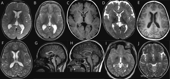
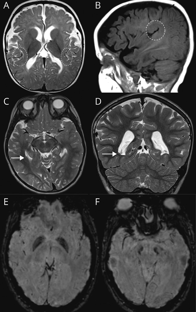
Results: We define AP-4-HSP as a disorder of gray and white matter and demonstrate that abnormal myelination is common and that metrics of reduced white matter volume correlate with severity of motor symptoms. We identify a common diagnostic imaging signature consisting of (1) a thin splenium of the corpus callosum, (2) an absent or thin anterior commissure, (3) characteristic signal abnormalities of the forceps minor ("ears of the grizzly sign"), and (4) periventricular white matter abnormalities. The presence of 2 or more of these findings has a sensitivity of ∼99% for detecting AP-4-HSP; the combination of all 4 is found in ∼45% of cases. Compared to other HSPs with a thin corpus callosum, the absent anterior commissure appears to be specific to AP-4-HSP. Our analysis identified a subset of patients with polymicrogyria, underscoring the role of AP-4 in early brain development. These patients displayed a higher prevalence of seizures and status epilepticus, many at a young age.

Discussion: Our findings define the MRI spectrum of AP-4-HSP, providing opportunities for early diagnosis, identification of individuals at risk for complications, and a window into the role of the AP-4 complex in brain development and neurodegeneration.


Figures

Figure 1
Figure 1. AP-4-HSP Is a Disorder of Gray and White Matter
(A) T1-weighted sagittal image at the level of the midline (patient with AP4B1-related hereditary spastic paraplegia [HSP]: AP4B1 [NM_001253852.3]: c.1345A>T [p.Arg449Ter]/c.1160_1161delCA [p.Thr387ArgfsTer30]; age: 11 months). Underdevelopment of the corpus callosum is one of the defining features of adaptor protein complex 4–associated HSP (AP-4-HSP), present in 94% of patients. This includes prominent thinning of the splenium and a decreased anterior-posterior diameter. (B) T2-weighted axial image demonstrating severe reduction in white matter volume, particularly in the posterior, periventricular white matter that, in combination with the thinning of the posterior corpus callosum, gives rise to ex vacuo ventriculomegaly in a colpocephalic configuration (patient with AP4B1-related HSP: AP4B1 [NM_001253852.3]: c.664delC [p.Leu222CysfsTer31]/c.664delC [p.Leu222CysfsTer31]; age: 6 years). (C) T2-weighted axial image demonstrating mild reduction in white matter volume as is the case in the majority of patients (patient with AP4B1-related HSP: AP4B1 [NM_001253852.3]: c.530_531insA [p.Asn178GlufsTer20]/c.114-2A>C; age: 4 years). (D) T1-weighted sagittal image showing cerebellar atrophy, which is overall a rare finding, present in only 3% of cases and mainly in patients with advanced disease (patient with AP4B1-associated HSP: AP4B1 [NM_001253852.3]: c.1177C>T [p.Arg393Ter]/c.1177C>T [p.Arg393Ter]; age: 19 years).
Figure 2
Figure 2. AP-4-HSP Presents With Abnormal Periventricular White Matter
(A, B) T2-weighted axial image showing delayed myelination at age 1.5 years (A) with evidence of continued myelination at age 9 years (B) (patient with AP4E1-related hereditary spastic paraplegia [HSP]: AP4E1 [NM_007347.5]: c.652_653delGA [p.Asp218CysfsTer13]/c.652_653delGA [p.Asp218CysfsTer13]; age: 9 years). (C, D) T1-weighted axial image (C) and T2-weighted axial image (D) showing hypointense signal on T1 and hyperintense signal on T2 images in the forceps minor consistent with the ears of the lynx sign, found in 11% of cases (patient with AP4S1-associated SPG52: AP4S1 [NM_007077.4]: c.289C>T [p.Arg97Ter]/c.289C>T [p.Arg97Ter]; age: 12 months). (E, F) T1-weighted axial image (E) and T2-weighted axial image (F) showing short and round T1 hypointense and T2 hyperintense signal in the forceps minor. Based on this appearance and in reference to the ears of the lynx sign, we term this the ears of the grizzly sign, which is present in 48% of cases (patient with AP4B1-related HSP: AP4B1 [NM_001253852.3]: c.664delC [p.Leu222CysfsTer31]/c.664delC [p.Leu222CysfsTer31]; age: 6 years). (G–J) T1-weighted sagittal (G, H) image demonstrating an absent anterior commissure in adaptor protein complex 4–associated HSP (AP-4-HSP), contrasting findings in other forms of complex HSP with a thin corpus callosum such as SPG15. (I, J) The anterior commissure is confirmed to be absent in the axial plane on T2-weighted axial images. Overall a thin or absent anterior commissure is found in 96% of AP-4-HSP cases (G and I: patient with AP4S1-related HSP: AP4S1 [NM_007077.4]: c.138+3_138+6delAAGT/c.138+3_138+6delAAGT; age: 12 years; H and J: patient with SPG15: ZFYVE26 [NM_015346.4]: c.4312C>T [p.Arg1438Ter]/c.837T>G [p.Tyr279Ter]; age: 19 years).
Figure 3
Figure 3. Malformations of Cortical Development in AP-4-HSP
(A, B) T2-weighted axial (A) and T1-weighted sagittal (B) images demonstrating perisylvian polymicrogyria, found in up to 25% of cases (shown is a patient with AP4S1-related hereditary spastic paraplegia [HSP]: AP4S1 [NM_007077.4]: c.294+1G>T/c.294+1G>T). (C, D) Periventricular nodular heterotopia in 1 patient with adaptor protein complex 4–associated HSP (AP-4-HSP) (patient with AP4B1-related HSP: AP4B1 [NM_001253852.3]: c.530_531insA [p.Asn178GlufsTer20]/c.114-2A>C; age: 4 years). (E, F) Susceptibility-weighted axial images showing evidence of iron accumulation in the globus pallidus (E) and substantia nigra (F) in a patient with AP4M1-related HSP, previously reported by Roubertie et al.30 in 2018 (patient with AP4M1-related HSP: AP4M1 [NM_004722.4]: c.916C>T [p.Arg306Ter]/c.916C>T [p.Arg306Ter]).
Figure 4
Figure 4. Radioclinical Correlations in AP-4-HSP
Several metrics of white matter (WM) volume inversely correlate with Spastic Paraplegia Rating Scale (SPRS) scores as an indicator of motor impairment and associated complications. This includes (A) the thickness of the splenium of the corpus callosum (Pearson correlation coefficient: r2 = 0.14, p = 0.02) and (B) the width of the periatrial WM (Pearson correlation coefficient: r2 = 0.11, p = 0.03). (C) No such correlation is found for the fronto-occipital horn ratio (FOHR) as a surrogate of ventriculomegaly, indicating that this metric lacks sensitivity (Pearson correlation coefficient: r2 = 0.02, p = 0.32). By contrast, indicators of gray matter volume such as (D) the thickness of the precentral gyrus or (E) diameter of the head of the caudate did not correlate with SPRS scores. (F) Venn diagram of key MRI findings in adaptor protein complex 4–associated hereditary spastic paraplegia (AP-4-HSP). Key imaging findings in AP-4-HSP include (1) reduced thickness of the splenium of the corpus callosum, (2) an absent or abnormally thin anterior commissure (AC), (3) abnormal signal of the forceps minor, and (4) periventricular WM (PVWM) signal abnormalities. All 4 features were present in 45% (33/73) of cases. A thin splenium and an absent or thin anterior commissure covered ∼84% (61/73) and more than 1 of the 4 findings was present in the large majority of cases (72/73; 99%).

References

    1. Shribman S, Reid E, Crosby AH, Houlden H, Warner TT. Hereditary spastic paraplegia: from diagnosis to emerging therapeutic approaches. Lancet Neurol. 2019;18(12):1136-1146. - PubMed
    1. Erichsen AK, Koht J, Stray-Pedersen A, Abdelnoor M, Tallaksen CM. Prevalence of hereditary ataxia and spastic paraplegia in southeast Norway: a population-based study. Brain. 2009;132(Pt 6):1577-1588. - PubMed
    1. Ebrahimi-Fakhari D, Teinert J, Behne R, et al. . Defining the clinical, molecular and imaging spectrum of adaptor protein complex 4-associated hereditary spastic paraplegia. Brain. 2020;143(10):2929-2944. - PMC - PubMed
    1. Ebrahimi-Fakhari D, Behne R, Davies AK, Hirst J. AP-4-associated hereditary spastic paraplegia. In: Adam MP, Ardinger HH, Pagon RA, et al., eds. GeneReviews®. University of Washington; 2018. - PubMed
    1. Jin SC, Lewis SA, Bakhtiari S, et al. . Mutations disrupting neuritogenesis genes confer risk for cerebral palsy. Nat Genet. 2020;52(10):1046-1056. - PMC - PubMed

Publication types

MeSH terms

Substances