. 2021 Sep 28;118(39):e2017460118.
doi: 10.1073/pnas.2017460118.

Extracellular matrix protein N-glycosylation mediates immune self-tolerance in Drosophila melanogaster

Affiliations

Extracellular matrix protein N-glycosylation mediates immune self-tolerance in Drosophila melanogaster

Nathan T Mortimer et al. Proc Natl Acad Sci U S A. .

Abstract

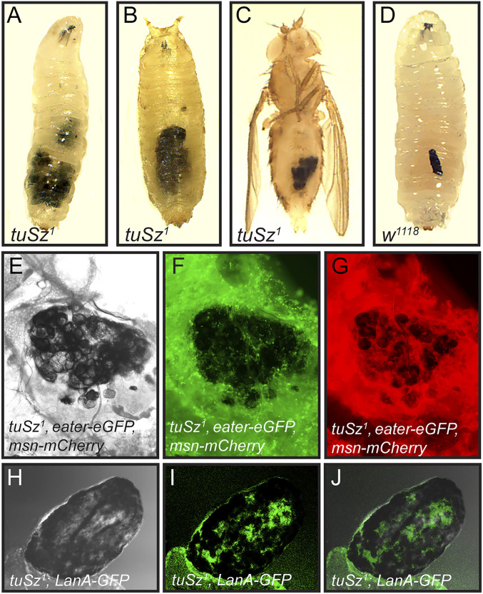
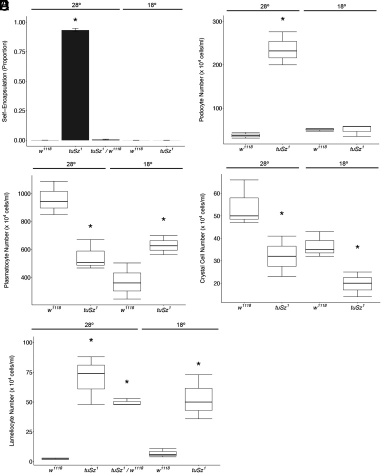
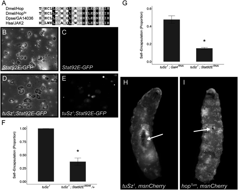
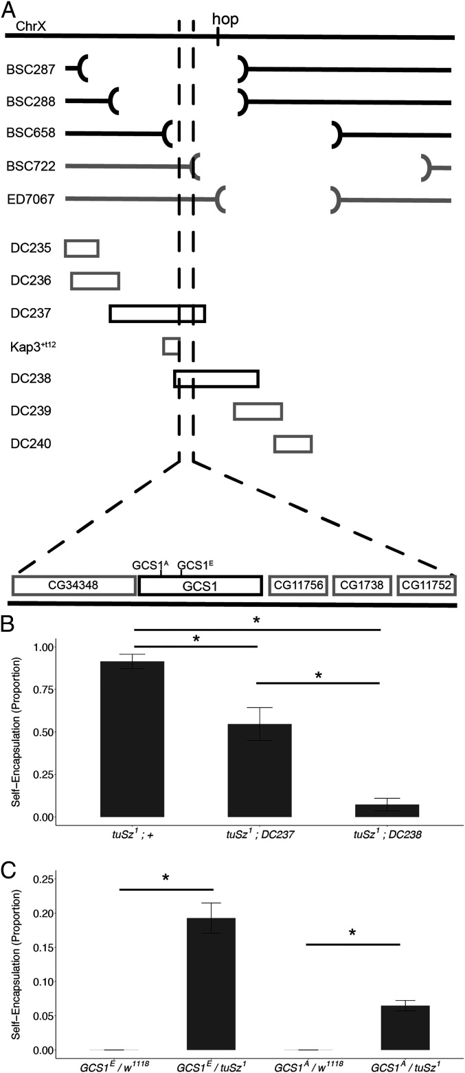
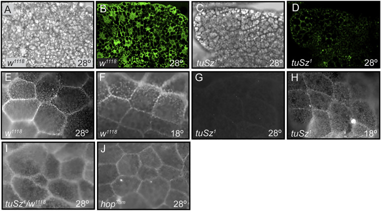
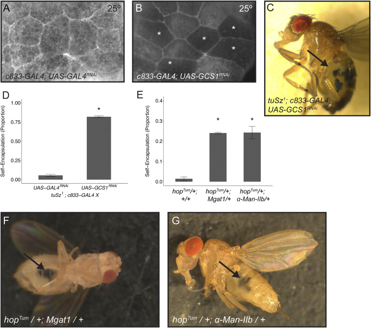
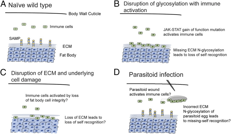
In order to respond to infection, hosts must distinguish pathogens from their own tissues. This allows for the precise targeting of immune responses against pathogens and also ensures self-tolerance, the ability of the host to protect self tissues from immune damage. One way to maintain self-tolerance is to evolve a self signal and suppress any immune response directed at tissues that carry this signal. Here, we characterize the Drosophila tuSz1 mutant strain, which mounts an aberrant immune response against its own fat body. We demonstrate that this autoimmunity is the result of two mutations: 1) a mutation in the GCS1 gene that disrupts N-glycosylation of extracellular matrix proteins covering the fat body, and 2) a mutation in the Drosophila Janus Kinase ortholog that causes precocious activation of hemocytes. Our data indicate that N-glycans attached to extracellular matrix proteins serve as a self signal and that activated hemocytes attack tissues lacking this signal. The simplicity of this invertebrate self-recognition system and the ubiquity of its constituent parts suggests it may have functional homologs across animals.

Keywords: autoimmunity; innate immunity; protein N-glycosylation; self-recognition; self-tolerance.


Conflict of interest statement

The authors declare no competing interest.

Figures

Fig. 1.
Fig. 1.
The tuSz self-encapsulation phenotype. (A–C) In tuSz1 mutants at 28 °C, posterior fat body tissues are melanized during the third-instar larval stage (A), and the melanization persists through the pupal (B) and adult stages (C). The self-encapsulation phenotype is morphologically similar to the encapsulation of a Leptopilina clavipes (parasitoid wasp) egg by a w1118 control larva (D). Brightfield (E), eGFP (F), and mCherry (G) images of fat bodies dissected from tuSz1, eater-eGFP, msn-mCherry larvae demonstrate that plasmatocytes (marked by eGFP expression) and lamellocytes (marked by mCherry expression) interact with fat body tissue that is undergoing melanization. Brightfield (H), GFP (I), and merged (J) images of fat bodies dissected from tuSz1; LanA-GFP larvae show that self-encapsulated fat body tissue nevertheless maintains intact ECM (marked by GFP expression).
Fig. 2.
Fig. 2.
Phenotypic characterization of tuSz flies. (A) Penetrance of the self-encapsulation phenotype of w1118 (control), tuSz1, and tuSz1/w1118 heterozygous flies raised at the indicated temperature. *P < 0.05 compared to w1118 at 28 °C. (B–E) Plasmatocyte number (B), lamellocyte number (C), podocyte number (D), and crystal cell number (E) of the indicated genotypes at the indicated temperatures. *P < 0.05 compared to w1118 at each temperature. P values for A and C were determined by Dunnett’s test. P values for B, D, and E were determined by Welch’s two-sample t test.
Fig. 3.
Fig. 3.
The role of JAK-STAT signaling in tuSz1 mutants. (A) An amino acid sequence alignment within the JH2 domain of Hop and the Hop orthologs GA14036 from Drosophila pseudoobscura and JAK2 from humans shows the missense D. melanogaster HopSz allele. (B–E) Brightfield (B) and GFP (C) images of immune cells from Stat92E-GFP larvae raised at 28 °C show no JAK-STAT pathway activity. Brightfield (D) and GFP (E) images of immune cells from tuSz1; Stat92E-GFP larvae raised at 28 °C show JAK-STAT pathway activity. (F) Penetrance of the self-encapsulation phenotype in tuSz1 and tuSz1; Stat92E06346/+ flies raised at 28 °C. *P < 0.05 compared to tuSz1. (G) Penetrance of the self-encapsulation phenotype in tuSz1; msn-GAL4; UAS-GAL4RNAi and tuSz1; msn-GAL4; UAS-Stat92ERNAi flies raised at 28 °C. *P < 0.05 compared to tuSz1; msn-GAL4; UAS-GAL4RNAi. P values were determined by generalized linear models. (H and I) Lamellocytes (marked by mCherry expression) aggregate around large self-encapsulations in tuSz1, msn-mCherry larvae (arrow in H) but are dispersed throughout hopTum, msn-mCherry larvae (I). Tissue self-encapsulations are not seen in hopTum, msn-mCherry larvae, but small melanized nodules are common (arrow in I).
Fig. 4.
Fig. 4.
Mapping the hypothesized tuSz1 self-tolerance mutation. (A) Schematic demonstrating the mapping of the tuSz1 recessive mutation on the X chromosome. Lines are listed by strain name. The hop gene locus is indicated on the chromosome. (Top) Interrupted segments illustrate the location of genome deletions in the indicated Drosophila strains. Black lines indicate deletions that fail to complement the tuSz1 self-encapsulation phenotype. Gray lines indicate complementing deletions. (Middle) Blocks represent duplicated regions of the X chromosome. Black blocks indicate duplications that rescue the tuSz1 self-encapsulation phenotype, and gray blocks indicate failure to rescue. (Bottom) The deletion and duplication lines indicate a small locus for the tuSz1 SAMP mutation. Candidate genes are shown. (B) Penetrance of the self-encapsulation phenotype of tuSz1 flies in combination with the indicated duplication raised at 28 °C. *P < 0.05 for the indicated comparisons. (C) Penetrance of the self-encapsulation phenotype of tuSz1 heterozygous flies in combination with w1118 controls or the indicated GCS1 allele raised at 28 °C. *P < 0.05 for the indicated comparisons. P values for B and C were determined by Dunnett’s test.
Fig. 5.
Fig. 5.
Fat body GCS1 expression and protein N-glycosylation. (A–D) Posterior fat body tissue dissected from early third-instar w1118 (A and B) and tuSz1 (C and D) larvae raised at 28 °C prior to tissue melanization. (A and C) Brightfield images of dissected fat body tissue demonstrating the absence of tissue melanization. (B and D) Corresponding fluorescent images of α-GCS1 antibody staining. (E–J) Magnified posterior fat body tissue dissected from early third-instar larvae and stained by FITC-WGA to assay protein N-glycosylation. w1118 fat body from larvae raised at 28 °C (E) or 18 °C (F) is positive for FITC-WGA staining. tuSz1 fat body tissue is negative for FITC-WGA at 28 °C (G) but positive at 18 °C (H). tuSz1/w1118 heterozygous (I) and hopTum (J) larvae raised at 28 °C are positive for FITC-WGA staining, indicating that hop mutations do not affect fat body ECM protein N-glycosylation.
Fig. 6.
Fig. 6.
Loss of GCS1 leads to loss of self-tolerance. (A and B) Posterior fat body tissue dissected from larvae raised at 25 °C and stained with FITC-WGA to assay protein N-glycosylation. Control c833-GAL4; UAS-GAL4RNAi tissue is positive for FITC-WGA staining (A). c833-GAL4; UAS-GCS1RNAi shows a mosaic loss of FITC-WGA staining, where * marks fat body cells with decreased staining (B). (C) tuSz1; c833-GAL4; UAS-GCS1RNAi flies raised at 25 °C show melanized self-encapsulations (indicated by the arrow). (D) Penetrance of the self-encapsulation phenotype in control tuSz1; c833-GAL4; UAS-GAL4RNAi and tuSz1; c833-GAL4; UAS-GCS1RNAi flies raised at 25 °C. *P < 0.05 compared to tuSz1; c833-GAL4; UAS-GAL4RNAi. (E) Penetrance of the self-encapsulation phenotype in hopTum/+, hopTum/+; Mgat1KG02444/+, and hopTum/+; α-Man-IIbMI09613/+ flies raised at 28 °C. *P < 0.05 compared to hopTum/+. (F) hopTum/+; Mgat1KG02444/+ flies raised at 28 °C show melanized self-encapsulations (indicated by the arrow). (G) hopTum/+; α-Man-IIbMI09613/+ flies raised at 28 °C show melanized self-encapsulations (indicated by the arrow). P values (D and E) were determined by generalized linear models.
Fig. 7.
Fig. 7.
Models describing the necessity for two independent signals in fly encapsulation responses. (A) Homeostasis is maintained in naïve wild-type larvae. (B) In tuSz1 mutant larvae, immune cells are inappropriately activated by JAK-STAT pathway activation due to the hopSz gain-of-function mutation. The loss of protein N-glycosylation in posterior fat body tissue due to the GCS1Sz mutation leads to loss of self-tolerance and tissue encapsulation. (C) In the model of self-tolerance described by Kim and Choe (55), the coupled phenotypes of loss of cell integrity and loss of ECM integrity are sufficient to disrupt self-tolerance. (D) Immune cells are activated following parasitoid wasp infection, presumably due to the wound-mediated activation of JAK-STAT signaling. SAMP-presenting host tissues are protected from encapsulation, and wasp eggs may be targeted for encapsulation because they are missing the ECM N-glycosylation SAMP.

References

    1. Danilova N., The evolution of immune mechanisms. J. Exp. Zoolog. B Mol. Dev. Evol. 306, 496–520 (2006). - PubMed
    1. Janeway C. A. Jr, Medzhitov R., Innate immune recognition. Annu. Rev. Immunol. 20, 197–216 (2002). - PubMed
    1. Medzhitov R., Janeway C. A. Jr, Decoding the patterns of self and nonself by the innate immune system. Science 296, 298–300 (2002). - PubMed
    1. Babcock D. T., et al. ., Circulating blood cells function as a surveillance system for damaged tissue in Drosophila larvae. Proc. Natl. Acad. Sci. U.S.A. 105, 10017–10022 (2008). - PMC - PubMed
    1. Cooper M. D., Alder M. N., The evolution of adaptive immune systems. Cell 124, 815–822 (2006). - PubMed

Publication types

MeSH terms

LinkOut - more resources