. 2021 Sep 28;118(39):e2106369118.
doi: 10.1073/pnas.2106369118.

Norrie disease protein is essential for cochlear hair cell maturation

Affiliations

Norrie disease protein is essential for cochlear hair cell maturation

Yushi Hayashi et al. Proc Natl Acad Sci U S A. .

Abstract

Mutations in the gene for Norrie disease protein (Ndp) cause syndromic deafness and blindness. We show here that cochlear function in an Ndp knockout mouse deteriorated with age: At P3-P4, hair cells (HCs) showed progressive loss of Pou4f3 and Gfi1, key transcription factors for HC maturation, and Myo7a, a specialized myosin required for normal function of HC stereocilia. Loss of expression of these genes correlated to increasing HC loss and profound hearing loss by 2 mo. We show that overexpression of the Ndp gene in neonatal supporting cells or, remarkably, up-regulation of canonical Wnt signaling in HCs rescued HCs and cochlear function. We conclude that Ndp secreted from supporting cells orchestrates a transcriptional network for the maintenance and survival of HCs and that increasing the level of β-catenin, the intracellular effector of Wnt signaling, is sufficient to replace the functional requirement for Ndp in the cochlea.

Keywords: Norrie disease; Wnt signaling; cochlear hair cells.


Conflict of interest statement

The authors declare no competing interest.

Figures
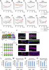
Fig. 1.
Fig. 1.
Ndp KO mice exhibit progressive hearing loss and OHC death. (A) ABR thresholds were increased in male KO (XY) mice at 1 mo (WT: n = 14, KO: n = 10; P < 0.0001, ANOVA; post hoc tests at 5.66, 8.00, 11.33, and 45.25 kHz). (B) ABR threshold elevations in KO mice were more severe at 2 mo (WT: n = 10, KO: n = 9; P < 0.0001, ANOVA; post hoc tests at all frequencies except 45.25 kHz). (C and D) DPOAE thresholds in KO mice were unchanged at 1 mo (C, WT: n = 14, KO: n = 10; P < 0.0001, ANOVA) but were elevated at 2 mo (D, WT: n = 10, KO: n = 9; P < 0.0001, ANOVA; post hoc tests at 8.00 and 11.33 kHz). (E) ABR thresholds were increased across frequencies in female KO (XX) mice and at high frequencies in heterozygous (X+X, Hetero) mice at 1 mo (WT: n = 5, Hetero: n = 7, KO: n = 6; P < 0.0001, ANOVA; post hoc tests at 8.00, 11.33, 16.00, 22.65, 32.00, and 45.25 kHz in KO mice and at 45.25 kHz in heterozygous mice). (F) ABR thresholds were further elevated across frequencies in female KO mice and at 32.00 and 45.25 kHz in heterozygous mice at 2 mo (WT: n = 4, Hetero: n = 7, KO: n = 5; P < 0.0001, ANOVA; post hoc tests at 5.66, 8.00, 11.33, 16.00, 22.65, 32.00, and 45.25 kHz in KO mice and at 32.00 and 45.25 kHz in heterozygous mice). (G and H) DPOAE thresholds in KO mice were elevated at 1 mo (G, WT: n = 5, Hetero: n = 7, KO: n = 6; P < 0.0001, ANOVA; post hoc tests at 11.33 and 32.00 kHz in KO mice) and were further elevated at 2 mo in KO mice (H, WT: n = 4, Hetero: n = 7, KO: n = 5; P < 0.0001, ANOVA; post hoc tests at 8.00 and 11.33 kHz in KO mice). (A–H) Error bars represent SEs. *P < 0.05, **P < 0.01, ***P < 0.001, ****P < 0.0001 by Fisher’s least significant difference method. (I) Diagrams of the organ of Corti including HCs, supporting cells, and blood vessels (arrowheads) (Upper: section view, YZ plane; Lower: surface view, XY plane; greater epithelial ridge: GER, inner sulcus: ISu, inner border cell: IBC, pillar cell: PC, Deiters’ cell: DC, Hensen’s cell: HeC, Claudius’ cell: ClC, inner hair cell: IHC, and outer hair cell: OHC). (J) In the WT, HCs were positive for Myo7a at the base of the cochlea in both nuclear and cuticular plate layers. Computational sections showed Myo7a-positive HCs (PCs and DCs are positive for Sox2, a supporting cell marker). (K) In the KO, many OHCs at the base of the cochlea were negative for Myo7a as viewed in the nuclear layer, with weak positive immunoreactivity in the cuticular plate layer, and some OHCs were missing (arrows). Computational sections showed Myo7a-negative OHCs in the base. (J and K) (Scale bars in whole mounts and computational sections indicate 50 and 10 μm, respectively.) (L and M) Significant loss of OHCs (L) and decreased numbers of Myo7a-positive OHCs (M) were observed at the base of the KO (n = 4) compared to the WT (n = 4) cochlea. No loss of OHCs was observed at the apex. Cell numbers are given as cells/high-power field (hpf). (N and O) No loss of IHCs was observed in the KO cochlea (N), but a tendency toward decreased numbers of Myo7a-positive IHCs in the base and a significant decrease in Myo7a-positive IHCs in the apex in the KO (n = 4) as compared to the WT (n = 4) cochlea were observed (O). (L–O) Error bars represent SEs. *P < 0.05, **P < 0.001, ***P < 0.0001.
Fig. 2.
Fig. 2.
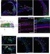
Expression pattern of Ndp and Fzd4 mRNA in the cochlea at P28. (A) Ndp expression was observed widely under the IHCs and OHCs, in the inner sulcus (ISu), outer sulcus (OSu), and spiral ligament (SL) in a cryosection view of the cochlea (Left). High-power view of the organ of Corti (Middle) shows staining of Ndp in supporting cells (SCs), including DCs, HeCs, and ClCs. Cd31(arrowheads) is expressed in endothelial cells lining the capillaries under the organ of Corti. High-power view of the lateral wall (Right) shows Ndp expression in the SL and Cd31-positive endothelial cells (arrows) in the vessels of the stria vascularis (SV). (B) Diagram of the organ of Corti with the confocal planes of the SC and HC layers indicated by dashed lines. The SC layer shows Ndp expression in the inner sulcus (ISu), DCs, and HeCs, and the HC layer shows Ndp expression in the inner sulcus (ISu) and ClCs. (C) In situ hybridization showed Fzd4 mRNA expression in HCs, IBCs, DCs, HeCs, ClCs, and endothelial cells under the organ of Corti (arrowhead). Fzd4 mRNA and DAPI are shown alone (Upper) and merged with Myo7a and Cd31 (Lower). (D) Fzd4 mRNA was expressed in endothelial cells of the SV and SL. Arrowheads and arrows indicate Cd31-positive endothelial cells in the SV and the spiral ligament (SL), respectively. Fzd4 mRNA and DAPI alone (Left) and merged with Cd31 (Right). (Scale bars, 50 μm.)
Fig. 3.
Fig. 3.
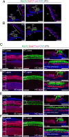
Myo7a-negative HCs in the Ndp KO cochlea. (A and B) Cd31 immunostaining (red) indicates the locations of capillaries in the organ of Corti (arrowheads) and lateral wall in cryosections (Left) from the WT (A) and KO (B) cochlea at P3. Arrowheads indicate endothelial cells in a high-power view (Middle) of the organ of Corti. Arrows indicate endothelial cells in a high-power view (Right) of the stria vascularis (SV). There were no obvious changes in endothelial cell morphology. (C and D) A WT cochlear sensory epithelium had Sox2-positive (red) supporting cells (Left, SC layer) and Myo7a-positive (green) HCs (Middle, HC layer) in surface views and computational sections (Right) at the base (C) and apex (D) of the cochlea. (E and F) An Ndp KO cochlear sensory epithelium had normal supporting cells (Left, SC layer), but both surface views (Middle) and computational sections (Right) showed Myo7a-negative HCs (arrowheads) at the base of the cochlea. (Scale bars, 50 μm.)
Fig. 4.
Fig. 4.
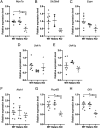
Altered expression of HC markers in the Ndp KO cochlea. (A–H) Expression of genes critical for HC development and function assessed by qRT-PCR in sorted HCs from the Atoh1-GFP;Ndp KO mouse cochlea. ANOVA followed by post hoc Bonferoni test revealed that expression levels of Myo7a, Pou4f3, and Gfi1 were significantly decreased in HCs from the KO cochlea as compared to the WT and heterozygous cochlea (P = 0.0052 for Myo7a, P = 0.0037 for Pou4f3, and P = 0.0076 for Gfi1). Error bars represent SEs. *P < 0.01.
Fig. 5.
Fig. 5.
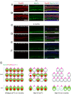
Pou4f3 expression is lost in HCs in Ndp KO cochlea. (A and B) Pou4f3 expression was assessed by immunohistochemistry in the WT and KO cochlea at P3. HCs were positive for Pou4f3 in the base of the WT cochlea (A). Pou4f3-negative HCs (arrowheads) were observed in the base of the KO cochlea (B). (C–F) Pou4f3 expression pattern was evaluated in the WT and KO cochlea at 2 mo. Both IHC and OHC nuclear layers of the base are shown. All IHCs and OHCs in the base of the WT cochlea were positive for Pou4f3 (C and D). Most of IHCs and OHCs in the base of the KO cochlea showed weak or no expression of Pou4f3 (E and F). (Scale bars in AF, 50 μm.) (G) Diagram shows the progressive loss of Myo7a from the cytoplasm and Pou4f3 from the nucleus during HC maturation in the Ndp KO mouse cochlea. By 2 mo, the KO mouse shows significant OHC loss. Expression of espin (Espn) in the stereociliary bundles and prestin (Pres) in the OHC membrane is maintained.
Fig. 6.
Fig. 6.
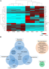
Differential expression analysis of Atoh1-GFP–positive cells in the Ndp WT and KO cochlea. (A) Differentially expressed genes from RNA-Seq (adjusted P < 0.05) show segregation between WT and Ndp KO. (B) Genes with adjusted P < 0.05 were analyzed in the Venn diagrams. Groups with significant changes include those regulating transcription (Pdzd2, Nr4a3, and Setd7), angiogenesis (Nampt and Edil3), ECM (Col6a4, Coch, and Edil3), cytoskeleton (Nyap2 and Vav1), cochlear development (Adam19), neuronal disease (Wdr93, Il1rapl1, Inpp5d, Cnrip1, and Slc17a7), and ER stress (Rrbp1). Of these, Nr4a3, Setd7, Edil3, Vav1, and Adam19, shown in the Center of the diagram, were Wnt related, and in addition, other significantly changed genes, Adcyap1r1, Sla, Cntnap4, Entpd3, and Map3k8 were correlated with Wnt signaling.
Fig. 7.
Fig. 7.
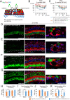
Forced Ndp expression in supporting cells after birth preserves HCs and rescues cochlear function in Ndp KO mice. (A) Diagram indicates the secretion of Ndp from Sox2-positive supporting cells and the greater epithelial ridge (GER)/inner sulcus (ISu) in the Ndp KO (XY);Sox2-CreER;Z/Norrin;tdTomato (Tm) ear and the mechanism for Ndp effects on HCs. (B) ABR thresholds showed recovery at 2 mo in Ndp KO (XY);Sox2-CreER;Z/Norrin;Tm mice (n = 5) as compared to Ndp KO (XY);Sox2-CreER;Tm (n = 7) and other controls (Ndp KO (XY);Z/Norrin;Tm (n = 2) and Ndp KO (XY);Tm (n = 2); P < 0.0001, ANOVA). Post hoc tests using Fisher’s least significant difference (LSD) method revealed significant differences at all frequencies except 45.25 kHz. (C) DPOAE thresholds in Ndp KO (XY);Sox2-CreER;Z/Norrin;Tm mice at 2 mo had recovered compared to the Ndp KO (XY);Sox2-CreER;Tm and other controls (Ndp KO (XY);Z/Norrin;Tm and Ndp KO (XY);Tm; P < 0.0001, ANOVA). Post hoc tests using Fisher’s LSD method revealed significant differences at 11.33, 16.00, 22.65, and 32.00 kHz. (B and C) Error bars represent SEs. *P < 0.05, **P < 0.01, ***P < 0.001, ****P < 0.0001 by Fisher’s LSD method. (D) Most OHCs and some IHCs (arrowheads) were negative for Myo7a in the base of the cochlea of Ndp KO (XY);Sox2-CreER;Tm mice (arrows indicate Myo7a-positive OHCs). Myo7a-negative OHCs are also seen (*) in computational sections (Right). (E) All but a few (^) IHCs were negative for Myo7a in the apex (arrowheads) as confirmed in computational sections (Right). (F) After forced expression of Ndp in Sox2-positive supporting cells and the greater epithelial ridge (GER)/inner sulcus (ISu) of the Ndp KO (XY);Sox2-CreER;Z/Norrin;Tm cochlea, many OHCs survived and retained Myo7a expression (arrows). Computational sections (Right) exhibited OHCs negative (*) and positive (arrows) for Myo7a. (G) IHCs expressing Myo7a were observed in the apex of the cochlea. (Scale bars in DG, 50 μm.) (H and I) A significant decrease in the number of OHCs (H) and Myo7a-positive OHCs (I) at the base of the cochlea in the Ndp KO (XY);Sox2-CreER;Tm (n = 3) was restored in the Ndp KO (XY);Sox2-CreER;Z/Norrin;Tm (n = 3) cochlea. (J and K) No loss of IHCs (J) was observed in the Ndp KO (XY);Sox2-CreER;Tm cochlea, but a significant decrease in Myo7a-positive IHCs (K) in the base and apex of the cochlea in the Ndp KO (XY);Sox2-CreER;Tm (n = 3) was restored in the Ndp KO (XY);Sox2-CreER;Z/Norrin;Tm cochlea (n = 3). (H–K) Error bars represent SEs. *P < 0.05, **P < 0.01 by Student’s t test.
Fig. 8.
Fig. 8.
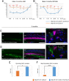
Stabilization of β-catenin in HCs rescues cochlear function in Ndp KO mice. (A and B) Ndp KO mice in which β-catenin was overexpressed in HCs by deletion of exon 3 of β-catenin using an Atoh1-Cre (Ndp KO (XY);Atoh1-Cre;β-cateninflox(Exon3)+; n = 5) showed significantly lower ABR (A) and DPOAE (B) thresholds (P < 0.0001, ANOVA) than control mice lacking Atoh1-Cre (Ndp KO (XY);β-cateninflox(Exon3)/+; n = 7). The line with stabilized β-catenin is referred to as Ndp KO (XY);Atoh1-Cre;Exon3 and the control is referred to as Ndp KO (XY);Exon3. Post hoc tests using Fisher’s least significant difference (LSD) method revealed significant difference at 5.66, 8.00, 11.33, 16.00, and 22.65 kHz in the ABR and 11.33, 16.00, 22.65, and 32.00 kHz in the DPOAE. (C) Ndp KO (XY);Exon3 mice showed loss of OHCs and loss of Myo7a in surviving OHCs (*) in the base of the cochlea. IHCs were positive for Myo7a. (D) Ndp KO (XY);Atoh1-Cre;Exon3 mice exhibited continued Myo7a immunoreactivity in OHCs (arrows) at the base of the cochlea. (Scale bars in C and D, 50 μm.) (E and F) The Ndp KO (XY);Atoh1-Cre;Exon3 mice (n = 4) showed significantly more surviving (E) and Myo7a-positive (F) OHCs than the Ndp KO (XY);Exon3 mice (n = 5; Student’s t test). (A, B, E, and F) Error bars represent SEs. *P < 0.05, **P < 0.01, ***P < 0.001, and ****P < 0.0001 by Fisher’s LSD method or Student’s t test.

References

    1. Ye X., Wang Y., Nathans J., The Norrin/Frizzled4 signaling pathway in retinal vascular development and disease. Trends Mol. Med. 16, 417–425 (2010). - PMC - PubMed
    1. Halpin C., Sims K., Twenty years of audiology in a patient with Norrie disease. Int. J. Pediatr. Otorhinolaryngol. 72, 1705–1710 (2008). - PubMed
    1. Luhmann U. F., et al. ., Role of the Norrie disease pseudoglioma gene in sprouting angiogenesis during development of the retinal vasculature. Invest. Ophthalmol. Vis. Sci. 46, 3372–3382 (2005). - PubMed
    1. Ke J., et al. ., Structure and function of Norrin in assembly and activation of a Frizzled 4-Lrp5/6 complex. Genes Dev. 27, 2305–2319 (2013). - PMC - PubMed
    1. Jin T., George Fantus I., Sun J., Wnt and beyond Wnt: Multiple mechanisms control the transcriptional property of beta-catenin. Cell. Signal. 20, 1697–1704 (2008). - PubMed

Publication types

MeSH terms

LinkOut - more resources