Primary Cardiac Angiosarcoma Accompanying Cardiac Tamponade
- PMID: 34544954
- PMCID: PMC9038456
- DOI: 10.2169/internalmedicine.8250-21
Primary Cardiac Angiosarcoma Accompanying Cardiac Tamponade
Abstract
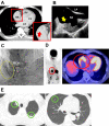
A de novo cardiac malignant tumor is rare and sometimes challenging to diagnose. We encountered a 67-year-old man without any medical history complaining of dyspnea on effort. On admission, his hemodynamics were deteriorated due to cardiac tamponade, which was improved by percutaneous drainage of 1,200 mL pericardial effusion, showing 11.0 g/dL of hemoglobin. We suspected primary cardiac malignancy following multidisciplinary tests, and a cardiac biopsy via sternotomy demonstrated the definitive diagnosis of primary malignant tumor (angiosarcoma) infiltrating the right atrial myocardium. We initiated weekly paclitaxel therapy. Further studies are warranted to establish the optimal diagnostic and therapeutic strategy for de novo cardiac malignancy.
Keywords: cardiogenic shock; heart failure; hemodynamics; pericardial effusion.
Conflict of interest statement
Figures


References
-
- Reynen K. Frequency of primary tumors of the heart. Am J Cardiol 77: 107, 1996. - PubMed
-
- Selkane C, Amahzoune B, Chavanis N, et al. . Changing management of cardiac myxoma based on a series of 40 cases with long-term follow-up. Ann Thorac Surg 76: 1935-1938, 2003. - PubMed
-
- Molina JE, Edwards JE, Ward HB. Primary cardiac tumors: experience at the University of Minnesota. Thorac Cardiovasc Surgeon 38 (Suppl 2): 183-191, 1990. - PubMed
-
- Truong PT, Jones SO, Martens B, et al. . Treatment and outcomes in adult patients with primary cardiac sarcoma: the British Columbia Cancer Agency experience. Ann Surg Oncol 16: 3358-3365, 2009. - PubMed
-
- Pearman JL, Wall SL, Chen L, Rogers JH. Intracardiac echocardiographic-guided right-sided cardiac biopsy: case series and literature review. Catheter Cardiovasc Interv. Forthcoming. - PubMed

