. 2021 Sep 20;12(1):5533.
doi: 10.1038/s41467-021-25844-5.

Identification of limb-specific Lmx1b auto-regulatory modules with Nail-patella syndrome pathogenicity

Affiliations

Identification of limb-specific Lmx1b auto-regulatory modules with Nail-patella syndrome pathogenicity

Endika Haro et al. Nat Commun. .

Abstract

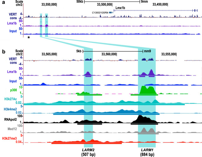
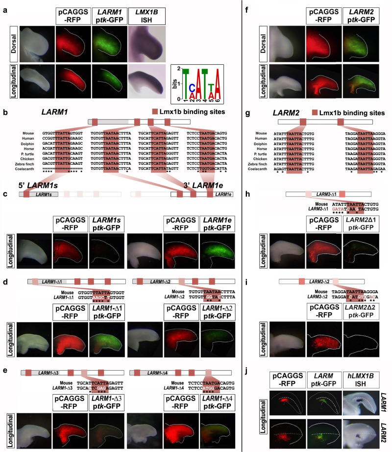
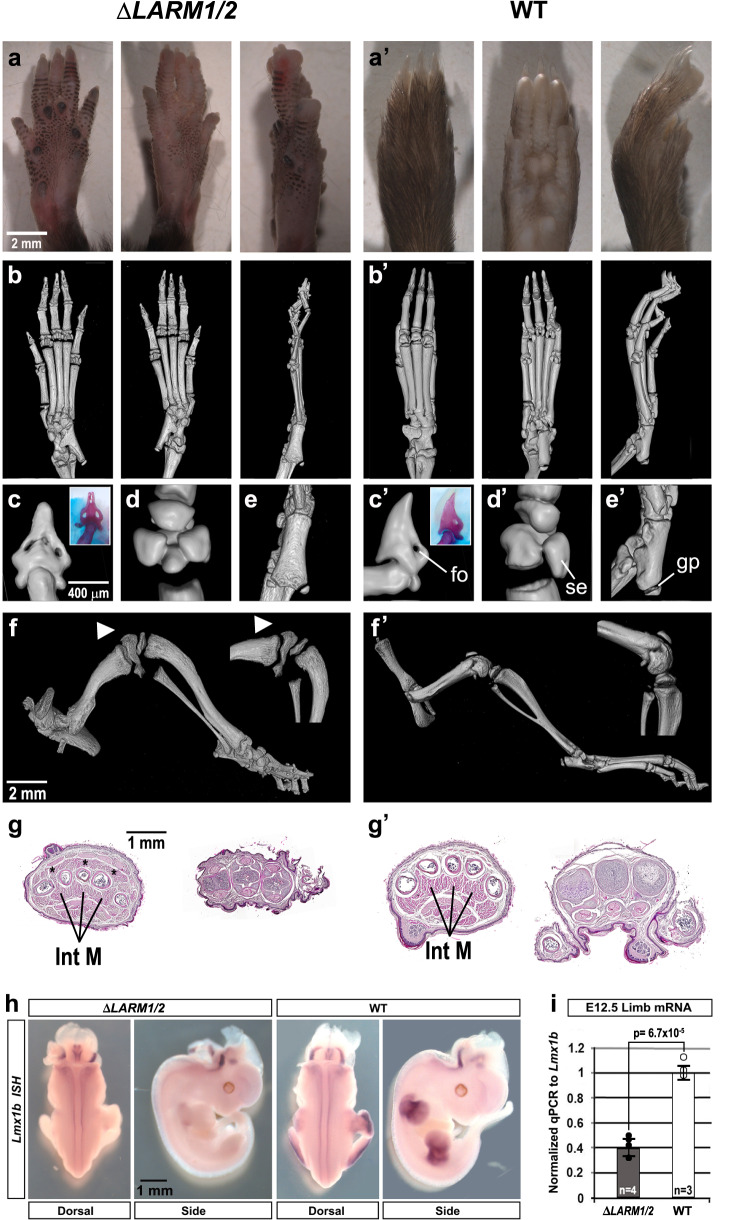
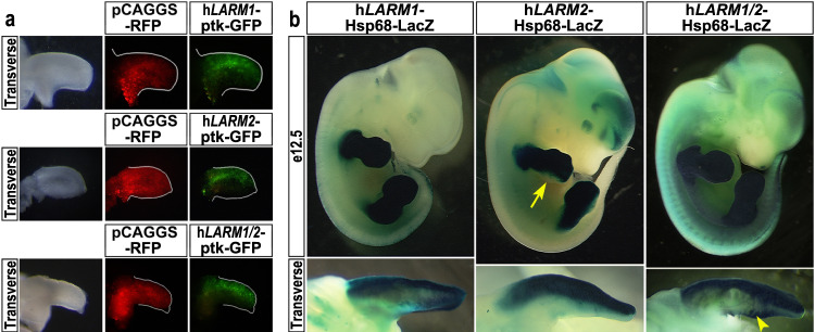
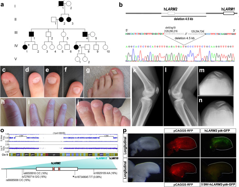
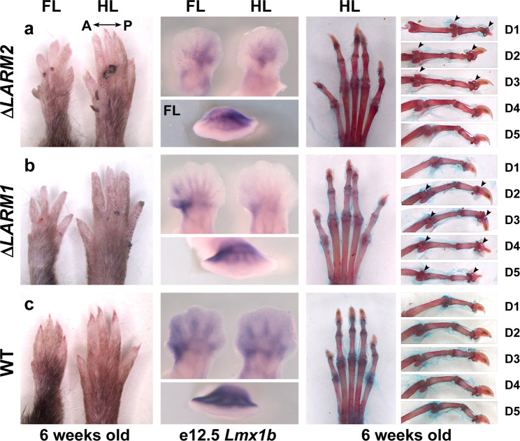
LMX1B haploinsufficiency causes Nail-patella syndrome (NPS; MIM 161200), characterized by nail dysplasia, absent/hypoplastic patellae, chronic kidney disease, and glaucoma. Accordingly in mice, Lmx1b has been shown to play crucial roles in the development of the limb, kidney and eye. Although one functional allele of Lmx1b appears adequate for development, Lmx1b null mice display ventral-ventral distal limbs with abnormal kidney, eye and cerebellar development, more disruptive, but fully concordant with NPS. In Lmx1b functional knockouts (KOs), Lmx1b transcription in the limb is decreased nearly 6-fold, indicating autoregulation. Herein, we report on two conserved Lmx1b-associated cis-regulatory modules (LARM1 and LARM2) that are bound by Lmx1b, amplify Lmx1b expression with unique spatial modularity in the limb, and are necessary for Lmx1b-mediated limb dorsalization. These enhancers, being conserved across vertebrates (including coelacanth, but not other fish species), and required for normal locomotion, provide a unique opportunity to study the role of dorsalization in the fin to limb transition. We also report on two NPS patient families with normal LMX1B coding sequence, but with loss-of-function variations in the LARM1/2 region, stressing the role of regulatory modules in disease pathogenesis.


Conflict of interest statement

The authors declare no competing interests.

Figures

Fig. 1
Fig. 1. LARM1 and LARM2 are conserved, bound by Lmx1b, and associated with active chromatin marks.
a UCSC genome screenshot displaying the Lmx1b locus and the associated cis-regulatory modules LARM1 and LARM2 (highlighted in blue) showing vertebrate conservation (VERT cons), Lmx1b binding (Lmx1b-targeted ChIP-seq) and input. b Magnification of the putative enhancer region displaying the overlap with active enhancer-associated regulatory marks present in limb buds. From top to bottom: vertebrate conservation (VERT cons), ChIP-seq tracks for Lmx1b, control Input limb DNA, p300, histone 3 acetylation at lysine 27 (H3K27Ac), histone 3 dimethylation at lysine 4 (H3K4me2), RNA polymerase II, Med 12, and histone 3 trimethylation at lysine 27 (H3K7me3). Note that LARM1 consists of two conserved peaks, both of which are recognized by Lmx1b-targeted ChIP-seq. Upstream of LARM2, another conserved region is present (asterisk). This potential cis-regulatory module is also associated with the 9430024E24Rik gene, but does not appear to be bound by Lmx1b, as shown in the Lmx1b-targeted ChIP-seq track (see also Supplementary Fig. 2).
Fig. 2
Fig. 2. The Lmx1b binding sites are necessary for dorsal LARM1 and LARM2 activity.
LARM1 (ae and j) and LARM2 (fj) reporter activity in chick wing buds 48 hrs after electroporation. Each assay/experiment includes: bright-field view of the electroporated limbs, RFP fluorescence image (red) reflecting transfection efficiency, and GFP fluorescent image (green) showing enhancer activity. Longitudinal views illustrate activity along the dorsoventral axis (dorsal on top). a LARM1 activity is restricted to the dorsal mesoderm (n = 22) coincident with Lmx1b expression (Lmx1b ISH for comparison). Inset showing the TMATWA consensus DNA binding motif for Lmx1b. b Conserved Lmx1b binding sites (LBS) are shaded clay-red in LARM1 schematics and sequences. An asterisk indicates sequence variations across species. c Analysis of the isolated 5’LARM1 element (LARM1s) does not convey enhancer activity (n = 4), while the isolated 3’LARM1 element (LARM1e) is active in both dorsal and ventral mesoderm (n = 16). d Left panel. Site-directed mutagenesis of the 5′ LARM1s LBS in the full LARM1 construct (LARM11, n = 5) expands the activity into the ventral mesoderm indicating that the LBS is necessary for restriction of dorsal activity (three different site-directed mutants were generated to ensure that a new permissive/gain of function LBS was not created— Supplementary Fig. 1). d Right panel and e Disruption of any of the LBS in the 3′ LARM1e leads to a marked reduction in enhancer activity (LARM12, n = 7; LARM13, n = 5; LARM14, n = 5). f LARM2 activity is restricted to the dorsal mesoderm (n = 13) coincident with LMX1B expression (shown in (a)). g Two highly conserved LBS are present in LARM2 (shaded clay-red as in (b)). h, i Disruption of either LBS leads to a loss in LARM2 activity (LARM2-Δ1, n = 5; LARM2- Δ2, n = 6). Nucleotides altered by site-directed mutagenesis are indicated in white and by an asterisk below the sequence. Dorsal or longitudinal views of the limbs are indicated on the left. j Ectopic expression of human LMX1B in the ventral mesoderm drives activity of the co-transfected LARM1 or LARM2-reporter constructs (LARM1 n = 4; LARM2 n = 4). The human LMX1B probe used to demonstrate LMX1B expression by in situ hybridization does not cross-react with the dorsal expression of chicken LMX1B.
Fig. 3
Fig. 3. Mice lacking the LARM region exhibit a double ventral limb phenotype.
a-a’ Dorsal, ventral and lateral gross morphology of hindlimbs (forelimb morphology in Supplementary Fig. 3). b-b’ microCT scan views of a 3-week-old ΔLARM1/2 homozygous mouse hindlimbs showing footpad development and the absence of nails and hair in the dorsal autopod compared to wild type (WT). ce Magnified views of the digit tips (inset alizarin red staining of the distal phalanx) showing symmetrical ventral features, i.e., bony ventral foramen (fo) associated with the toe pad, symmetrical ventral sesamoid bones (se) of the metatarsal-phalangeal joint, and the ventrally oriented growth plate (gp) of the proximal tali display dorsoventral symmetry in the absence of the LARM region. Compare with the normal dorsoventral asymmetry of wild-type controls (c´–e´). In images ce’ dorsal is to the left. f-f’ The patella is absent in ΔLARM1/2 mice (hindlimb lateral view, white arrowheads). g-g´ Transverse sections of the autopod show duplicated flexor tendons and intrinsic muscles (Int M) (asterisk) in the ΔLARM1/2 mouse (n = 3, wild type n = 2). h In situ hybridization of Lmx1b expression in limb buds is below detection in animals lacking the LARM region, while expression in the neural tube is equivalent. i Comparative RT-qPCR analyses of Lmx1b mRNA levels in the whole hindlimb bud of e12.5 ΔLARM1/2 (black dots) and WT (white dots) embryos. The level of expression of the mutant is 40% of the control (set to 1). P = 6.7 × 10−5 (two-tailed, unpaired t-593 test, error bars represent standard deviation) (control limb n = 3, mutant limbs n = 4). Source data for the RT-qPCR are provided as Supplementary Data 3.
Fig. 4
Fig. 4. The human LARM region also exhibits dorsal enhancer activity.
a Human LARM constructs electroporated into chick wing buds display dorsally restricted expression (hLARM1, n = 13; hLARM2, n = 7; hLARM1/2 region, n = 3). b Similarly, transgenic mice containing the human LARM sequences linked to a LacZ reporter demonstrate dorsally accentuated activity. All three LARM1 transgenic embryos display limb-restricted, dorsally accentuated activity. LARM2 transgenic embryos exhibit tight dorsally restricted activity in the limb (5/5). LacZ staining is reduced-to-absent in the posterior distal autopod mesoderm (5th digit region, yellow arrow). Transgenic embryos containing both hLARM1/2 (~8 kb, hLARM1/2) also show activity restricted to the dorsal limb (7/7). However, focal ventral activity at the zeugopod/autopod junction is also evident (yellow arrowhead).
Fig. 5
Fig. 5. Clinical features of LARM loss-of-function.
a Pedigree of a family with a LARM deletion. b The 4.5 kb region deleted removes all of LARM2. cg, kn Phenotypic images of individual IV-7. cd Koïlonychia of thumb and 2nd finger. ef Triangular lunulae of 3rd and 4th fingers. g Nail dysplasia of the hallux showing longitudinal striations. hj Phenotypic images of individual IV-1. h Koïlonychia of thumb. i Hypoplastic nails, ungueal dysplasia of 2nd finger. j Ungueal dysplasia of right foot predominating on 1st and 5th toes. kn Knee X-rays showing bilateral hypoplasia of the patella. o Schematic of the patient’s chromosome 9 showing large segments of the chromosome with loss of heterozygosity (LOH), i.e., homozygosity, when comparing the allele frequencies to the Log R ratio of the alleles. One of the homozygous regions includes the LARM-LMX1B locus. The homozygous hLARM2 sequence showing the five single nucleotide variations (SNVs). The asterisk indicates the rare (0.08%) sequence in LARM2. The LMX1B binding sites are indicated as clay-red boxes. p Using site-directed mutagenesis, we generated a human LARM2 construct containing the patient´s 5 SNVs; following electroporation into embryonic chick wings, the patient-LARM2 sequence showed markedly reduced activity (n = 6; compare with the activity of the common hLARM2 sequence in Fig. 4a). Interestingly, mutation of only the single SNV within the conserved LARM2 region did not alter LARM2 activity.
Fig. 6
Fig. 6. Individual deletions of LARM1 and LARM2 reveal posterior and anterior contributions, respectively, to dorsalization.
a Homozygous ΔLARM2 mouse forelimb (FL) and hindlimb (HL) autopods showing loss of dosalization exclusively in the anterior aspect at 6 weeks (left), reduced anterior expression of Lmx1b in e12.5 limb buds (middle) and ventral-ventral morphology specifically in anterior 1–3 digits (right). b Homozygous ΔLARM1 mouse showing loss of dorsalization that is accentuated posteriorly at 6 weeks (left), reduced posterior expression of Lmx1b in e12.5 limb buds (middle) and ventral-ventral morphology specifically in digits 2–5 (right). Note the progressive loss of dorsalization towards digit 5 (e.g., increasing size of duplicated ventral sesamoid bones). c Equivalent views of a wild-type littermate for comparison. Arrowheads highlight the duplicated ventral sesamoid bones and distal phalanges.

References

    1. Riddle RD, et al. Induction of the LIM homeobox gene Lmx1 by WNT7a establishes dorsoventral pattern in the vertebrate limb. Cell. 1995;83:631–640. doi: 10.1016/0092-8674(95)90103-5. - DOI - PubMed
    1. Vogel A, Rodriguez C, Warnken W, Izpisua Belmonte JC. Dorsal cell fate specified by chick Lmx1 during vertebrate limb development. Nature. 1995;378:716–720. doi: 10.1038/378716a0. - DOI - PubMed
    1. Chen H, et al. Limb and kidney defects in Lmx1b mutant mice suggest an involvement of LMX1B in human Nail patella syndrome. Nat. Genet. 1998;19:51–55. doi: 10.1038/ng0598-51. - DOI - PubMed
    1. McIntosh I, et al. Mutation analysis of LMX1B gene in Nail-patella syndrome patients. Am. J. Hum. Genet. 1998;63:1651–1658. doi: 10.1086/302165. - DOI - PMC - PubMed
    1. Dreyer SD, et al. LMX1B transactivation and expression in Nail patella syndrome. Hum. Mol. Genet. 2000;9:1067–1074. doi: 10.1093/hmg/9.7.1067. - DOI - PubMed

Publication types