. 2021 Dec;46(13):2330-2339.
doi: 10.1038/s41386-021-01168-2. Epub 2021 Sep 20.

Release of endogenous dynorphin opioids in the prefrontal cortex disrupts cognition

Affiliations

Release of endogenous dynorphin opioids in the prefrontal cortex disrupts cognition

Antony D Abraham et al. Neuropsychopharmacology. 2021 Dec.

Abstract

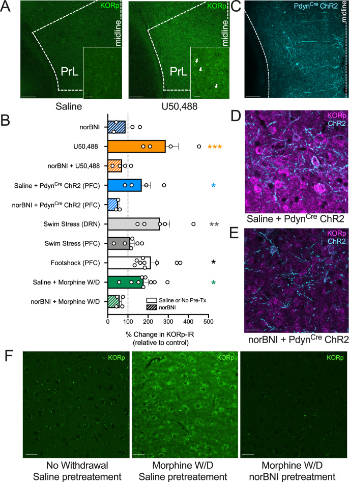
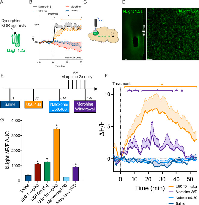
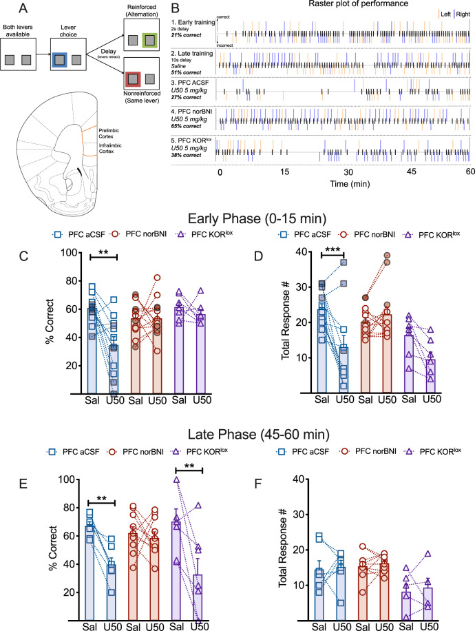
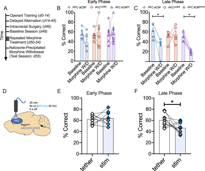
Following repeated opioid use, some dependent individuals experience persistent cognitive deficits that contribute to relapse of drug-taking behaviors, and one component of this response may be mediated by the endogenous dynorphin/kappa opioid system in neocortex. In C57BL/6 male mice, we find that acute morphine withdrawal evokes dynorphin release in the medial prefrontal cortex (PFC) and disrupts cognitive function by activation of local kappa opioid receptors (KORs). Immunohistochemical analyses using a phospho-KOR antibody confirmed that both withdrawal-induced and optically evoked dynorphin release activated KOR in PFC. Using a genetically encoded sensor based on inert KOR (kLight1.2a), we revealed the in vivo dynamics of endogenous dynorphin release in the PFC. Local activation of KOR in PFC produced multi-phasic disruptions of memory processing in an operant-delayed alternation behavioral task, which manifest as reductions in response number and accuracy during early and late phases of an operant session. Local pretreatment in PFC with the selective KOR antagonist norbinaltorphimine (norBNI) blocked the disruptive effect of systemic KOR activation during both early and late phases of the session. The early, but not late phase disruption was blocked by viral excision of PFC KORs, suggesting an anatomically dissociable contribution of pre- and postsynaptic KORs. Naloxone-precipitated withdrawal in morphine-dependent mice or optical stimulation of pdynCre neurons using Channelrhodopsin-2 disrupted delayed alternation performance, and the dynorphin-induced effect was blocked by local norBNI. Our findings describe a mechanism for control of cortical function during opioid dependence and suggest that KOR antagonism could promote abstinence.


Conflict of interest statement

The authors declare no competing interests.

Figures

Fig. 1
Fig. 1. KOR agonists, optogenetic stimulation of PFC dynorphin neurons, or environmental stressors increase KOR phosphorylation in the PFC.
A Pharmacological validation of phospho-KOR antibody. Representative images (10×, 40× inset) show increased fluorescent signal from phospho-KOR (S369 site) immunoreactivity (KORp-IR) following KOR agonist (U50,488; 10 mg/kg) treatment, compared to saline. Dashed lines show boundaries of the prelimbic prefrontal cortex. Scale bar for 10× image shows 150 μm, scale bar for 40× image (inset) shows 30 μm. White arrows indicate KORp-IR+ cell. B Conditions for KOR activation in the prefrontal cortex. All groups were normalized to their corresponding control group. A one-way ANOVA showed an overall significant effect of treatment (F(9, 42) = 5.597, p < 0.0001). Fisher’s LSD post-hoc test was used for planned comparisons between groups that were run in the same cohorts and normalized to their own saline or no-stress controls. KOR agonist (10 mg/kg U50,488; n = 4) significantly increased KORp-IR compared to norBNI treatment alone (p = 0.0006). Pretreatment with norBNI (n = 4) blocked the increase in KORp-IR following U50,488 treatment (p = 0.0002). These groups were normalized to saline-treated controls (n = 4). Optogenetic stimulation (n = 4) of PFC dynorphin neurons (normalized to EYFP control; n = 2) significantly increased KORp IR, which was blocked by norBNI pretreatment prior to optogenetic stimulation (n = 3; p = 0.0468), demonstrating the presence of a local PFC dynorphin/kappa opioid receptor circuit. Repeated forced swim stress, a stressor known to cause dynorphin release in the brain, significantly increased (p = 0.0024) KORp IR (normalized to no-stress controls n = 5 dorsal raphe nucleus (DRN); n = 6 PFC) in the DRN (n = 6) compared to the prefrontal cortex (PFC; stress n = 6) in the same mice. This showed that swim stress did not cause the release of dynorphin in the prefrontal cortex. In contrast, repeated footshock (0.3 mA shock each min, for 15 min) increased KORp IR in the PFC compared to control mice (one sample t-test; p = 0.0036). Naloxone (1 mg/kg)-precipitated withdrawal (W/D) following chronic experimenter-administered morphine (10 mg/kg, 4 days twice per day, one morphine injection 2 h prior to naloxone on day 5) treatment (normalized to chronic saline treatment n = 8) also increased KORp IR in PFC (n = 9), which was blocked by pretreatment with norBNI (n = 5) when given 24 h prior to 1st morphine injection and 24 h prior to naloxone injection (p = 0.012). Error bars indicate SEM. *p < 0.05; **p < 0.01; ***p < 0.0001. C Representative image for ChR2 expression in PFC dynorphin neurons. Prodynorphin Cre (pdynCre) mice were injected in the PFC with Channelrhodopsin-2 (ChR2; AAV1-DIO-ChR2). Image shows ChR2 (cyan) expression in pdyn-Cre+ neurons in the PFC. 10× image; Scale bar shows 150 μm. D Representative image for ChR2 expression and KORp IR in PFC dynorphin neurons with saline pretreatment. Image shows 40× image of ChR2 (cyan) and KORp IR (magenta) in mice treated with saline prior to optical stimulation. For optical stimulation, mice were tethered to a fiberoptic patchcord and received 473 nm light (10 mW @ fiber tip) over 30 min (Duty cycle: 5 s on @ 20 Hz, 5 s off). Within 10 min after the end of optical stimulation, brains were perfused for tissue processing; 40× image; scale bar shows 20 μm. E Representative image for ChR2 and KORp IR in PFC dynorphin neurons with norBNI pretreatment. Pretreatment with norBNI (10 mg/kg; 24 h prior to stimulation) blocked ChR2-mediated increase in KORp IR; 40× image, scale bar shows 20 μm. F Representative images for morphine withdrawal (W/D) KORp IR. Saline treatment after repeated morphine treatment (left) did not produce withdrawal and did not increase KORp IR. Naloxone-precipitated morphine withdrawal increased KORp IR in the PFC during withdrawal, which was blocked by norBNI (10 mg/kg) pretreatment; 40× image; scale bars show 20 μm.
Fig. 2
Fig. 2. A dynorphin ligand sensor (kLight 1.2a) detects the time course of drug withdrawal-induced dynorphin release in the PFC in vivo.
A kLight 1.2a diagram. kLight 1.2a is composed of an inert KOR protein fused to a circular green fluorescent protein that responds to conformational changes in KOR when bound to dynorphin or other KOR ligands. B KOR ligands increase kLight1.2a fluorescence. U50,488 (n = 6 cells) and dynorphin (n = 4) treatment significantly increased fluorescence (ΔF/F) during the 30-min imaging session starting at 5.75 min (U50,488) and 1.25 min (dynorphin B) following drug treatment. This increase persisted until the end of the imaging session. There was a significant interaction between drug treatment and time (F(180, 1380) = 5.930, p < 0.0001) and Dunnett’s post hoc showed significant (p < 0.05) differences between dynorphin and U50,488 compared to vehicle (n = 12). There was no significant effect of morphine treatment (n = 5) on kLight fluorescence. C kLight1.2a procedure. KORCre mice (n = 5) were unilaterally injected in the PFC with AAV-kLight1.2a and an optical fiber for photometry was implanted above the injection target region. D Representative images for kLight1.2a. kLight1.2a (green) expression was confirmed in mice after recordings. Fiber track for PFC implant visualized in 10× image (left), and 63× (right) image of expression shown on the right. Scale bar for 10× shows 100 μm and scale bar for 63× shows 20 μm. E Experimental timeline. kLight1.2a fluorescence was measured using fiber photometry (30 μW at fiber tip; 331 Hz; wavelength 470/405 nm). Following a 5-min baseline recording period, mice were injected with saline or U50,488 (1, 5, and 10 mg/kg) and allowed to freely explore a novel context. Naloxone (10 mg/kg) pretreatment occurred following a baseline period and 30 min later kLight1.2a response to 10 mg/kg U50,488 was recorded. For morphine withdrawal, mice underwent repeated morphine (10 mg/kg) injection procedure, and were tested for baseline fluorescence prior to naloxone (1 mg/kg) precipitated withdrawal. Each mouse received each condition on separate days. F kLight1.2a detects endogenous dynorphin release in the PFC during morphine withdrawal. Change in fluorescence (ΔF/F) over a 60-min period is shown (solid line). SEM. is shown above the mean with a filled area. A two-way ANOVA (drug × time) showed a significant interaction between treatment and time (F(3565, 14260) = 3.776, p < 0.0001). Dunnett’s post-hoc tests showed that U50,488 (10 mg/kg) injection significantly increased kLight fluorescence from 3.3 to 54.5 min compared to saline injection. Pretreatment with the nonselective opioid antagonist naloxone (10 mg/kg) blocked 10 mg/kg U50,488-mediated increases in kLight fluorescence. During the 60-min following 1 mg/kg naloxone administration to precipitate withdrawal in morphine-dependent mice, there was a significant increase in kLight fluorescence from 5.3 to 8.0, 9.0 to 13.0, 15.3 to 37.8, 39.0 to 41.5, and 43.5 to 44.8 min. This showed that kLight detected endogenous dynorphin release within the PFC of mice undergoing morphine withdrawal and that a 10 mg/kg dose of naloxone blocked U50488-mediated increases in kLight fluorescence. G Dose-dependent effects on kLight fluorescence. Area under peak value was compared between saline and all other conditions with a one-way ANOVA. There was a significant effect of treatment (F(5, 21378) = 561.6, p < 0.0001), and Dunnett’s post hoc showed that saline treatment was significantly different (p < 0.0001) compared to morphine withdrawal or U50,488 (1, 5, and 10 mg/kg). There was no difference between saline treatment and the naloxone/U50,488 group. Error bands above the mean indicate SEM. *p < 0.05.
Fig. 3
Fig. 3. KOR activation disrupts delayed alternation performance in early and late phases of the test session.
A Delayed alternation procedure. C57BL/6 male mice were food restricted and trained to complete lever alternations for reinforcement (detailed description in Methods). At the start of a trial, both levers were extended. Lever choice initiated a 10-s delay period during which both levers remained retracted and unavailable. When levers were reinserted, mice chose the alternate lever for food pellet reinforcement. Choosing the same lever as the previous response was recorded as an incorrect response and was not reinforced. After a 20 s intertrial interval, levers were reinserted to allow mice to start the next trial. The diagram of a hemi-coronal section outlines the PFC region (infralimbic and prelimbic cortex) targeted for injection; all placements were confirmed postmortem. B Raster plots of performance of a representative mouse during a 60-min trial session. Ticks (black) on the center line show trial initiating responses. Ticks above the center line show correct (reinforced) responses and ticks below show incorrect (nonreinforced) responses. Lever choice is shown by orange (left lever) or blue (right lever) ticks. Line 1 shows performance of a PFC aCSF mouse early in delayed alternation training (with a 2-s delay). Mice rarely alternated levers early in training, leading to a significant number of response errors. Raster line 2 shows improved performance in the same mouse after several training sessions, and saline injection (IP) prior to the delayed alternation session did not significantly alter performance in the task. Raster line 3 shows that treatment with the KOR agonist U50,488 (5 mg/kg, IP) in the same mouse decreased the percent of correct responses and decreased total response number during the delayed alternation session. Line 4 shows that bilateral local injection of the KOR antagonist (norBNI; 0.5 μg/0.2 μL) in the PFC blocked the disruptive effects of systemically administered U50,488 on both total response number and percent correct responses. Line 5 shows that U50,488 treatment decreased the correct responses in mice with postsynaptic KOR deletion (KORlox/lox mice injected with 0.2 μL AAV5-Cre-eYFP bilaterally in the PFC; PFC KORlox) during the late phase of the session, but not during the early phase of the session. C Postsynaptic KORs are required for early phase agonist-mediated decreases in correct response % in delayed alternation. After training for 3 weeks, aCSF (n = 12) or norBNI (n = 14) was bilaterally injected into PFC (3–5 days prior to testing), and the first 15 min of performance were compared between saline and KOR agonist (U50,488) treatment days. There was a significant interaction between group and agonist treatment F(2, 30) = 5.600, p = 0.0086. Sidak’s post-hoc test showed a significant difference between U50 and Saline treatment in the PFC aCSF mice (p = 0.0003), but not in PFC norBNI or PFC KORlox mice (n = 7). Gray borders and filled symbols indicate mice that received 30 min, rather than 60 min, training and testing sessions. D Systemic KOR agonist significantly decreases total responses in PFC aCSF mice. Total number of alternation responses was significantly decreased in PFC aCSF mice during the first 15 min of the session (Drug × Group Interaction: F(2, 30) = 8.582, p = 0.0011; Sidak’s post hoc: p = 0.002). There was a nonsignificant trend toward a decrease in response number in the PFC KORlox group (p = 0.0921). Gray borders and filled symbols indicate mice that received 30 min, rather than 60 min, training and testing sessions. E Postsynaptic KOR deletion does not block late phase KOR-mediated decreases in delayed alternation performance. KOR activation (PFC aCSF n = 7; norBNI n = 10; KORlox n = 7) significantly decreased percent correct in the delayed alternation task (F(2, 21) = 4.761, p = 0.0197) during the last 15 min of the session (late phase; 45–60 min) in PFC aCSF (Sidak’s p = 0.0171) and PFC KORlox (p = 0.0011) mice. F KOR activation did not affect total response number during the late phase of a delayed alternation session. There was no significant effect of drug on total response number following KOR activation. Dashed lines connect responses of individual mice receiving saline 1 day prior to the U50,488 trial session. Error bars indicate SEM. *p < 0.05, **p < 0.01; ***p < 0.0001.
Fig. 4
Fig. 4. Dynorphin release in the PFC disrupts delayed alternation performance.
A Experimental timeline. Mice received training in delayed alternation until reaching stable performance, then were intracranially injected with aCSF or norBNI. Floxed KOR mice were injected prior to training. Following recovery, mouse performance in a baseline delayed alternation session was assessed. Mice then received morphine (10 mg/kg) for 4 days, and on the fifth day received naloxone (1 mg/kg) prior to entry into the operant chamber. B There was no significant effect of morphine withdrawal on performance during the early phase of the task in the aCSF (n = 7), norBNI (n = 6), or floxed KOR (n = 7) groups. C Morphine withdrawal disrupts delayed alternation performance after naloxone treatment. In the 30–45 min window following naloxone treatment, there was a significant decrease in percent correct responding during a delayed alternation session in the aCSF group (n = 5), and the disruptive effect was not blocked by postsynaptic KOR deletion (n = 5). However, local norBNI pretreatment (n = 5) in the PFC blocked the disruptive effect of morphine withdrawal on delayed alternation performance (main effect of drug; F(2, 12) = 4.628; p = 0.0324); Sidak’s post hoc compared to baseline: PFC aCSF (p = 0.0211), PFC norBNI (p = 0.959), KORlox (p = 0.0191). D PFC pdynCre optical stimulation procedure. PdynCre mice were injected with ChR2 in the PFC and were trained in delayed alternation. On baseline and test days, mice were tethered to an optical patchcord for 30-min prior to a delayed alternation session and received either no stimulation (tether) or optical stimulation (stim) on separate days. E Optical stimulation of PFC pdynCre neurons does not disrupt early phase delayed alternation. Optical stimulation had no significant effect on percent correct in delayed alternation in pdynCre mice (n = 5) during the early phase of the test session. F Optical stimulation of PFC pdynCre neurons disrupts late phase delayed alternation performance. Compared to a tether session, photostimulation significantly decreased the percent of correct responses 45–60 min after stimulation (t8 = 2.33, p = 0.048). Error bars indicate SEM. *p < 0.05.

References

    1. Millan MJ, Agid Y, Brüne M, Bullmore ET, Carter CS, Clayton NS, et al. Cognitive dysfunction in psychiatric disorders: characteristics, causes and the quest for improved therapy. Nat Rev Drug Discov. 2012;11:141–68. - PubMed
    1. Arnsten AFT. Stress signalling pathways that impair prefrontal cortex structure and function. Nat Rev Neurosci. 2009;10:410–22. - PMC - PubMed
    1. Bruchas MR, Chavkin C. Kinase cascades and ligand-directed signaling at the kappa opioid receptor. Psychopharmacology. 2010;210:137–47. - PMC - PubMed
    1. Knoll AT, Carlezon WA. Dynorphin, stress, and depression. Brain Res. 2010;1314:56–73. - PMC - PubMed
    1. Maqueda AE, Valle M, Addy PH, Antonijoan RM, Puntes M, Coimbra J, et al. Salvinorin-A induces intense dissociative effects, blocking external sensory perception and modulating interoception and sense of body ownership in humans. Int J Neuropsychopharmacol. 2015;18:pyv065. - PMC - PubMed

Publication types