

4D imaging of fetal right ventricle-feasibility study and a review of the literature
- PMID: 34545461
- PMCID: PMC8888475
- DOI: 10.1007/s10554-021-02407-9
4D imaging of fetal right ventricle-feasibility study and a review of the literature
Abstract
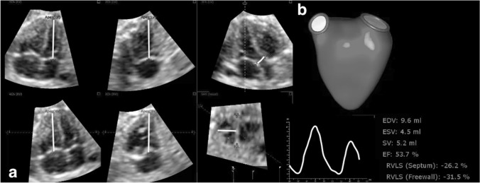
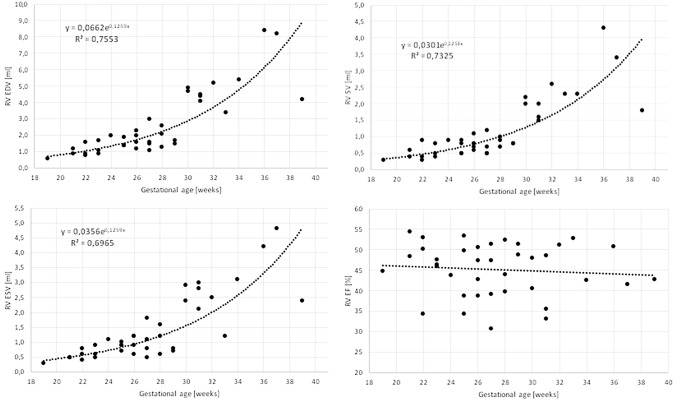
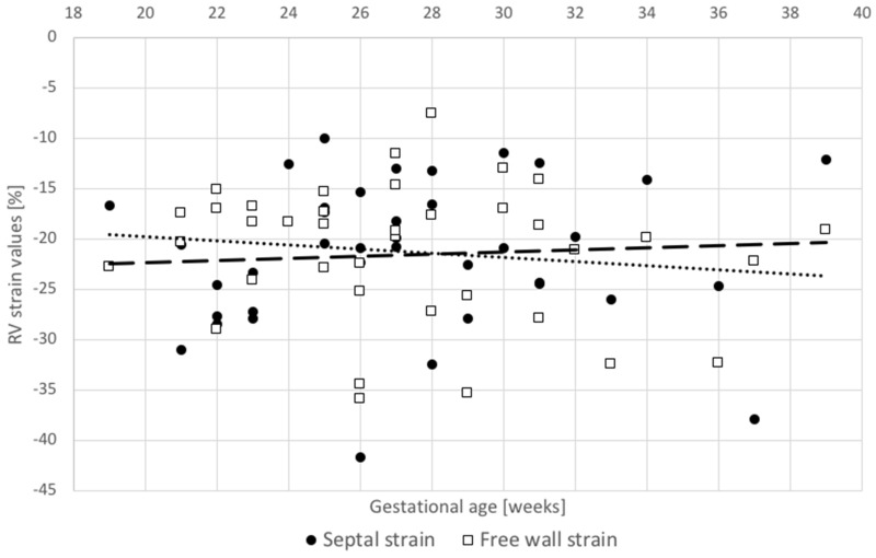
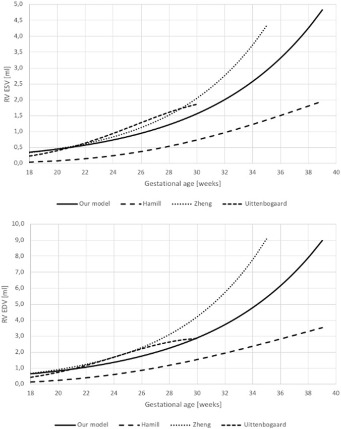
Functional analysis of the fetal cardiovascular system is crucial for the assessment of fetal condition. Evaluation of the right ventricle with standard 2D echocardiography is challenging due to its complex geometry and irregular muscle fibers arrangement. Software package TOMTEC 4D RV-Function is an analysis tool which allows assessment of right ventricular function based on volumetric measurements and myocardial deformation. The aim of this study was to determine the feasibility of this method in fetal echocardiography. The retrospective study was conducted in the high-flow Referral Center for Fetal Cardiology. We recorded 4D echocardiographic sequences of 46 fetuses with normal hearts. Following parameters were calculated: end-diastolic volume (EDV), end-systolic volume (ESV), stroke volume (SV) and ejection fraction (EF), right ventricle longitudinal free-wall (RVLS free-wall) and septal strain (RVLS septum). Tei index was calculated as a standard measure or RV function for comparison. 4D assessment was feasible in 38 out of 46 fetuses (83%). RV volumetric parameters-EDV, ESV and SV-increased exponentially with gestational age. Functional parameters-RV Tei index, EF and strains-were independent of gestational age. Mean EF was 45.2% (± 6%), RV free-wall strain was - 21.2% and RV septal strain was - 21.5%. There was a statistically significant correlation between septal and free-wall strains (r = 0.51, p = 0.001) as well as between EF and RV free-wall strain (r = - 0.41, p = 0.011). 4D RV assessment is feasible in most fetuses. Its clinical application should be further investigated in larger prospective studies.
Keywords: 4D echocardiography; Fetal cardiac function; Fetal echocardiography; Myocardial strain; Right ventricle; Right ventricular volume.
© 2021. The Author(s).
Conflict of interest statement
Authors declare that there is no conflict of interest.
Figures
References
-
- Donofrio MT, Moon-Grady AJ, Hornberger LK, Copel JA, Sklansky MS, Abuhamad A, Cuneo BF, Huhta JC, Jonas RA, Krishnan A, Lacey S, Lee W, Michelfelder EC, Sr, Rempel GR, Silverman NH, Spray TL, Strasburger JF, Tworetzky W, Rychik J, American Heart Association Adults With Congenital Heart Disease Joint Committee of the Council on Cardiovascular Disease in the Young and Council on Clinical Cardiology, Council on Cardiovascular Surgery and Anesthesia, and Council on Cardiovascular and Stroke Nursing Diagnosis and treatment of fetal cardiac disease: a scientific statement from the American heart association. Circulation. 2014;129(21):2183–242. doi: 10.1161/01.cir.0000437597.44550.5d. - DOI - PubMed
-
- Medvedofsky D, Addetia K, Patel AR, Sedlmeier A, Baumann R, Mor-Avi V, Lang RM. Novel approach to three-dimensional echocardiographic quantification of right ventricular volumes and function from focused views. J Am Soc Echocardiogr. 2015;28(10):1222–1231. doi: 10.1016/j.echo.2015.06.013. - DOI - PubMed
-
- Lang RM, Badano LP, Mor-Avi V, Afilalo J, Armstrong A, Ernande L, Flachskampf FA, Foster E, Goldstein SA, Kuznetsova T, Lancellotti P. Recommendations for cardiac chamber quantification by echocardiography in adults: an update from the American society of echocardiography and the European association of, cardiovascular imaging. Eur Heart J Cardiovasc Imaging. 2016;17(4):412. doi: 10.1093/ehjci/jew041. - DOI - PubMed
Publication types
MeSH terms
LinkOut - more resources
Full Text Sources
