. 2022 Apr;530(5):792-803.
doi: 10.1002/cne.25245. Epub 2021 Nov 15.

DARPP-32 distinguishes a subset of adult-born neurons in zebra finch HVC

Affiliations

DARPP-32 distinguishes a subset of adult-born neurons in zebra finch HVC

Jake V Aronowitz et al. J Comp Neurol. 2022 Apr.

Abstract

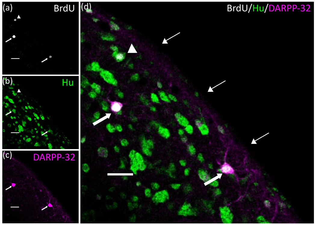
Adult male zebra finches (Taeniopygia guttata) continually incorporate adult-born neurons into HVC, a telencephalic brain region necessary for the production of learned song. These neurons express activity-dependent immediate early genes (e.g., zenk and c-fos) following song production, suggesting that these neurons are active during song production. Half of these adult-born HVC neurons (HVC NNs) can be backfilled from the robust nucleus of the arcopallium (RA) and are a part of the vocal motor pathway underlying learned song production, but the other half do not backfill from RA, and they remain to be characterized. Here, we used cell birth-dating, retrograde tract tracing, and immunofluorescence to demonstrate that half of all HVC NNs express the phosphoprotein DARPP-32, a protein associated with dopamine receptor expression. We also demonstrate that DARPP-32+ HVC NNs are contacted by tyrosine hydroxylase immunoreactive fibers, suggesting that they receive catecholaminergic input, have transiently larger nuclei than DARPP-32-neg HVC NNs, and do not backfill from RA. Taken together, these findings help characterize a group of HVC NNs that have no apparent projections to RA and so far have eluded positive identification other than HVC NN status.

Keywords: HVC; dopamine; neurogenesis; song; zebra finch.


Conflict of interest statement

CONFLICT OF INTERESTS

The authors declare no competing financial interests.

Figures

FIGURE 1
FIGURE 1
Cellular organization of male zebra finch HVC. Schematic of HVC showing known populations of HVC neurons. Yellow circles are BrdU+ nuclei meant to distinguish those generated during our BrdU injections from those generated on days we did not inject BrdU, and therefore are BrdU−. The question mark represents the approximately 50% of HVC NNs that cannot be backfilled from RA
FIGURE 2
FIGURE 2
Confocal photomicrograph of HVC in sagittal section showing representative Hu and DARPP-32 labeling. (a) Hu+ neurons. (b) Neurons labeled with a monoclonal antibody (ab40801) against DARPP-32 that recognizes and binds to DARPP-32 independent of its phosphorylation state. (c) Merged image of both channels. There are fewer DARPP-32+ cells in HVC compared to the underlying HVC shelf region, which, like much of the surrounding nidopallium, labels much more robustly for DARPP-32 than within HVC (Singh & Iyengar, 2019). White dotted line delineates the borders of HVC. 23.8X, scale bar = 100 μm
FIGURE 3
FIGURE 3
Twenty-eight-day old HVC NNs express the phosphoprotein DARPP-32. Confocal photomicrograph of a single optical plane of section of the same field of view in dorsal HVC showing two DARPP-32+ NNs (thick arrows) and one DARPP-32-neg HVC NN at 28 dpi (white arrowhead). (a) BrdU labeled nuclei. (b) Neurons labeled for the neuron-specific protein Hu. (c) DARPP-32+ neurons. (d) Merged image of all three channels; thick arrows indicate triple-labeled BrdU+/Hu+/DARPP-32+ HVC NNs at 28 dpi. Arrowhead indicates a double labeled BrdU+/Hu+/DARPP-32-neg HVC NN. Thin arrows indicate the dorsal surface of HVC. 18× magnification. Scale bars = 25 μm
FIGURE 4
FIGURE 4
DARPP-32 expression in HVC NNs changes as a function of neuronal age. (a) The density of HVC NNs as a function of days following the final BrdU injection. Dark gray bars represent BrdU+/Hu+/DARPP-32+ HVC NNs, and light gray bars represent BrdU+/Hu+/DARPP-32-neg HVC NNs. The total height of the stacked bars equals the average density of HVC NNs per cubic millimeter. We observed a significant decline in the density of HVC NNs between 14 and 21 dpi (t(8) = 2.57, p = .03, independent samples t-test). After 21 dpi, we observed no group differences in the density of HVC NNs. Error bars represent SEMs. Group sizes for all groups are n = 5 except for 28 dpi, n = 6; 45 dpi, n = 7; 180 dpi, n = 4. (b) To determine the fraction of HVC NNs that express DARPP-32 at each timepoint sampled, we divided the density of DARPP-32+ HVC NNs (BrdU+/Hu+/DARPP-32+) by the density of all HVC NNs (BrdU+/Hu+); these values are presented here as a derivation of 4A. These data show that the fraction of HVC NNs that express DARPP-32 increases to its peak at 28 dpi (50.4 ± 1.6%), then declines to 26.8 ± 7.3% at 45 dpi. At all later timepoints sampled, the fraction of HVC NNs expressing DARPP-32 was not significantly different from the 45 dpi timepoint, indicating that the number of DARPP-32+ HVC NNs in a given cohort of NNs is stable from 45 dpi onward. Error bars represent SEMs. Significance markers are as follows: p<.05 = *; p<.001 = *** and these markers are consistent throughout the manuscript
FIGURE 5
FIGURE 5
DARPP-32+ HVC NNs do not backfill from RA at 32 dpi. (a) Timeline of HVC-RA backfill experiments. (b) Simplified schematic of the song system in sagittal section showing some of the brain regions involved in song production. The retrograde tracer Fluorogold (FG) was injected into RA to backfill HVC-RA neurons. (c) Maximum intensity projection of a Z-stack showing backfilled FG+, RA-projecting neurons in HVC (18×; scale bar = 100 μm). Dotted white box delineates the area in HVC examined at higher magnification in panels (d) through (h) (18×, scale bar = 100 μm). (d–h) Confocal photomicrograph showing DARPP-32+ HVC NNs (white arrows) that did not backfill from RA. (d) BrdU+ nuclei. (e) Hu+ neurons. (f) DARPP-32+ neurons. (g) FG+, RA-projecting neurons. (h) Merged image of all four channels showing DARPP-32+ HVC NNs that do not backfill from RA (white arrows), surrounded by Hu+/FG+/DARPP-32-neg HVC-RA neurons (white arrowheads). 50×, scale bar = 25 μm. (i) 125/126 DARPP-32+ HVC NNs we examined were FG-negative. 32- to 34-day old DARPP-32+ HVC NNs do not backfill from RA. 53.6% of HVC NNs examined (green; 199/371) did not project to RA, while 46.4% did project to RA (yellow; 172/371 neurons), as measured by whether or not the somata were FG+. Of the 46.4% of RA-projecting HVC NNs, only 1/172 was DARPP-32+, suggesting that DARPP-32+ HVC NNs do not backfill from RA at 32 dpi
FIGURE 6
FIGURE 6
DARPP-32+ HVC NNs are contacted by tyrosine hydroxylase fibers. (a and c) Confocal photomicrographs of the same DARPP-32+ HVC NN at two different optical sections of the Z-stack, showing two different putative TH+ (green) synaptic contacts. (b and d) High magnification orthogonal views of the putative synaptic contacts (white arrows) shown in the dotted white boxes in images (a) and (c). White arrows indicate the points of putative contact between TH+ fibers and DARPP-32+ HVC NN. Scale bars: 5 μm: (b and d), 25 μm (a and c). Magnification: 75.2× (a and c), 140× (b and d)
FIGURE 7
FIGURE 7
Mean nuclear diameter of HVC NNs over the first 45 days post-BrdU injection. The mean nuclear diameter of DARPP-32+ HVC NNs (dark gray bars) was significantly larger than that of DARPP-32-neg HVC NNs (light gray bars) at 21 and 28 dpi. However, at 45 dpi, there was no significant difference between the mean nuclear diameter of the two subtypes of HVC NNs. Statistics revealed that both subtypes of new neurons demonstrated a decline in nuclear diameter with increasing neuronal age. Data points represent individual animal means. The values embedded in each bar represent the number of nuclei measured at each timepoint. Error bars represent SEMs. Significance markers are as follows: p<.05 = *; p<.01 = **; p<.001 = *** and these markers are consistent throughout the manuscript

References

    1. Akutagawa E, Konishi M (2010). New brain pathways found in the vocal control system of a songbird. The Journal of Comparative Neurology, 518(15), 3086–3100. 10.1002/cne.22383 - DOI - PubMed
    1. Alvarez-Buylla A, Theelen M, & Nottebohm F (1988). Birth of projection neurons in the higher vocal center of the canary forebrain before, during, and after song learning. Proceedings of the National Academy of Sciences of the United States of America, 85, 8722–8726. - PMC - PubMed
    1. Alvarez-Borda B, Haripal B, Nottebohm F (2004). Timing of brain-derived neurotrophic factor exposure affects life expectancy of new neurons. Proceedings of the National Academy of Sciences, 101(11), 3957–3961. 10.1073/pnas.0308118101 - DOI - PMC - PubMed
    1. Appeltants D, Absil P, Balthazart J, & Ball GF (2000). Identification of the origin of catecholaminergic inputs to HVc in canaries by retrograde tract tracing combined with tyrosine hydroxylase immunocytochemistry. Journal of Chemical Neuroanatomy, 18, 117–133 - PubMed
    1. Appeltants D, Ball GF, & Balthazart J (2001). The distribution of tyrosine hydroxylase in the canary brain: Demonstration of a specific and sexually dimorphic catecholaminergic innervation of the telencephalic song control nuclei. Cell and Tissue Research, 304, 237–259. - PubMed

Publication types

Substances