. 2021 Sep 2;62(12):22.
doi: 10.1167/iovs.62.12.22.

CD40 Expressed in Endothelial Cells Promotes Upregulation of ICAM-1 But Not Pro-Inflammatory Cytokines, NOS2 and P2X7 in the Diabetic Retina

Affiliations

CD40 Expressed in Endothelial Cells Promotes Upregulation of ICAM-1 But Not Pro-Inflammatory Cytokines, NOS2 and P2X7 in the Diabetic Retina

Jin-Sang Yu et al. Invest Ophthalmol Vis Sci. .

Abstract

Purpose: CD40 is an upstream inducer of inflammation in the diabetic retina. CD40 is upregulated in retinal endothelial cells in diabetes. The purpose of this study was to determine whether expression of CD40 in endothelial cells is sufficient to promote inflammatory responses in the retina of diabetic mice.

Methods: Transgenic mice with CD40 expression restricted to endothelial cells (Trg-CD40 EC), transgenic control mice (Trg-Ctr), B6, and CD40-/- mice were made diabetic using streptozotocin. Leukostasis was assessed using FITC-conjugated ConA. Pro-inflammatory molecule expression was examined by real-time PCR, immunohistochemistry, ELISA, or flow cytometry. Release of ATP was assessed by ATP bioluminescence.

Results: Diabetic B6 and Trg-CD40 EC mice exhibited increased retinal mRNA levels of ICAM-1, higher ICAM-1 expression in endothelial cells, and increased leukostasis. These responses were not detected in diabetic mice that lacked CD40 (CD40-/- and Trg-Ctr). Diabetic B6 but not Trg-CD40 EC mice upregulated TNF-α, IL-1β, and NOS2 mRNA levels. CD40 stimulation in retinal endothelial cells upregulated ICAM-1 but not TNF-α, IL-1β, or NOS2. CD40 ligation did not trigger ATP release by retinal endothelial cells or pro-inflammatory cytokine production in bystander myeloid cells. In contrast to diabetic B6 mice, diabetic Trg-CD40 EC mice did not upregulate P2X7 mRNA levels in the retina.

Conclusions: Endothelial cell CD40 promotes ICAM-1 upregulation and leukostasis. In contrast, endothelial cell CD40 does not lead to pro-inflammatory cytokine and NOS2 upregulation likely because it does not activate purinergic-mediated pro-inflammatory molecule expression by myeloid cells or induce expression of these pro-inflammatory molecules in endothelial cells.


Conflict of interest statement

Disclosure: J.-S. Yu, None; J. Daw, None; J.-A.C. Portillo, None; C.S. Subauste, None

Figures

Figure 1.
Figure 1.
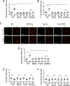
CD40 expression in endothelial cells from diabetic mice promotes upregulation of ICAM-1 and leukostasis in the retina. (A) Double transgenic (Trg-CD40 EC) mice express CD40 in endothelial cells. The cartoon depicts the binary tetracycline (Tet) repressible system. Immunohistochemistry for CD40 in the retinas of B6, CD40−/−, single transgenic (Trg-Ctr), or double transgenic (Trg-CD40 EC) mice. Sections were incubated with biotinylated anti-CD40 mAb followed by incubation with streptavidin-Alexa Fluor 647 and co-staining with Tomato lectin-DyLight 488 (X200). Bar, 10 µm. Four animals per group were analyzed. Results are representative of three independent experiments. (B) At 2 months of diabetes, retinas from diabetic B6, CD40−/−, Trg-Ctr, and Trg-CD40 EC mice as well as from non-diabetic controls were collected and used for mRNA extraction. The mRNA levels of ICAM-1 were assessed by real time quantitative PCR using 18S rRNA as internal control. One non-diabetic B6 mouse was given an arbitrary value of one and data are expressed as fold-increase compared to this animal. There were seven to nine animals per group. (C) At 2 months of diabetes, retinal sections were incubated with anti-ICAM-1 mAb plus either Tomato Lectin. GCL = Ganglion cell layer; IPL = Inner plexiform layer; INL = Inner nuclear layer. Scale bar, 10 µm. 6 mice/group. (D) At 2 months of diabetes, adherent leukocytes in the retinal vasculature of diabetic and non-diabetic control mice were quantified by labeling with Concanavalin A. *P < 0.05; **P < 0.01; ***P < 0.001 by ANOVA.
Figure 2.
Figure 2.
CD40 expression in endothelial cells from diabetic mice does not lead to upregulation of TNF-α, IL-1β,and NOS2 in the retina. (A, B) At 2 months of diabetes, retinas from diabetic B6, CD40−/−, Trg-Ctr, and Trg-CD40 EC mice as well as from non-diabetic controls were collected and used for mRNA extraction. The mRNA levels of TNF-α and IL-1β were assessed as above. There were seven to nine animals per group. (C) At 2 months of diabetes, retinal sections were incubated with antibodies against TNF-α and anti-Iba-1 (expressed in microglia/macrophages). Scale bar, 10 µm. There were six mice per group. (DF) The mRNA levels of NOS2, IL-6, and CXCL1 at 2 months of diabetes. **P < 0.01; ***P < 0.001 by ANOVA. n.s. = not significant.
Figure 3.
Figure 3.
CD40-activated retinal endothelial cells do not secrete TNF-α and IL-1β or upregulate NOS2. (A) Human retinal endothelial cells were transduced with EGFP-encoding empty retroviral vector (MIEG3) or with CD40-encoding retroviral vector (MIEG3-CD40). Dot plot shows expression of EGFP and CD40. (B) Endothelial cells were incubated with or without CD154 and ICAM-1 expression was examined by flow cytometry at 24 hours. (C) CD40-expressing retinal endothelial cells were incubated with or without CD154 and concentrations of TNF-α and IL-1β were measured at different time points of in vitro culture. In addition, concentration of CCL2 was examined at 0 and 24 hours. Results are presented as mean ± standard deviation (SD) of triplicate wells and are representative of three independent experiments. (D) Human retinal endothelial cells were incubated in low glucose or high glucose conditions for 7 days. CD40 expression was examined by flow cytometry. Retinal endothelial cells were incubated with or without CD154. ICAM-1 expression was examined by flow cytometry at 24 hours. Concentrations of TNF-α and IL-1β were measured by ELISA at 4 hours and 24 hours, respectively, and were found to be <7.8 pg/mL and < 0.5 pg/mL, respectively. (E) CD40-expressing retinal endothelial cells (RECs) or retinal Müller cells (RMCs) were incubated with or without CD154. Expression of NOS2 and actin were examined at 24 hours by immunoblot. Results are presented as mean ± SD of triplicate samples and are representative of three independent experiments. ***P < 0.001 by Student's t-test.
Figure 4.
Figure 4.
CD40-activated retinal endothelial cells do not secrete ATP or induce production of pro-inflammatory cytokines by monocytic cells. (A) CD40-expressing retinal endothelial cells were incubated with or without CD154 and concentrations of extracellular ATP were measured at different time points. Release of extracellular ATP by CD40+ human Muller cells is included for comparison. (B, C), CD40-expressing retinal endothelial cells were incubated with CD40 monocytic cells (MonoMac6) with or without CD154. TNF-α B and IL-1β C were measured in supernatants by ELISA. Results are presented as mean ± SD of triplicate wells and are representative of three independent experiments. ***P < 0.001 by Student's t-test.
Figure 5.
Figure 5.
CD40 expression in endothelial cells from diabetic mice does not lead to upregulation of P2X7 in the retina. At 2 months of diabetes, retinas from diabetic B6, CD40−/−, Trg-Ctr, and Trg-CD40 EC mice as well as from non-diabetic controls were collected and used for mRNA extraction. The mRNA levels were assessed as above. There were seven to nine animals per group. **P < 0.01; ***P < 0.001 by ANOVA.

References

    1. Bourne RR, Stevens GA, White RA, et al. .. Causes of vision loss worldwide, 1990-2010: a systematic analysis. Lancet Glob Health. 2013; 1: e339–e349. - PubMed
    1. Giacco F, Brownlee M.. Oxidative stress and diabetic complications. Circ Res. 2010; 107: 1058–1070. - PMC - PubMed
    1. Eid S, Sas KM, Abcouwer SF, et al. .. New insights into the mechanisms of diabetic complications: role of lipids and lipid metabolism. Diabetologia. 2019; 62: 1539–1549. - PMC - PubMed
    1. Tang J, Kern TS.. Inflammation in diabetic retinopathy. Prog Retin Eye Res. 2011; 30: 343–358. - PMC - PubMed
    1. Antonetti DA, Klein R, Gardner TW.. Diabetic retinopathy. N Eng J Med. 2012; 366: 1227–1239. - PubMed

Publication types

MeSH terms