. 2021 Oct;35(10):e21927.
doi: 10.1096/fj.202101066R.

Disease mechanisms of X-linked cone dystrophy caused by missense mutations in the red and green cone opsins

Affiliations

Disease mechanisms of X-linked cone dystrophy caused by missense mutations in the red and green cone opsins

Ping Zhu et al. FASEB J. 2021 Oct.

Abstract

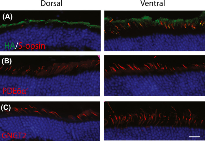
Cone photoreceptors are responsible for the visual acuity and color vision of the human eye. Red/green cone opsin missense mutations N94K, W177R, P307L, R330Q, and G338E have been identified in subjects with congenital blue cone monochromacy or color-vision deficiency. Studies on disease mechanisms due to these cone opsin mutations have been previously carried out exclusively in vitro, and the reported impairments were not always consistent. Here we expressed these mutants via AAV specifically in vivo in M-opsin knockout mouse cones to investigate their subcellular localization, the pathogenic effects on cone structure, function, and cone viability. We show that these mutations alter the M-opsin structure, function, and localization. N94K and W177R mutants appeared to be misfolded since they localized exclusively in cone inner segments and endoplasmic reticulum. In contrast, P307L, R330Q, and G338E mutants were detected predominately in cone outer segments. Expression of R330Q and G338E, but not P307L opsins, also partially restored expression and correct localization of cone PDE6α' and cone transducin γ and resulted in partial rescue of M-cone-mediated light responses. Expression of W177R and P307L mutants significantly reduced cone viability, whereas N94K, R330Q, and G338E were only modestly toxic. We propose that although the underlying biochemical and cellular defects caused by these mutants are distinct, they all seem to exhibit a dominant phenotype, resembling autosomal dominant retinitis pigmentosa associated with the majority of rhodopsin missense mutations. The understanding of the molecular mechanisms associated with these cone opsin mutants is fundamental to developing targeted therapies for cone dystrophy/dysfunction.

Keywords: blue cone monochromacy; cone dystrophy; cone opsin; disease mechanism; photoreceptors.


Conflict of interest statement

WWH and the University of Florida have a financial interest in the use of AAV therapies, and WWH owns equity in a company (AGTC Inc.) that might, in the future, commercialize some aspects of this work.

Figures

FIGURE 1
FIGURE 1
Expression and function of cone opsin mutants. (A) Location of mutant residues in human green opsin (OPN1MW, PDB ID: 1KPW; drawn with PyMOL, Molecular Graphics System, Version 1.2r3pre, Schrödinger, LLC). N94 is located in helix II, W177 in helix IV, P307 in helix VII, and R330 and G338 in C‐terminal α‐helix. (B) Representative immunoblot of AAV‐mediated expression of cone opsin mutants in Opn1mw–/– mice at 2 months of postinjection. Anti‐M‐opsin (red); α‐tubulin (loading control, green). Retinas from C57BL/6J and Opn1mw–/– mice were used as positive and negative controls, respectively. (C) Representative ERG traces of R330Q and G338E opsins at 1.4 log cd s/m2 under 530 nm wavelength light. (D) Individual and averaged M‐cone ERG responses from each mutant opsin injected Opn1mw–/– eye. Average represents the mean ± SD of b‐wave amplitudes recorded at 1.4 log cd s/m2 (N = 8 for each group, *p < .0001). Untreated Opn1mw–/– and wild‐type mice served as controls
FIGURE 2
FIGURE 2
Subcellular localization of N94K opsin, PDE6α’, and GNGT2 in injected Opn1mw–/– retinas. (A) N94K expression was detected by an anti‐HA antibody (green). N94K opsin was misfolded and localized predominately in cone inner segments and ER (arrows) in both dorsal and ventral retinas. The rough ER (arrows), where cone opsins are synthesized, was shown as green circles surrounding the cone nucleus by HA staining. S‐opsin expression (red) was mainly detected in cone outer segments in the ventral area. (B) PDE6α’ was barely detectible in the dorsal area, while it was expressed normally and localized correctly in cone outer segments in the ventral area of the same eye, where S‐opsin was present. (C) GNGT2 was not detected in the dorsal area, while it was expressed normally and localized in cone outer segments in the ventral area of the same eye. Scale bar: 20 µm
FIGURE 3
FIGURE 3
Subcellular localization of W177R opsin, PDE6α’, and GNGT2 in injected Opn1mw–/– retinas. (A) W177R (green) was misfolded and localized exclusively to cone inner segments and ER (arrows) in both the dorsal and ventral retina. (B) PDE6α’ was barely detectible in the dorsal area while it was expressed normally and localized to cone outer segments in the ventral area of the same eye, where S‐opsin was present. (C) GNGT2 was not detected in the dorsal area, while it was expressed normally and localized to cone outer segments in ventral area. Scale bar: 20 µm
FIGURE 4
FIGURE 4
Subcellular localization of P307L opsin, PDE6α’, and GNGT2 in injected Opn1mw–/– retinas. (A) P307L opsin (green) was expressed in cone outer segments in both dorsal and ventral retinas and colocalized with endogenous S‐opsin in the ventral area. (B) PDE6α’ expression was barely detectible in the dorsal retina, while it was expressed normally and localized in cone outer segments in the ventral area. (C) GNGT2 was also not detected in the dorsal area while expressed normally and localized to cone outer segments in the ventral area. Scale bar: 20 µm
FIGURE 5
FIGURE 5
Subcellular localization of R330Q opsin, PDE6α’, and GNGT2 in injected Opn1mw–/– retinas. (A) R330Q opsin (green) was expressed in cone outer segments in both dorsal and ventral retinas. (B) PDE6α’ expression was partially restored in cone outer segments in the dorsal retina. (C) GNGT2 expression was also partially restored in cone outer segments in the dorsal retina. Scale bar: 20 µm
FIGURE 6
FIGURE 6
Subcellular localization of G338E opsin, PDE6α’, and GNGT2 in injected Opn1mw–/– retinas by immunohistochemistry. (A) G338E opsin (green) was expressed in cone outer segments in both dorsal and ventral retinas. (B) PDE6α’ expression was partially restored in cone outer segments in the dorsal retina. (C) GNGT2 expression was also partially restored in cone outer segments in the dorsal retina. Scale bar: 20 µm
FIGURE 7
FIGURE 7
Evaluation of cone viability in Opn1mw–/– retinas expressing each cone opsin mutant. (A) Representative images were taken from dorsal areas of retinal flat mounts of Opn1mw–/– mice injected with each cone opsin mutant at 5 months of postinjection. Retinal whole mounts were stained with PNA. (B) Quantification of PNA staining from dorsal and ventral areas of retinal wholemounts of Opn1mw–/– eyes injected with different opsin mutants. Seven‐month‐old uninjected Opn1mw–/– and wild‐type mice were used as controls. Each bar represents PNA‐positive cells counted within a 0.01mm2 surface area of the retina from eight images taken from four different mice. Data show average ± SD. *p < .05, when compared with untreated controls

References

    1. Curcio CA, Allen KA, Sloan KR, et al. Distribution and morphology of human cone photoreceptors stained with anti‐blue opsin. J Comp Neurol. 1991;312:610‐624. - PubMed
    1. Curcio CA, Sloan KR, Kalina RE, Hendrickson AE. Human photoreceptor topography. J Comp Neurol. 1990;292:497‐523. - PubMed
    1. Mustafi D, Engel AH, Palczewski K. Structure of cone photoreceptors. Prog Retin Eye Res. 2009;28:289‐302. - PMC - PubMed
    1. Nathans J, Thomas D, Hogness DS. Molecular genetics of human color vision: the genes encoding blue, green, and red pigments. Science. 1986;232:193‐202. - PubMed
    1. Ayyagari R, Kakuk LE, Coats CL, et al. Bilateral macular atrophy in blue cone monochromacy (BCM) with loss of the locus control region (LCR) and part of the red pigment gene. Mol Vis. 1999;5:13. - PubMed

Publication types

LinkOut - more resources