Review
. 2021 Sep 16;81(18):3775-3785.
doi: 10.1016/j.molcel.2021.08.030.

Metabolic channeling: predictions, deductions, and evidence

Affiliations
Review

Metabolic channeling: predictions, deductions, and evidence

Vidhi Pareek et al. Mol Cell. .

Abstract

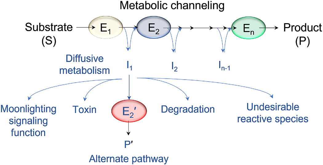
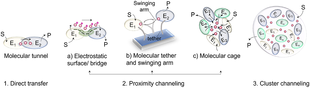
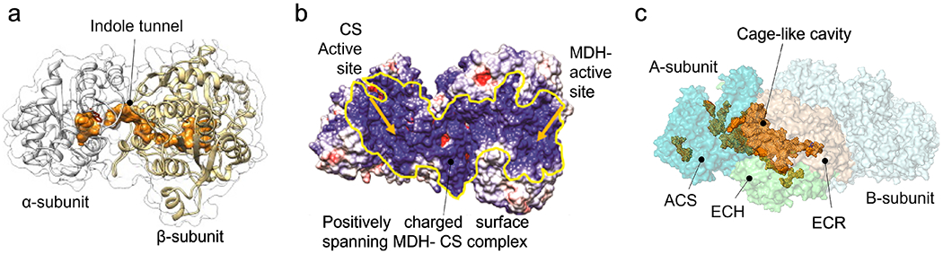
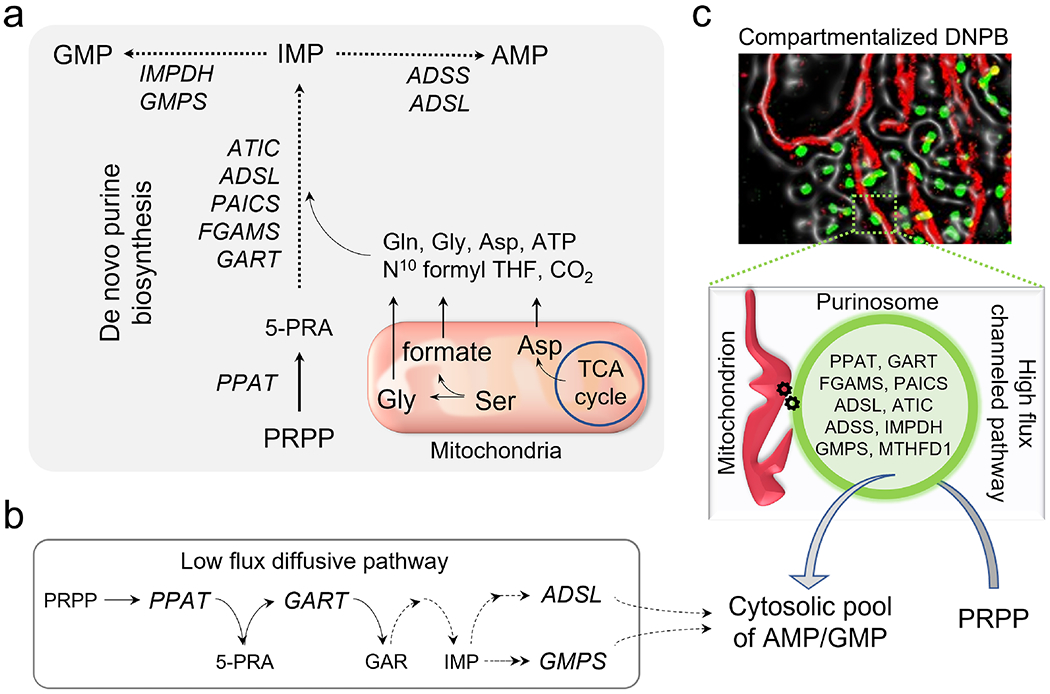
With the elucidation of myriad anabolic and catabolic enzyme-catalyzed cellular pathways crisscrossing each other, an obvious question arose: how could these networks operate with maximal catalytic efficiency and minimal interference? A logical answer was the postulate of metabolic channeling, which in its simplest embodiment assumes that the product generated by one enzyme passes directly to a second without diffusion into the surrounding medium. This tight coupling of activities might increase a pathway's metabolic flux and/or serve to sequester unstable/toxic/reactive intermediates as well as prevent their access to other networks. Here, we present evidence for this concept, commencing with enzymes that feature a physical molecular tunnel, to multi-enzyme complexes that retain pathway substrates through electrostatics or enclosures, and finally to metabolons that feature collections of enzymes assembled into clusters with variable stoichiometric composition. Lastly, we discuss the advantages of reversibly assembled metabolons in the context of the purinosome, the purine biosynthesis metabolon.

Keywords: membrane-less compartmentalization; metabolic channeling; metabolon; molecular tunnel; purinosome.


Conflict of interest statement

Declaration of interests The authors declare no competing interests.

Figures

Figure 1.
Figure 1.. Metabolic channeling in cascade reactions.
Unlike diffusive metabolism, metabolic channeling leads to intermediate sequestration, thus preventing accumulation of toxic, unstable, reactive intermediates, and /or depletion of intermediates by utilization in alternative pathways (P’). For intermediates with a signaling function, metabolic channeling could modulate such functions by regulating the abundance of free intermediates. Channeling leads to effective utilization of intermediates and higher pathway fluxes in response to cellular metabolic requirements. Abbreviation: E, enzymes, with successive enzymes in a pathway cascade numbered from E1 to En.
Figure 2.
Figure 2.. Different modes of metabolic channeling.
(1) Channeling via direct transfer by formation of contiguous molecular tunnels connecting two active sites. (2) Proximity channeling aided by (a) electrostatic interactions of the intermediate with the protein surfaces; (b) tethering the enzymes and a shared cofactor, such that the co-factor can swing back and forth and be effectively shared between the two active sites; and (c) formation of homo/ hetero oligomeric structures with a cavity that acts to increase the intermediate retention and accessibility for the cascade reactions. (3) Cluster channeling by formation of a metabolon. Abbreviations: S, substrate, P, product, E, enzymes, with successive enzymes in a pathway cascade numbered from E1 to En.
Figure 3.
Figure 3.. Examples of direct and proximity channeling.
Illustrations showing (a) indole tunnel in tryptophan synthase (adapted from (Fleming, Schupfner et al. 2018)). Indole is produced in the α-subunits and is directly channeled to the β-subunits by sequestration and the indole tunnel; (b) the charged surface of the MDH-CS complex enables electrostatic binding of the intermediate, OAA, and its delivery to the CS active site (adapted from (Bulutoglu, Garcia et al. 2016)). Yellow arrows indicate the location of the two active sites; (c) molecular cage-like cavity in the propionyl-CoA synthase (PDB ID 6EQO) connecting the active sites of acyl-CoA synthetase (ACS, teal), enoyl-CoA hydratase (ECH, limegreen) enoyl-CoA reductase (ECR, wheat) domains of A-subunit are shown. B-subunit is shown in lightcyan. Figure created in PyMOL 1.7.4.5 and cavity dimensions were determined by PCASA 1.1 (http://g6altair.sci.hokudai.ac.jp/g6/service/pocasa/).
Figure 4.
Figure 4.. Purine synthesis and its dependency on mitochondrial metabolism.
(a)De novo purine biosynthesis (DNPB) converts phosphoribosyl pyrophosphate (PRPP) to inosine monophosphate (IMP) which is bifurcated into the products adenosine monophosphate (AMP) and guanosine monophosphate (GMP) employing nine enzymes: amidophosphoribosyl transferase (PPAT), trifunctional phosphoribosylglycinamide formyltransferase (GART), phoshoribosylformylglycinimidine transferase (PFAS/ FGAMS), bifunctional phosphoribosylaminoimidazole carboxylase and phosphoribosyl aminoimidazole succinocarboxamide synthase (PAICS), bifunctional adenylosuccinate lyase (ADSL), bifunctional 5-aminoimidazole-4-carboxamide nucleotide formyltransferase/ IMP cyclohydroxylase (ATIC), adenylosuccinate synthetase (ADSS), IMP dehydrogenase (IMPDH), GMP synthetase (GMPS). Generation of the pathway substrates glycine (Gly), aspartic acid (Asp), and cofactor N 10formyl-tetrahydrofolate (N10 formyl THF) depends on mitochondrial metabolism. Solid arrows: single reaction step, dashed arrows: multiple steps in the cascade. (b) Under purine depletion, HeLa cells perform diffusive DNPB with only a low flux contribution. Abbreviations: 5-phosphoribosylamine (5-PRA) and glycinamide ribonucleotide (GAR). (c) Channeled synthesis utilizing mitochondria (labeled in red) associated purinosomes (labeled in green) visualized by fluorescence imaging in HeLa cells is the major contributor to the overall DNPB flux. Note the inclusion in the purinosome of methylenetetrahydrofolate dehydrogenase/cyclohydrolase (cytosolic isoform MTHFD1). The factors responsible for stabilizing purinosomes on mitochondrial membranes are not yet known.

References

    1. Ahmed SA, Ruvinov SB, Kayastha AM, and Miles EW (1991). Mechanism of mutual activation of the tryptophan synthase alpha and beta subunits. Analysis of the reaction specificity and substrate-induced inactivation of active site and tunnel mutants of the beta subunit. J Biol Chem 266, 21548–21557. - PubMed
    1. Alber BE, and Fuchs G (2002). Propionyl-coenzyme A synthase from Chloroflexus aurantiacus, a key enzyme of the 3-hydroxypropionate cycle for autotrophic CO2 fixation. J Biol Chem 277, 12137–12143. - PubMed
    1. Amaro RE, Sethi A, Myers RS, Davisson VJ, and Luthey-Schulten ZA (2007). A Network of Conserved Interactions Regulates the Allosteric Signal in a Glutamine Amidotransferase. Biochemistry 46, 2156–2173. - PubMed
    1. Anderson KS, Kim AY, Quillen JM, Sayers E, Yang X-J, and Miles EW (1995). Kinetic Characterization of Channel Impaired Mutants of Tryptophan Synthase (*). Journal of Biological Chemistry 270, 29936–29944. - PubMed
    1. Anderson KS, Miles EW, and Johnson KA (1991). Serine modulates substrate channeling in tryptophan synthase. A novel intersubunit triggering mechanism. J Biol Chem 266, 8020–8033. - PubMed

Publication types

LinkOut - more resources