. 2021 Nov;51(11):2665-2676.
doi: 10.1002/eji.202149302. Epub 2021 Sep 28.

A surrogate cell-based SARS-CoV-2 spike blocking assay

Affiliations

A surrogate cell-based SARS-CoV-2 spike blocking assay

Wolfgang Schuh et al. Eur J Immunol. 2021 Nov.

Abstract

To monitor infection by the severe acute respiratory syndrome coronavirus 2 (SARS-CoV-2) and successful vaccination against coronavirus disease 2019 (COVID-19), the kinetics of neutralizing or blocking anti-SARS-CoV-2 antibody titers need to be assessed. Here, we report the development of a quick and inexpensive surrogate SARS-CoV-2 blocking assay (SUBA) using immobilized recombinant human angiotensin-converting enzyme 2 (hACE2) and human cells expressing the native form of surface SARS-CoV-2 spike protein. Spike protein-expressing cells bound to hACE2 in the absence or presence of blocking antibodies were quantified by measuring the optical density of cell-associated crystal violet in a spectrophotometer. The advantages are that SUBA is a fast and inexpensive assay, which does not require biosafety level 2- or 3-approved laboratories. Most importantly, SUBA detects blocking antibodies against the native trimeric cell-bound SARS-CoV-2 spike protein and can be rapidly adjusted to quickly pre-screen already approved therapeutic antibodies or sera from vaccinated individuals for their ACE2 blocking activities against any emerging SARS-CoV-2 variants.

Keywords: COVID-19; SARS-CoV-2; Spike protein; Surrogate blocking assay; hACE2.


Conflict of interest statement

DM and HMJ have filed a patent application for SUBA. All other authors have no commercial or financial conflict of interests.

Figures

Figure 1
Figure 1
Design of the surrogate SARS‐CoV‐2 spike blocking assay (SUBA). Note that 96‐well plates are coated with soluble recombinant human ACE2. Ramos B cells or HEK293T cells expressing constitutively or tetracycline‐inducibly the SARS‐CoV‐2 spike protein are attached to the well in the absence or presence of anti‐spike protein antibodies. Nonbound cells are removed, bound cells are fixed and stained with crystal violet. Fixed crystal violet is solubilized and quantified in a spectrophotometer.
Figure 2
Figure 2
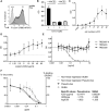
Determination of SUBA parameters. (A) Stably spike‐expressing Ramos‐null B cells (RSp) or Ramos‐null B cells (R) were stained with the recombinant anti‐SARS CoV‐2‐spike‐RBD binding antibody TRES224, followed by secondary AF647‐conjugated anti‐human IgG antibody and analyzed by flow cytometry. Live cells were pregated based on FSC/SSC characteristics. Numbers indicate the mean fluorescence intensity (×103). (B) 105 stably spike‐expressing Ramos‐null B cells (RSp) or Ramos‐null B cells (R) were allowed to attach to hACE2‐ or noncoated plates, fixed and stained with crystal violet. Crystal violet was solubilized and OD570nm was measured. Data are depicted as mean ± SD of one experiment with three technical replicates. Representative of at least ten experiments. (C) Titration of the cell number per 96 well with stably SARS‐CoV‐2‐spike expressing Ramos‐null B cells (RSp) and background (Ramos‐null B cells) subtracted. Data are presented as mean ± SD of three experiments performed with each with three technical replicates. (D) Titration of the hACE2 coating concentration (μg/mL) with stably SARS‐CoV‐2‐spike‐expressing Ramos‐null B cells (RSp; 105/well) and background (Ramos‐null B cells) subtracted. Data are presented as mean ± SD of three experiments performed with each three technical replicates. (E) Blockage of RSp cell binding to hACE2 with the RBD‐specific TRES224 antibody. 105 RSp cells were allowed to attach to hACE2‐coated plates in the presence of increasing concentrations of the anti‐RBD neutralizing antibody TRES224, an anti‐SARS‐CoV‐2‐spike N‐terminal domain (NTD) antibody (TRES328), and an isotype‐matched control antibody (TRES567‐II), fixed and stained with crystal violet. Crystal violet was solubilized and OD570nm was measured. The black line represents the control (without antibody). Data are represented as mean ± SD of four measurements from two experiments with two technical replicates/experiments. (F) The anti‐RBD neutralizing antibody TRES224 was tested in a Pseudovirus‐neutralization assay (mean ± SD of four replicates) and in SUBA (two experiments performed with each three technical replicates, mean ± SD). Data were fitted by nonlinear regression and IC50 values as well as Hill slopes are depicted.
Figure 3
Figure 3
Analysis of spike‐blocking activity in human sera. (A) Spike‐expressing Ramos‐null B cells (RSp; 105/well) were allowed to attach to hACE2‐coated wells in the presence or absence of sera of purified SARS‐CoV‐2 blocking antibodies or sera from SARS‐CoV‐2‐infected individuals, with (+) or without (−) symptoms, from three families (F1–F3) with at least one SARS‐CoV‐2 PCR‐positive family member (PCR test: +). Plates were washed, fixed, stained with crystal violet, solubilized, and OD was measured. The background (Ramos‐null B cells) was subtracted. Data are presented as mean % binding relative to control (RSp cells in the absence of serum or blocking antibodies) of three measurements performed with three technical replicates each. nd: not determined. (B) The binding assay was performed with increasing dilutions of sera with no (control), weak (serum P4), and strong blocking activity (serum F2.2). Spike‐expressing Ramos‐null B cells (RSp; 105/well) were allowed to attach to hACE2‐coated wells in the presence or absence of different dilutions or sera with previously defined weak or strong blocking activity. Data are presented as mean % binding relative to control (RSp cells in the absence of serum or blocking antibodies) performed in three technical replicates in one experiment. Red arrows indicate the dilutions where similar blocking activity was detected. (C) Sera from eight COVID‐19 negative and six COVID‐19 positive donors were subjected to SUBA in two technical replicates in one experiment. Data are depicted as mean ± 95% CI. Red arrows and numbers depict the lower limit of COVID‐19 negative sera. (D) Receiver operating characteristic (ROC) analysis of SUBA using the data obtained in (C) and additional serum dilutions. (E) Spike‐expressing Ramos‐null B cells (RSp; 105/well) were allowed to attach to hACE2‐coated wells in the presence of increasing concentrations of the monoclonal antibodies TRES224 or Regeneron R10933 diluted either with the assay medium or a human serum from an individual tested negative for anti‐SARS‐CoV‐2 antibodies. Data are presented as mean % binding relative to control (RSp cells in the absence of serum or blocking antibodies) performed in three technical replicates. Representative of two experiments.
Figure 4
Figure 4
Analysis of SARS‐CoV‐2 spike‐binding and ‐blocking antibodies. (A) Flow cytometric analysis of SARS‐CoV‐2 spike‐binding IgM, IgA, and IgG serum antibodies in patients (P1–P6) from different families with at least one PCR‐positive case of SARS‐CoV‐2 infection. P1 served as a negative control and did not contain serum antibodies recognizing the SARS‐CoV‐2 spike protein. HEK293T cells were transiently transfected with a plasmid encoding SARS‐CoV‐2 spike protein and stained with sera as described in Fig. S2. Numbers indicate the mean fluorescence intensities (MFI) of SARS‐CoV‐2‐Spike IgG (green), IgA (blue), and IgM (red). (B) SUBA assay to detect SARS‐CoV‐2‐blocking serum antibodies in sera from patients P1–P6. Data are presented as mean % RSp binding relative to control (RSp cells in the absence of serum or blocking antibodies) of three measurements performed in three technical replicates each. (C) Correlation between COVID‐19 symptoms and the presence of SARS‐CoV‐2 spike‐blocking antibodies in family members with at least one reported PCR‐positive SARS‐CoV‐2 infection. Typical symptoms were fever, head and body ache, diarrhea, exhaustion, cough, shortness of breath, and chest tightness. p = 0.0003 (unpaired t test).
Figure 5
Figure 5
COVID‐19 and vaccination responses against SARS‐CoV‐2 mutants measured by SUBA. (A) Flow cytometric analysis of Ramos cells expressing SARS‐CoV‐2 spike wild‐type (Wuhan), Alpha (B.1.1.7) or Beta (B.1.351) mutants with anti‐RBD Ab TRES224 and secondary AF647‐labeled anti‐human IgG antibody. Live cells were pregated based on FSC/SSC characteristics. Representative of three experiments with one sample per experiment. Numbers indicate the mean fluorescence intensities (MFI). (B) SUBA assay for Ramos cells expressing SARS‐CoV‐2 spike wild‐type (Wuhan, n = 14), Alpha (B.1.1.7, n = 11), or Beta (B.1.351, n = 11) mutants. Data are shown as mean ± SD from three experiments. (C) Wuhan spike‐expressing Ramos‐null B cells or Ramos‐null B cells expressing the Alpha (B.1.1.7) or Beta (B.1.351) spike mutants (each 105/well) were allowed to attach to hACE2‐coated wells in the presence of increasing concentrations of the monoclonal antibody TRES224 diluted with the assay medium. Data are presented as mean % binding relative to control (RSp cells in the absence of serum or blocking antibodies) performed in one experiment with two technical replicates. (D) Wuhan spike‐expressing Ramos‐null B cells or Ramos‐null B cells expressing the Alpha (B.1.1.7) or Beta (B.1.351) spike mutants (each 105/well) were allowed to attach to hACE2‐coated wells in the presence of decreasing dilutions of sera from COVID‐19 positive (F2.2) or negative (P1) donors and SUBA was performed. Data are presented as mean % binding relative to control (cells in the absence of serum or blocking antibodies) performed in two technical replicates. Representative of two experiments. (E) Timeline for blood sampling before and after Comirnaty vaccination from two volunteers (V1 and V2). (F and G) Wuhan spike‐expressing Ramos‐null B cells or Ramos‐null B cells expressing the Alpha (B.1.1.7) or Beta (B.1.351) spike mutants (each 105/well) were allowed to attach to hACE2‐coated wells in the presence of decreasing dilutions of sera from V1 and V2 donors and SUBA was performed. Data are presented as mean % binding ± SEM, relative to control (cells in the absence of serum or blocking antibodies) from two experiments performed with each two technical replicates, with values from the preimmune sera subtracted.

References

    1. Hu, B. , Guo, H. , Zhou, P. and Shi, Z.‐L. Characteristics of SARS‐CoV‐2 and COVID‐19. Nat. Rev. Microbiol. 2021. 19(3): 141–154. - PMC - PubMed
    1. Pezzini, A. and Padovani, A. Lifting the mask on neurological manifestations of COVID‐19. Nat. Rev. Neurol. 2020. 16(11): 636–644. - PMC - PubMed
    1. Yang, X. , Yu, Y. , Xu, J. , Shu, H. , Xia, J. , Liu, H. , Wu, Y. et al., Clinical course and outcomes of critically ill patients with SARS‐CoV‐2 pneumonia in Wuhan, China: a single‐centered, retrospective, observational study. Lancet Respir. Med. 2020. 8(5): 475–481. - PMC - PubMed
    1. Wajnberg, A. , Amanat, F. , Firpo, A. , Altman, D. R. , Bailey, M. J. , Mansour, M. , McMahon, M. et al., Robust neutralizing antibodies to SARS‐CoV‐2 infection persist for months. Science 2020. 370(6521): 1227–1230. - PMC - PubMed
    1. To, K. K.‐W. , Tsang, O. T.‐Y. , Leung, W.‐S. , Tam, A. R. , Wu, T.‐C. , Lung, D. C. , Yip, C. C.‐Y. et al., Temporal profiles of viral load in posterior oropharyngeal saliva samples and serum antibody responses during infection by SARS‐CoV‐2: an observational cohort study. Lancet Infect. Dis. 2020. 20(5): 565–574. - PMC - PubMed

Publication types

MeSH terms