

A Systematic Review of Imaging Studies in Olfactory Dysfunction Secondary to COVID-19
- PMID: 34548231
- PMCID: PMC8403662
- DOI: 10.1016/j.acra.2021.08.010
A Systematic Review of Imaging Studies in Olfactory Dysfunction Secondary to COVID-19
Abstract
Rationale and objectives: Hyposmia/anosmia is common among patients with coronavirus disease-2019 (COVID-19). Various imaging modalities have been used to assess olfactory dysfunction in COVID-19. In this systematic review, we sought to categorize and summarize the imaging data in COVID-19-induced anosmia.
Material and methods: Eligible articles were included after a comprehensive review using online databases including Google scholar, Scopus, PubMed, Web of science and Elsevier. Duplicate results, conference abstracts, reviews, and studies in languages other than English were excluded.
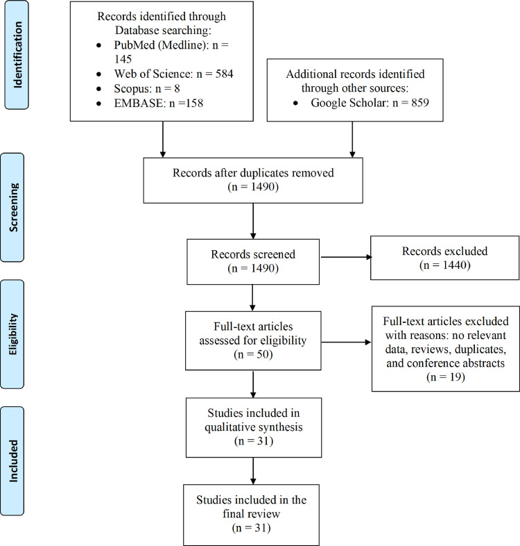
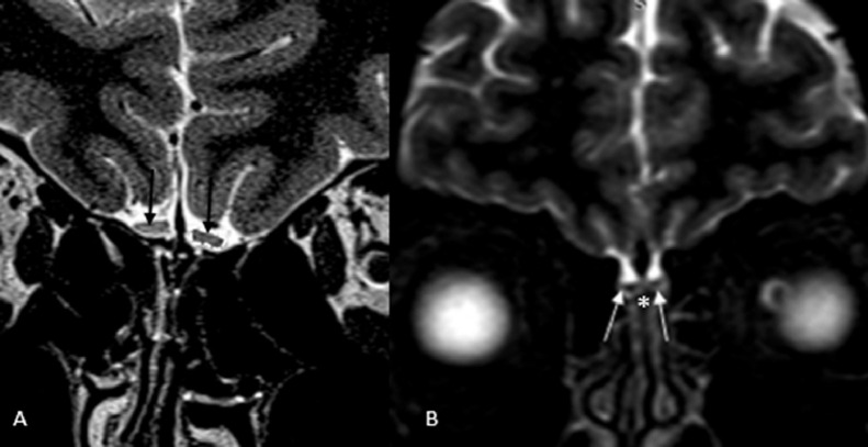
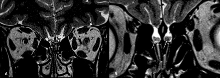
Results: In total, 305 patients undergoing MRI/functional MRI (177), CT of paranasal sinuses (129), and PET/CT or PET/MRI scans (14) were included. Out of a total of 218 findings reported on MRI, 80 were reported on early (≤ 1 month) and 85 on late (>1 month) imaging in relation to the onset of anosmia. Overall, OB morphology and T2-weighted or FLAIR signal intensity were normal in 68/218 (31.2%), while partial or complete opacification of OC was observed in 60/218 (27.5%). T2 hyperintensity in OB was detected in 11/80 (13.75%) and 18/85 (21.17%) on early and late imaging, respectively. Moreover, OB atrophy was reported in 1/80 (1.25%) on early and in 9/85 (10.58%) on late imaging. Last, among a total of 129 CT scans included, paranasal sinuses were evalualted in 88 (68.21%), which were reported as normal in most cases (77/88, [87.5%]).
Conclusion: In this systematic review, normal morphology and T2/FLAIR signal intensity in OB and OC obstruction were the most common findings in COVID-19-induced anosmia, while paranasal sinuses were normal in most cases. OC obstruction is the likely mechanism for olfactory dysfunction in COVID-19. Abnormalities in OB signal intensity and OB atrophy suggest that central mechanisms may also play a role in late stage in COVID-19-induced anosmia.
Keywords: COVID-19; anosmia; computed tomography; magnetic resonance imaging; olfactory dysfunction; positron emission tomography.
Copyright © 2021 The Association of University Radiologists. Published by Elsevier Inc. All rights reserved.
Figures
Similar articles
-
Olfactory Bulb MRI and Paranasal Sinus CT Findings in Persistent COVID-19 Anosmia.Acad Radiol. 2021 Jan;28(1):28-35. doi: 10.1016/j.acra.2020.10.006. Epub 2020 Oct 19. Acad Radiol. 2021. PMID: 33132007 Free PMC article.
-
Transient modifications of the olfactory bulb on MR follow-up of COVID-19 patients with related olfactory dysfunction.J Neuroradiol. 2022 Jun;49(4):329-332. doi: 10.1016/j.neurad.2022.03.003. Epub 2022 Mar 17. J Neuroradiol. 2022. PMID: 35306004 Free PMC article.
-
Paranasal sinuses computed tomography findings in anosmia of COVID-19.Am J Otolaryngol. 2020 Nov-Dec;41(6):102636. doi: 10.1016/j.amjoto.2020.102636. Epub 2020 Jul 3. Am J Otolaryngol. 2020. PMID: 32652405 Free PMC article.
-
Olfactory system measurements in COVID-19: a systematic review and meta-analysis.Neuroradiology. 2023 Jan;65(1):25-39. doi: 10.1007/s00234-022-03014-8. Epub 2022 Jul 18. Neuroradiology. 2023. PMID: 35843987 Free PMC article.
-
Objective Sensory Testing Methods Reveal a Higher Prevalence of Olfactory Loss in COVID-19-Positive Patients Compared to Subjective Methods: A Systematic Review and Meta-Analysis.Chem Senses. 2020 Dec 5;45(9):865-874. doi: 10.1093/chemse/bjaa064. Chem Senses. 2020. PMID: 33245136 Free PMC article.
Cited by
-
Intratympanic steroid treatments rescued recurrent hearing loss following COVID-19 vaccination and detection of an intralabyrinthine schwannoma.BMJ Case Rep. 2022 Jul 6;15(7):e249316. doi: 10.1136/bcr-2022-249316. BMJ Case Rep. 2022. PMID: 35793841 Free PMC article.
-
Persistent olfactory dysfunction after COVID-19 is associated with reduced perfusion in the frontal lobe.Acta Neurol Scand. 2022 Aug;146(2):194-198. doi: 10.1111/ane.13627. Epub 2022 Apr 25. Acta Neurol Scand. 2022. PMID: 35467007 Free PMC article.
-
Clinical and Imaging Evaluation of COVID-19-Related Olfactory Dysfunction.Am J Rhinol Allergy. 2023 Jul;37(4):456-463. doi: 10.1177/19458924231163969. Epub 2023 Mar 21. Am J Rhinol Allergy. 2023. PMID: 36945746 Free PMC article.
-
Anosmia in COVID-19 could be associated with long-term deficits in the consolidation of procedural and verbal declarative memories.Front Neurosci. 2022 Dec 9;16:1082811. doi: 10.3389/fnins.2022.1082811. eCollection 2022. Front Neurosci. 2022. PMID: 36570827 Free PMC article.
-
Treatments for Olfactory Dysfunction in COVID-19: A Systematic Review.Int Arch Otorhinolaryngol. 2024 May 25;28(4):e728-e743. doi: 10.1055/s-0044-1786046. eCollection 2024 Oct. Int Arch Otorhinolaryngol. 2024. PMID: 39464360 Free PMC article. Review.
References
-
- Politi LS, Salsano E, Grimaldi M. Magnetic resonance imaging alteration of the brain in a patient with coronavirus disease 2019 (COVID-19) and anosmia. JAMA Neurol. 2020;77(8):1028–1029. - PubMed
Publication types
MeSH terms
LinkOut - more resources
Full Text Sources
Medical
Miscellaneous
