Clinical Trial
. 2022 Jan;12(1):74-89.
doi: 10.1158/2159-8290.CD-21-0715. Epub 2021 Sep 21.

Efficacy and Safety of Patritumab Deruxtecan (HER3-DXd) in EGFR Inhibitor-Resistant, EGFR-Mutated Non-Small Cell Lung Cancer

Affiliations
Clinical Trial

Efficacy and Safety of Patritumab Deruxtecan (HER3-DXd) in EGFR Inhibitor-Resistant, EGFR-Mutated Non-Small Cell Lung Cancer

Pasi A Jänne et al. Cancer Discov. 2022 Jan.

Erratum in

Abstract

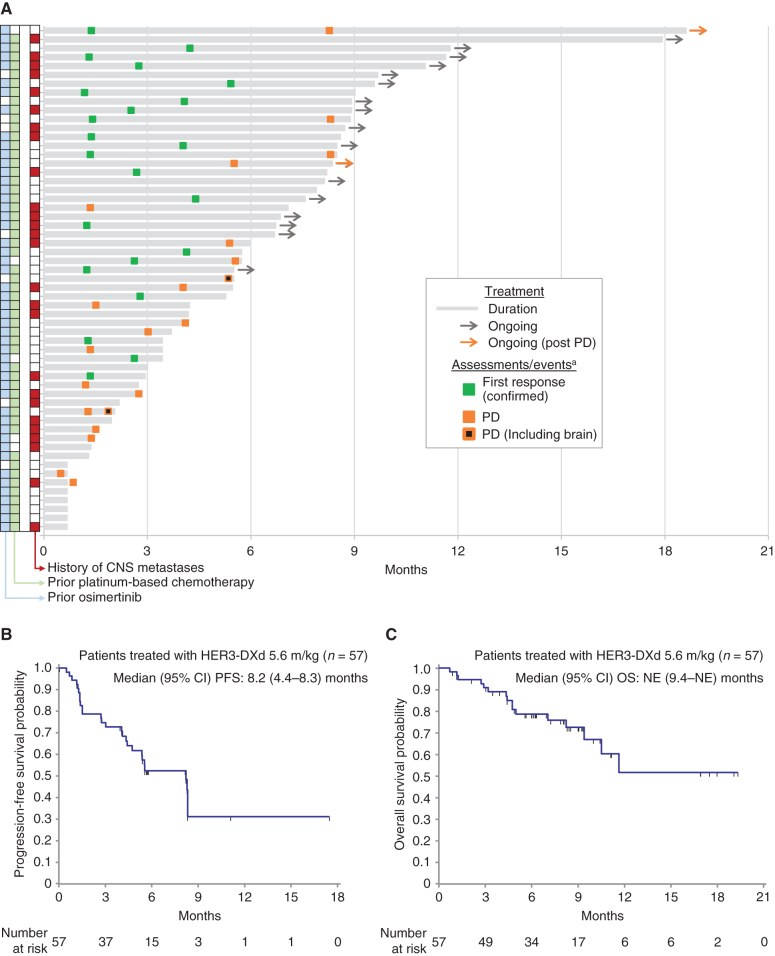
Receptor tyrosine-protein kinase ERBB3 (HER3) is expressed in most EGFR-mutated lung cancers but is not a known mechanism of resistance to EGFR inhibitors. HER3-DXd is an antibody-drug conjugate consisting of a HER3 antibody attached to a topoisomerase I inhibitor payload via a tetrapeptide-based cleavable linker. This phase I, dose escalation/expansion study included patients with locally advanced or metastatic EGFR-mutated non-small cell lung cancer (NSCLC) with prior EGFR tyrosine kinase inhibitor (TKI) therapy. Among 57 patients receiving HER3-DXd 5.6 mg/kg intravenously once every 3 weeks, the confirmed objective response rate by blinded independent central review (Response Evaluation Criteria in Solid Tumors v1.1) was 39% [95% confidence interval (CI), 26.0-52.4], and median progression-free survival was 8.2 (95% CI, 4.4-8.3) months. Responses were observed in patients with known and unknown EGFR TKI resistance mechanisms. Clinical activity was observed across a broad range of HER3 membrane expression. The most common grade ≥3 treatment-emergent adverse events were hematologic toxicities. HER3-DXd has clinical activity in EGFR TKI-resistant cancers independent of resistance mechanisms, providing an approach to treat a broad range of drug-resistant cancers. SIGNIFICANCE: In metastatic EGFR-mutated NSCLC, after disease progression on EGFR TKI therapy, treatment approaches include genotype-directed therapy targeting a known resistance mechanism or chemotherapy. HER3-DXd demonstrated clinical activity spanning known and unknown EGFR TKI resistance mechanisms. HER3-DXd could present a future treatment option agnostic to the EGFR TKI resistance mechanism.See related commentary by Lim et al., p. 16.This article is highlighted in the In This Issue feature, p. 1.


Figures

Figure 1. Best percentage change in the tumor sum of diameters (SoD) from baseline for the pooled HER3-DXd (5.6 mg/kg, i.v. once every 3 weeks) population. Tumor genomic alterations prior to treatment with HER3-DXd are provided for each patient. Six patients could not be evaluated for BOR due to lack of adequate post–baseline tumor assessment and are not shown; one patient had a BOR of NE due to achieving SD too early (<5 weeks) and is shown with hatched markings. Genomic analysis was performed centrally using Oncomine Comprehensive Assay v3 (Thermo Fisher Scientific). Results from local testing are included, if available, together with any additional mutations detected using GuardantOMNI assay of ctDNA in blood collected prior to treatment with HER3-DXd. For ctDNA analysis, a minor allelic frequency of ≥0.1% was used as a threshold for detection of mutations. aPatient had multiple tumor mutations comprising CDKN2A A143V; PIK3CA E542K, E545K, E726K; ERBB2 K200N; and ERBB3 Q847*, Q849*. cBOR, Confirmed BOR.
Figure 1.
Best percentage change in the tumor sum of diameters (SoD) from baseline for the pooled HER3-DXd (5.6 mg/kg, i.v. once every 3 weeks) population. Tumor genomic alterations prior to treatment with HER3-DXd are provided for each patient. Six patients could not be evaluated for BOR due to lack of adequate post–baseline tumor assessment and are not shown; one patient had a BOR of NE due to achieving SD too early (<5 weeks) and is shown with hatched markings. Genomic analysis was performed centrally using Oncomine Comprehensive Assay v3 (Thermo Fisher Scientific). Results from local testing are included, if available, together with any additional mutations detected using GuardantOMNI assay of ctDNA in blood collected prior to treatment with HER3-DXd. For ctDNA analysis, a minor allelic frequency of ≥0.1% was used as a threshold for detection of mutations. aPatient had multiple tumor mutations comprising CDKN2A A143V; PIK3CA E542K, E545K, E726K; ERBB2 K200N; and ERBB3 Q847*, Q849*. cBOR, Confirmed BOR.
Figure 2. Tumor response as assessed by BICR in patients treated with HER3-DXd at 5.6 mg/kg (n = 57). A, Swimmer plot showing treatment duration, first occurrence of confirmed tumor response, and progression. Prior treatment and history of CNS metastases are indicated for each patient. B, Kaplan–Meier plot of progression-free survival probability. C, Kaplan–Meier plot of overall survival probability. aMarkers show the time of the initial response for confirmed responses. Two patients continued treatment after progression. In one case (top of swimmer plot), the patient had an equivocal (small) lesion and continued on treatment, but the lesion was later assessed as unequivocal—the swimmer plot is marked PD at the date of the lesion's first appearance. In the other case, treatment continued because PD was determined by BICR but not by the local investigator (the study treatment discontinuation criteria were based on local tumor assessment).
Figure 2.
Tumor response as assessed by BICR in patients treated with HER3-DXd at 5.6 mg/kg (n = 57). A, Swimmer plot showing treatment duration, first occurrence of confirmed tumor response, and progression. Prior treatment and history of CNS metastases are indicated for each patient. B, Kaplan–Meier plot of progression-free survival probability. C, Kaplan–Meier plot of overall survival probability. aMarkers show the time of the initial response for confirmed responses. Two patients continued treatment after progression. In one case (top of swimmer plot), the patient had an equivocal (small) lesion and continued on treatment, but the lesion was later assessed as unequivocal—the swimmer plot is marked PD at the date of the lesion's first appearance. In the other case, treatment continued because PD was determined by BICR but not by the local investigator (the study treatment discontinuation criteria were based on local tumor assessment).
Figure 3. Pretreatment HER3 membrane expression and association with BOR. BOR was assessed by BICR in patients treated with HER3-DXd at 5.6 mg/kg (43 of 57 patients evaluable for HER3 membrane expression). A, Distribution of pretreatment HER3 membrane H-score (0–300). B, Pretreatment HER3 membrane H-score and association with time since last treatment with EGFR TKI. C and D, Pretreatment HER3 membrane H-score and confirmed BOR (cBOR).
Figure 3.
Pretreatment HER3 membrane expression and association with BOR. BOR was assessed by BICR in patients treated with HER3-DXd at 5.6 mg/kg (43 of 57 patients evaluable for HER3 membrane expression). A, Distribution of pretreatment HER3 membrane H-score (0–300). B, Pretreatment HER3 membrane H-score and association with time since last treatment with EGFR TKI. C and D, Pretreatment HER3 membrane H-score and confirmed BOR (cBOR).
Figure 4. Analyses of ctDNA changes. A, Proportions of confirmed BOR (cBOR; BICR per RECIST 1.1) by early clearance of ctDNA.a B, Kaplan–Meier plots showing PFS by early clearance of ctDNA.a C, Waterfall plot showing maximum change relative to baseline in the minor variant frequency (MVF) of EGFR-activating mutations in ctDNA and confirmed BOR by BICR (25 of 57 patients were evaluableb). aEarly clearance of ctDNA was defined as nondetectable plasma of both EGFR Ex19del and EGFR L858R at week 3 or week 6, where one or more allele comprising EGFR Ex19del or EGFR L858R was detectable at baseline. Serial ctDNA samples were collected from 45 of 57 patients, but 5 did not have evaluable ctDNA data for either week 3 or 6. In the 17 patients not evaluable for early clearance of ctDNA, responses were CR/PR, 29%; SD, 24%; PD, 17%; and NE, 35%. bFewer patients (25 of 57) were evaluable for relative change in ctDNA than for early clearance of ctDNA (40 of 57), as the former required ctDNA levels to be above the limit of quantification (MVF > 1%); the latter required levels to be above the limit of detection, which was lower (MVF > 0.02%). cPatients without GeneStrat data at C1D1 were analyzed if they had GuardantOMNI data at C1D1 when the MVF was >1%; this is because GeneStrat and GuardantOMNI data were concordant, except for the low MVF range (<1%). SoD, sum of diameters.
Figure 4.
Analyses of ctDNA changes. A, Proportions of confirmed BOR (cBOR; BICR per RECIST 1.1) by early clearance of ctDNA.aB, Kaplan–Meier plots showing PFS by early clearance of ctDNA.aC, Waterfall plot showing maximum change relative to baseline in the minor variant frequency (MVF) of EGFR-activating mutations in ctDNA and confirmed BOR by BICR (25 of 57 patients were evaluableb). aEarly clearance of ctDNA was defined as nondetectable plasma of both EGFR Ex19del and EGFR L858R at week 3 or week 6, where one or more allele comprising EGFR Ex19del or EGFR L858R was detectable at baseline. Serial ctDNA samples were collected from 45 of 57 patients, but 5 did not have evaluable ctDNA data for either week 3 or 6. In the 17 patients not evaluable for early clearance of ctDNA, responses were CR/PR, 29%; SD, 24%; PD, 17%; and NE, 35%. bFewer patients (25 of 57) were evaluable for relative change in ctDNA than for early clearance of ctDNA (40 of 57), as the former required ctDNA levels to be above the limit of quantification (MVF > 1%); the latter required levels to be above the limit of detection, which was lower (MVF > 0.02%). cPatients without GeneStrat data at C1D1 were analyzed if they had GuardantOMNI data at C1D1 when the MVF was >1%; this is because GeneStrat and GuardantOMNI data were concordant, except for the low MVF range (<1%). SoD, sum of diameters.

Comment in

References

    1. Zhang YL, Yuan JQ, Wang KF, Fu XH, Han XR, Threapleton Det al. The prevalence of EGFR mutation in patients with non-small cell lung cancer: a systematic review and meta-analysis. Oncotarget 2016;7:78985–93. - PMC - PubMed
    1. Soria JC, Ohe Y, Vansteenkiste J, Reungwetwattana T, Chewaskulyong B, Lee KHet al. Osimertinib in untreated EGFR-mutated advanced non-small-cell lung cancer. N Engl J Med 2018;378:113–25. - PubMed
    1. Karachaliou N, Fernandez-Bruno M, Paulina Bracht JW, Rosell R. EGFR first- and second-generation TKIs—there is still place for them in EGFR-mutant NSCLC patients. Transl Cancer Res 2019;8:S23–47. - PMC - PubMed
    1. Ramalingam SS, Vansteenkiste J, Planchard D, Cho BC, Gray JE, Ohe Yet al. Overall survival with osimertinib in untreated, EGFR-mutated advanced NSCLC. N Engl J Med 2020;382:41–50. - PubMed
    1. Rebuzzi SE, Alfieri R, La Monica S, Minari R, Petronini PG, Tiseo M. Combination of EGFR-TKIs and chemotherapy in advanced EGFR mutated NSCLC: review of the literature and future perspectives. Crit Rev Oncol Hematol 2020;146:102820. - PubMed

Publication types

MeSH terms