. 2022 Feb;12(2):484-501.
doi: 10.1158/2159-8290.CD-21-0601. Epub 2021 Sep 21.

Mesenchymal Lineage Heterogeneity Underlies Nonredundant Functions of Pancreatic Cancer-Associated Fibroblasts

Affiliations

Mesenchymal Lineage Heterogeneity Underlies Nonredundant Functions of Pancreatic Cancer-Associated Fibroblasts

Erin J Helms et al. Cancer Discov. 2022 Feb.

Abstract

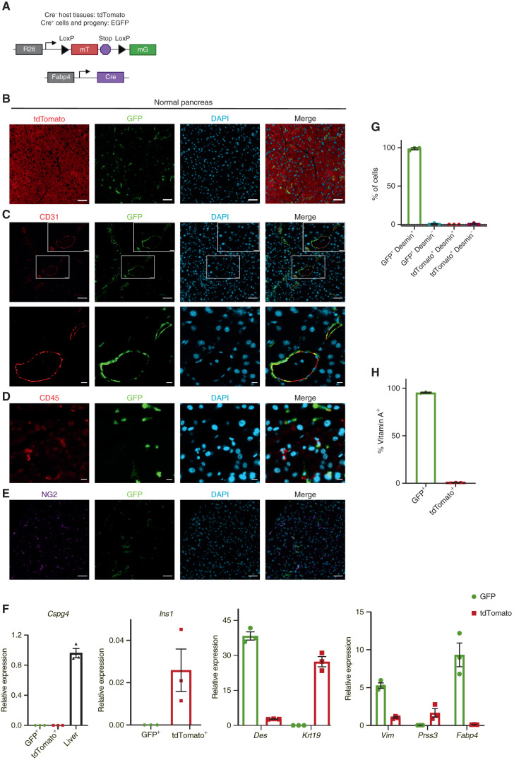
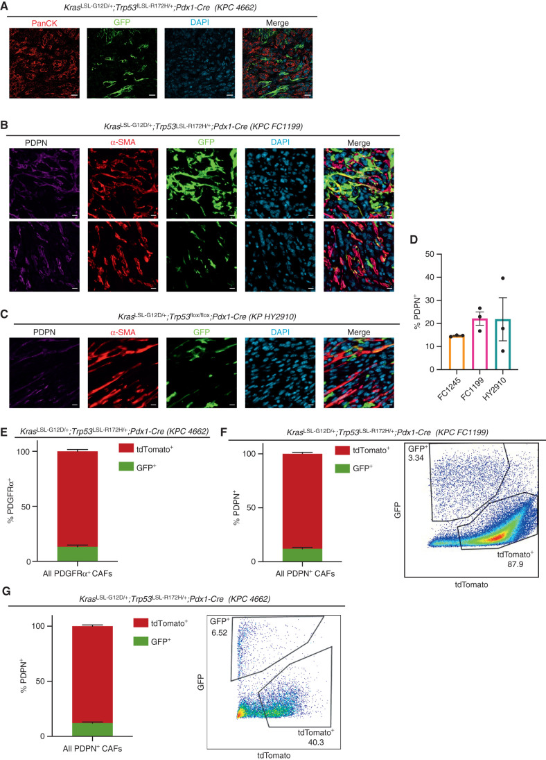
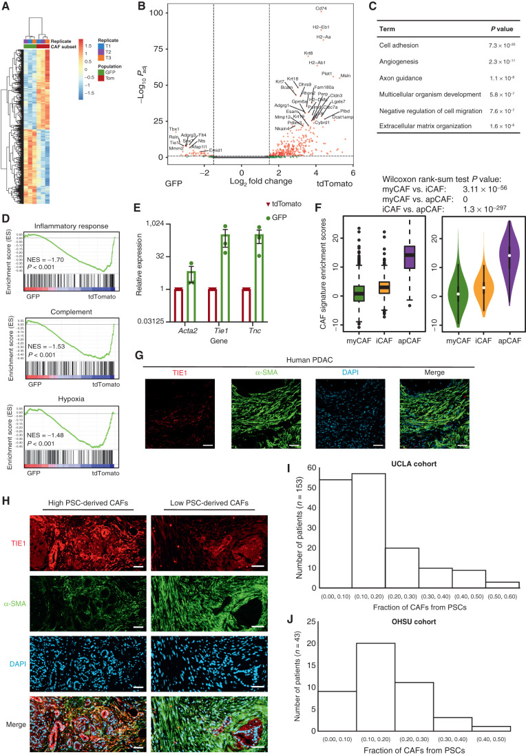
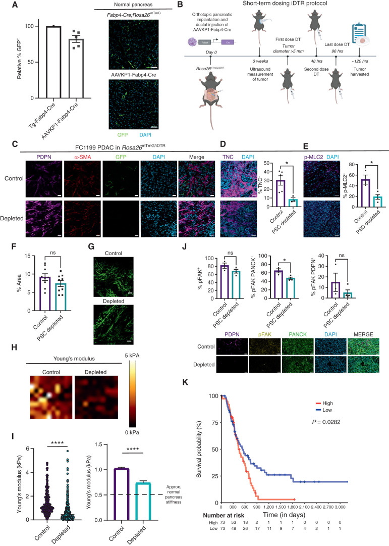
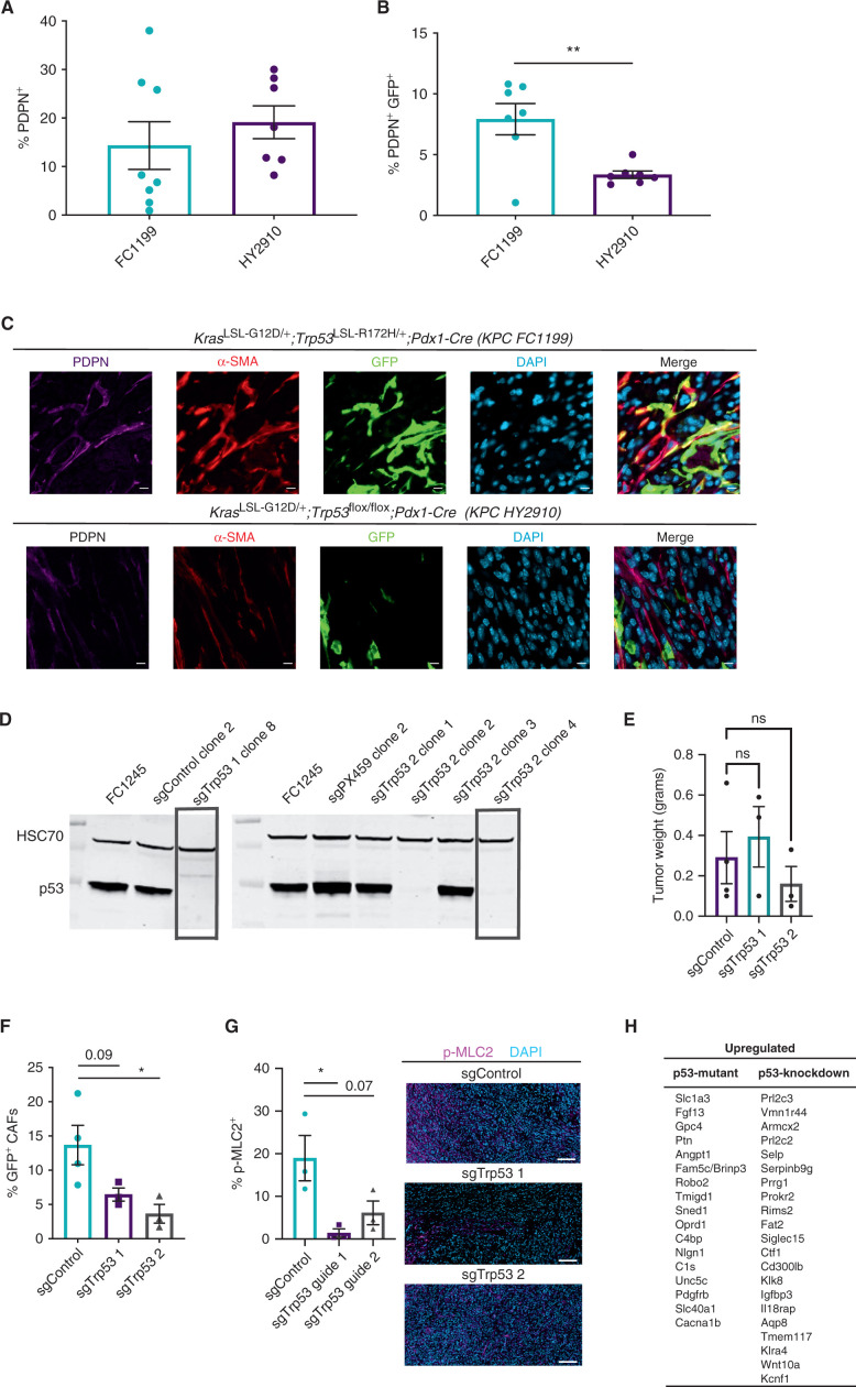
Cancer-associated fibroblast (CAF) heterogeneity is increasingly appreciated, but the origins and functions of distinct CAF subtypes remain poorly understood. The abundant and transcriptionally diverse CAF population in pancreatic ductal adenocarcinoma (PDAC) is thought to arise from a common cell of origin, pancreatic stellate cells (PSC), with diversification resulting from cytokine and growth factor gradients within the tumor microenvironment. Here we analyzed the differentiation and function of PSCs during tumor progression in vivo. Contrary to expectations, we found that PSCs give rise to a numerically minor subset of PDAC CAFs. Targeted ablation of PSC-derived CAFs within their host tissue revealed nonredundant functions for this defined CAF population in shaping the PDAC microenvironment, including production of specific extracellular matrix components and tissue stiffness regulation. Together, these findings link stromal evolution from distinct cells of origin to transcriptional heterogeneity among PDAC CAFs and demonstrate unique functions for CAFs of a defined cellular origin. SIGNIFICANCE: By tracking and ablating a specific CAF population, we find that a numerically minor CAF subtype from a defined cell of origin plays unique roles in establishing the pancreatic tumor microenvironment. Together with prior studies, this work suggests that mesenchymal lineage heterogeneity and signaling gradients diversify PDAC CAFs.See related commentary by Cukierman, p. 296.This article is highlighted in the In This Issue feature, p. 275.


Figures

Figure 1. Fabp4-Cre marks stellate cells specifically and pervasively within normal pancreas tissue. A, Schematic of the alleles used to label and track PSCs in vivo. B, Representative image of normal pancreas tissue from Fabp4-Cre;Rosa26mTmG mice (n = 7) showing rare GFP+ cells within a predominantly tdTomato+ tissue. Scale bar, 50 μm. C, Representative image of normal pancreas tissue from Fabp4-Cre;Rosa26mTmG mice (n = 3) showing CD31+ endothelial cells and GFP+ cells, some of which are adjacent to vessels. Top images: scale bar, 50 μm; bottom images: scale bar, 10 μm. D, Representative image of normal pancreas tissue from Fabp4-Cre;Rosa26mTmG mice (n = 3) showing CD45+ leukocytes and GFP+ cells. Scale bar, 10 μm. E, Representative image of normal pancreas tissue from Fabp4-Cre;Rosa26mTmG mice (n = 3) showing NG2+ pericytes and GFP+ cells. Scale bar, 50 μm. F, qPCR for the indicated genes in GFP+ and tdTomato+ cells isolated from normal pancreas tissue from Fabp4-Cre;Rosa26mTmG mice by FACS (n = 3, with each replicate pooled from two mice), including markers of pericytes (Cspg4; liver is a positive control), stellate cells and potentially other mesenchymal cells (Des), ductal cells (Krt19), mesenchymal cells (Vim), acinar cells (Prss3), and β cells (Ins1); Fabp4 was included as a control. Data were normalized to 36b4 and are presented as mean ± SEM. G, Quantification of GFP+ cells and tdTomato+ cells out of total, desmin+ PSCs isolated by density centrifugation from normal pancreas tissue in Fabp4-Cre;Rosa26mTmG mice (n = 3) and analyzed by immunofluorescence microscopy. Data are presented as mean ± SEM. H, Flow cytometry results depicting GFP+ and tdTomato+ cells among all vitamin A+ PSCs in normal pancreas tissue from Fabp4-Cre;Rosa26mTmG mice (n = 3). Data are presented as mean ± SEM.
Figure 1.
Fabp4-Cre marks stellate cells specifically and pervasively within normal pancreas tissue. A, Schematic of the alleles used to label and track PSCs in vivo. B, Representative image of normal pancreas tissue from Fabp4-Cre;Rosa26mTmG mice (n = 7) showing rare GFP+ cells within a predominantly tdTomato+ tissue. Scale bar, 50 μm. C, Representative image of normal pancreas tissue from Fabp4-Cre;Rosa26mTmG mice (n = 3) showing CD31+ endothelial cells and GFP+ cells, some of which are adjacent to vessels. Top images: scale bar, 50 μm; bottom images: scale bar, 10 μm. D, Representative image of normal pancreas tissue from Fabp4-Cre;Rosa26mTmG mice (n = 3) showing CD45+ leukocytes and GFP+ cells. Scale bar, 10 μm. E, Representative image of normal pancreas tissue from Fabp4-Cre;Rosa26mTmG mice (n = 3) showing NG2+ pericytes and GFP+ cells. Scale bar, 50 μm. F, qPCR for the indicated genes in GFP+ and tdTomato+ cells isolated from normal pancreas tissue from Fabp4-Cre;Rosa26mTmG mice by FACS (n = 3, with each replicate pooled from two mice), including markers of pericytes (Cspg4; liver is a positive control), stellate cells and potentially other mesenchymal cells (Des), ductal cells (Krt19), mesenchymal cells (Vim), acinar cells (Prss3), and β cells (Ins1); Fabp4 was included as a control. Data were normalized to 36b4 and are presented as mean ± SEM. G, Quantification of GFP+ cells and tdTomato+ cells out of total, desmin+ PSCs isolated by density centrifugation from normal pancreas tissue in Fabp4-Cre;Rosa26mTmG mice (n = 3) and analyzed by immunofluorescence microscopy. Data are presented as mean ± SEM. H, Flow cytometry results depicting GFP+ and tdTomato+ cells among all vitamin A+ PSCs in normal pancreas tissue from Fabp4-Cre;Rosa26mTmG mice (n = 3). Data are presented as mean ± SEM.
Figure 2. Stellate cells give rise to a numerically minor subset of PDAC CAFs. A, IHC staining of PDAC (KPC 4662) in Fabp4-Cre;Rosa26mTmG hosts (n = 5), with GFP in green and panCK (tumor cells) in red. Scale bar, 20 μm. B, IHC staining of PDAC (KPC FC1199) in Fabp4-Cre;Rosa26mTmG hosts (n = 3), stained for GFP, PDPN, and α-SMA. Scale bar, 10 μm. C, IHC staining of PDAC (KPflox/+C HY2910) in Fabp4-Cre;Rosa26mTmG hosts (n = 3), stained for GFP, PDPN, and α-SMA. Scale bar, 10 μm. D, Quantification of IHC staining for PDPN in the indicated transplantable PDAC models. E, Flow cytometry analysis of PDGFRα, GFP, and tdTomato in KPC 4662 tumors in Fabp4-Cre;Rosa26mTmG hosts (n = 5). Data are presented as mean ± SEM. For E–G, CAF frequency calculations excluded the tdTomato−GFP− fraction constituting transplanted PDAC cells. F, Flow cytometry analysis of PDPN, GFP, and tdTomato in KPC FC1199 tumors in Fabp4-Cre;Rosa26mTmG hosts (n = 8). Data are presented as mean ± SEM. G, Flow cytometry analysis of PDPN, GFP, and tdTomato in KPC 4662 tumors in Fabp4-Cre;Rosa26mTmG hosts (n = 3). Data are presented as mean ± SEM.
Figure 2.
Stellate cells give rise to a numerically minor subset of PDAC CAFs. A, IHC staining of PDAC (KPC 4662) in Fabp4-Cre;Rosa26mTmG hosts (n = 5), with GFP in green and panCK (tumor cells) in red. Scale bar, 20 μm. B, IHC staining of PDAC (KPC FC1199) in Fabp4-Cre;Rosa26mTmG hosts (n = 3), stained for GFP, PDPN, and α-SMA. Scale bar, 10 μm. C, IHC staining of PDAC (KPflox/+C HY2910) in Fabp4-Cre;Rosa26mTmG hosts (n = 3), stained for GFP, PDPN, and α-SMA. Scale bar, 10 μm. D, Quantification of IHC staining for PDPN in the indicated transplantable PDAC models. E, Flow cytometry analysis of PDGFRα, GFP, and tdTomato in KPC 4662 tumors in Fabp4-Cre;Rosa26mTmG hosts (n = 5). Data are presented as mean ± SEM. For E–G, CAF frequency calculations excluded the tdTomatoGFP fraction constituting transplanted PDAC cells. F, Flow cytometry analysis of PDPN, GFP, and tdTomato in KPC FC1199 tumors in Fabp4-Cre;Rosa26mTmG hosts (n = 8). Data are presented as mean ± SEM. G, Flow cytometry analysis of PDPN, GFP, and tdTomato in KPC 4662 tumors in Fabp4-Cre;Rosa26mTmG hosts (n = 3). Data are presented as mean ± SEM.
Figure 3. Mesenchymal lineage heterogeneity gives rise to transcriptional heterogeneity among PDAC CAFs. Heat map (A) and volcano plot (B) depicting differentially expressed genes in PSC-derived (GFP+) versus non–PSC-derived (tdTomato+) CAFs from KPC FC1199 PDAC in Fabp4-Cre;Rosa26mTmG hosts (n = 3), identified by RNA-seq. In B, gray circles indicate genes not different between the populations, green circles indicate genes with log2 fold change >1 or <−1, and pink circles indicate genes with the same log2 fold change criteria and P < 0.05. C, Gene ontology analysis identifying the top terms enriched in association with genes upregulated at least twofold in PSC-derived CAFs compared with non–PSC-derived CAFs. D, Gene set enrichment analysis showing pathways or processes with transcriptional signatures enriched in the tdTomato+ CAF population. NES, normalized enrichment score. E, qPCR for the indicated genes on PSC-derived and non–PSC-derived CAFs sorted from KPC FC1199 PDAC in Fabp4-Cre;Rosa26mTmG hosts (n = 3) by FACS. Data were normalized to 36b4 and are presented as mean ± SEM. F, Box (left) and violin (right) plots indicating enrichment scores for differentially expressed genes between GFP+ and tdTomato+ CAFs among myCAF-, iCAF-, and apCAF-associated genes per previously published small conditional RNA-seq results. G, Representative IHC staining of human PDAC for TIE1 and α-SMA (n = 5). Scale bar, 50 μm. H, Representative images from a human PDAC microarray after IHC staining for TIE1 and α-SMA (n = 153). Scale bar, 50 µm. I, Quantification of TIE1+α-SMA+ area out of total α-SMA+ area on each patient sample from the array. Different regions from the same patient were averaged together to yield one frequency per patient sample (four punches per patient, 612 total tumor regions analyzed, 153 plotted here after averaging for each patient). J, Quantification of TIE1+α-SMA+ area out of total α-SMA+ area using whole PDAC tissue sections (n = 43) from an independent patient cohort.
Figure 3.
Mesenchymal lineage heterogeneity gives rise to transcriptional heterogeneity among PDAC CAFs. Heat map (A) and volcano plot (B) depicting differentially expressed genes in PSC-derived (GFP+) versus non–PSC-derived (tdTomato+) CAFs from KPC FC1199 PDAC in Fabp4-Cre;Rosa26mTmG hosts (n = 3), identified by RNA-seq. In B, gray circles indicate genes not different between the populations, green circles indicate genes with log2 fold change >1 or <−1, and pink circles indicate genes with the same log2 fold change criteria and P < 0.05. C, Gene ontology analysis identifying the top terms enriched in association with genes upregulated at least twofold in PSC-derived CAFs compared with non–PSC-derived CAFs. D, Gene set enrichment analysis showing pathways or processes with transcriptional signatures enriched in the tdTomato+ CAF population. NES, normalized enrichment score. E, qPCR for the indicated genes on PSC-derived and non–PSC-derived CAFs sorted from KPC FC1199 PDAC in Fabp4-Cre;Rosa26mTmG hosts (n = 3) by FACS. Data were normalized to 36b4 and are presented as mean ± SEM. F, Box (left) and violin (right) plots indicating enrichment scores for differentially expressed genes between GFP+ and tdTomato+ CAFs among myCAF-, iCAF-, and apCAF-associated genes per previously published small conditional RNA-seq results. G, Representative IHC staining of human PDAC for TIE1 and α-SMA (n = 5). Scale bar, 50 μm. H, Representative images from a human PDAC microarray after IHC staining for TIE1 and α-SMA (n = 153). Scale bar, 50 µm. I, Quantification of TIE1+α-SMA+ area out of total α-SMA+ area on each patient sample from the array. Different regions from the same patient were averaged together to yield one frequency per patient sample (four punches per patient, 612 total tumor regions analyzed, 153 plotted here after averaging for each patient). J, Quantification of TIE1+α-SMA+ area out of total α-SMA+ area using whole PDAC tissue sections (n = 43) from an independent patient cohort.
Figure 4. Targeted ablation reveals unique roles for PSC-derived CAFs in regulation of the ECM and mechanosignaling. A, IHC staining and quantification of GFP+ cells in normal pancreas tissue from Fabp4-Cre;Rosa26mTmG mice and from Rosa26mTmG/iDTR mice 7 days after intraductal injection with AAVKP1-Fabp4-Cre (n = 5). Data are presented as mean ± SEM. Scale bar, 50 μm. B, Schematic of tumor modeling using intraductal injection of AAVKP1-Fabp4-Cre and orthotopic transplantation of KPC PDAC cells into Rosa26mTmG/iDTR hosts. C, IHC staining for GFP, PDPN, and α-SMA of KPC FC1199 PDAC in Rosa26mTmG/iDTR hosts with intraductal injection of AAVKP1-Fabp4-Cre, enrolled when tumors reached 5 to 6 mm in diameter and treated with PBS or DT for 5 days (n = 4). Scale bar, 20 μm. D, IHC staining for TNC of KPC FC1199 PDAC in AAVKP1-Fabp4-Cre–injected Rosa26mTmG/iDTR hosts, enrolled at 5 to 6 mm in tumor diameter and treated with PBS or DT for 5 days (n = 3). Scale bar, 50 μm. Data are presented as mean ± SEM. E, IHC staining for p-MLC2 of PDAC samples as described in D. Scale bar, 50 μm. Data are presented as mean ± SEM. Quantification (F) and images (G) of fibrillar collagen content analyzed by second harmonic generation with normalized intensity as a quantification of concentration in control and PSC-depleted PDAC (n = 3 per group). Scale bar, 42.5 μm. H, Force maps generated by atomic force microscopy (AFM) on KPC FC1199 PDAC in AAVKP1-Fabp4-Cre–injected Rosa26mTmG/iDTR hosts (n = 3 per treatment group, control: 1,063 data points, depleted: 717 data points), excised after 5 days of treatment with PBS or DT. I, Quantification of Young's modulus per AFM measurements on control and PSC-depleted PDAC as described in F. The dashed line on the graph to the right denotes the approximate stiffness of normal murine pancreas tissue. J, IHC staining for p-FAK (Y397) of control and PSC-depleted PDAC harvested after 5 days of depletion (n = 3). Scale bar, 50 μm. Data are presented as mean ± SEM. K, Kaplan–Meier plot depicting overall survival of patients with PDAC with high versus low expression of a PSC-derived CAF ECM gene signature comprising 99 genes (see Methods), plotting the upper versus lower quartile (n = 73 per arm). *, P < 0.05; ****, P < 0.0001 by unpaired t test; ns, not significant.
Figure 4.
Targeted ablation reveals unique roles for PSC-derived CAFs in regulation of the ECM and mechanosignaling. A, IHC staining and quantification of GFP+ cells in normal pancreas tissue from Fabp4-Cre;Rosa26mTmG mice and from Rosa26mTmG/iDTR mice 7 days after intraductal injection with AAVKP1-Fabp4-Cre (n = 5). Data are presented as mean ± SEM. Scale bar, 50 μm. B, Schematic of tumor modeling using intraductal injection of AAVKP1-Fabp4-Cre and orthotopic transplantation of KPC PDAC cells into Rosa26mTmG/iDTR hosts. C, IHC staining for GFP, PDPN, and α-SMA of KPC FC1199 PDAC in Rosa26mTmG/iDTR hosts with intraductal injection of AAVKP1-Fabp4-Cre, enrolled when tumors reached 5 to 6 mm in diameter and treated with PBS or DT for 5 days (n = 4). Scale bar, 20 μm. D, IHC staining for TNC of KPC FC1199 PDAC in AAVKP1-Fabp4-Cre–injected Rosa26mTmG/iDTR hosts, enrolled at 5 to 6 mm in tumor diameter and treated with PBS or DT for 5 days (n = 3). Scale bar, 50 μm. Data are presented as mean ± SEM. E, IHC staining for p-MLC2 of PDAC samples as described in D. Scale bar, 50 μm. Data are presented as mean ± SEM. Quantification (F) and images (G) of fibrillar collagen content analyzed by second harmonic generation with normalized intensity as a quantification of concentration in control and PSC-depleted PDAC (n = 3 per group). Scale bar, 42.5 μm. H, Force maps generated by atomic force microscopy (AFM) on KPC FC1199 PDAC in AAVKP1-Fabp4-Cre–injected Rosa26mTmG/iDTR hosts (n = 3 per treatment group, control: 1,063 data points, depleted: 717 data points), excised after 5 days of treatment with PBS or DT. I, Quantification of Young's modulus per AFM measurements on control and PSC-depleted PDAC as described in F. The dashed line on the graph to the right denotes the approximate stiffness of normal murine pancreas tissue. J, IHC staining for p-FAK (Y397) of control and PSC-depleted PDAC harvested after 5 days of depletion (n = 3). Scale bar, 50 μm. Data are presented as mean ± SEM. K, Kaplan–Meier plot depicting overall survival of patients with PDAC with high versus low expression of a PSC-derived CAF ECM gene signature comprising 99 genes (see Methods), plotting the upper versus lower quartile (n = 73 per arm). *, P < 0.05; ****, P < 0.0001 by unpaired t test; ns, not significant.
Figure 5. Tumor genotype with respect to p53 status influences stromal evolutionary routes. A, Flow cytometry analysis of PDPN+ cells in size-matched KPC FC1199 (p53 R172H, n = 8) and HY2910 (p53-null, n = 7) PDAC in Fabp4-Cre;Rosa26mTmG hosts. Data are presented as mean ± SEM. B, Flow cytometry analysis of PDPN, GFP, and tdTomato in the tumors described in A to quantify the percentage of CAFs derived from PSCs. Data are presented as mean ± SEM. C, IHC staining for GFP and PDPN on KPC FC1199 and HY2910 PDAC in Fabp4-Cre;Rosa26mTmG hosts (n = 3). Scale bar, 10 μm. D, Western blots for p53 and HSC70 (loading control) using whole-cell lysates from parental KPC FC1245 (p53 R172H) cells or derivative lines transfected with control plasmid or one of two sgTrp53 sequences. Boxes indicate clones selected for experimentation. E, Tumor weights at experimental endpoint from control and sgTrp53 PDAC in Fabp4-Cre;Rosa26mTmG hosts. F, Flow cytometry analysis of PDPN, GFP, and tdTomato in size-matched control (n = 4) and sgTrp53 (n = 3 per line) PDAC in Fabp4-Cre;Rosa26mTmG hosts. Data are presented as mean ± SEM. G, IHC staining for p-MLC2 in size-matched control and sgTrp53 PDAC in Fabp4-Cre;Rosa26mTmG hosts (n = 3). Scale bar, 100 μm. H, List of candidate secreted factors differentially expressed in p53-mutant versus p53-null PDAC cells. *, P < 0.05; **, P < 0.01 by one-way ANOVA; ns, not significant.
Figure 5.
Tumor genotype with respect to p53 status influences stromal evolutionary routes. A, Flow cytometry analysis of PDPN+ cells in size-matched KPC FC1199 (p53 R172H, n = 8) and HY2910 (p53-null, n = 7) PDAC in Fabp4-Cre;Rosa26mTmG hosts. Data are presented as mean ± SEM. B, Flow cytometry analysis of PDPN, GFP, and tdTomato in the tumors described in A to quantify the percentage of CAFs derived from PSCs. Data are presented as mean ± SEM. C, IHC staining for GFP and PDPN on KPC FC1199 and HY2910 PDAC in Fabp4-Cre;Rosa26mTmG hosts (n = 3). Scale bar, 10 μm. D, Western blots for p53 and HSC70 (loading control) using whole-cell lysates from parental KPC FC1245 (p53 R172H) cells or derivative lines transfected with control plasmid or one of two sgTrp53 sequences. Boxes indicate clones selected for experimentation. E, Tumor weights at experimental endpoint from control and sgTrp53 PDAC in Fabp4-Cre;Rosa26mTmG hosts. F, Flow cytometry analysis of PDPN, GFP, and tdTomato in size-matched control (n = 4) and sgTrp53 (n = 3 per line) PDAC in Fabp4-Cre;Rosa26mTmG hosts. Data are presented as mean ± SEM. G, IHC staining for p-MLC2 in size-matched control and sgTrp53 PDAC in Fabp4-Cre;Rosa26mTmG hosts (n = 3). Scale bar, 100 μm. H, List of candidate secreted factors differentially expressed in p53-mutant versus p53-null PDAC cells. *, P < 0.05; **, P < 0.01 by one-way ANOVA; ns, not significant.

Comment in

References

    1. Biffi G, Tuveson DA. Diversity and biology of cancer-associated fibroblasts. Physiol Rev 2021;101:147–76. - PMC - PubMed
    1. Helms E, Onate MK, Sherman MH. Fibroblast heterogeneity in the pancreatic tumor microenvironment. Cancer Discov 2020;10:648–56. - PMC - PubMed
    1. Sahai E, Astsaturov I, Cukierman E, DeNardo DG, Egeblad M, Evans RMet al. . A framework for advancing our understanding of cancer-associated fibroblasts. Nat Rev Cancer 2020;20:174–86. - PMC - PubMed
    1. Maitra A, Hruban RH. Pancreatic cancer. Annu Rev Pathol 2008;3:157–88. - PMC - PubMed
    1. Sousa CM, Biancur DE, Wang X, Halbrook CJ, Sherman MH, Zhang Let al. . Pancreatic stellate cells support tumour metabolism through autophagic alanine secretion. Nature 2016;536:479–83. - PMC - PubMed

Publication types