. 2022 Jan 1;82(1):130-141.
doi: 10.1158/0008-5472.CAN-21-2426. Epub 2021 Sep 21.

EGFR Inhibition Enhances the Cellular Uptake and Antitumor-Activity of the HER3 Antibody-Drug Conjugate HER3-DXd

Affiliations

EGFR Inhibition Enhances the Cellular Uptake and Antitumor-Activity of the HER3 Antibody-Drug Conjugate HER3-DXd

Heidi M Haikala et al. Cancer Res. .

Abstract

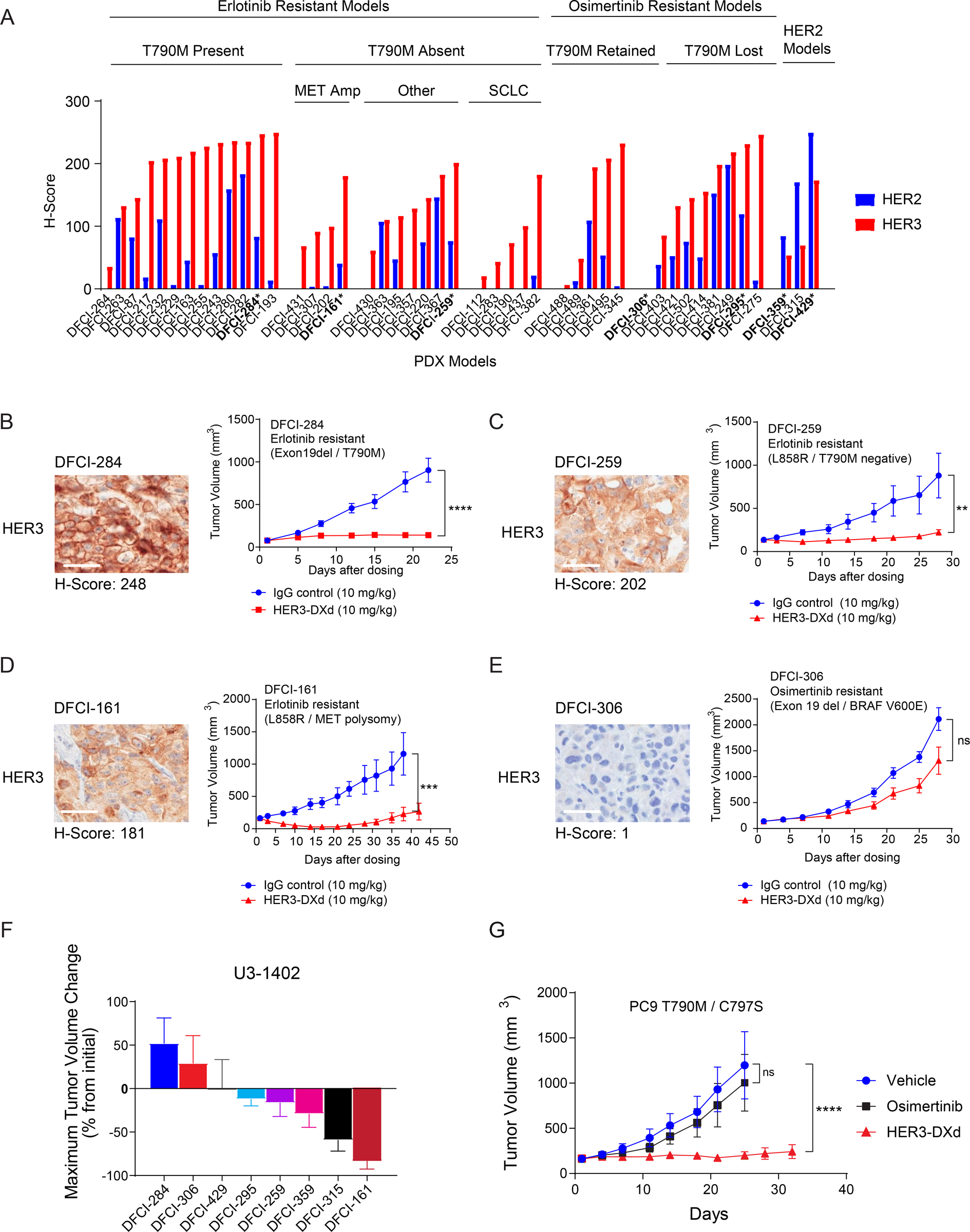
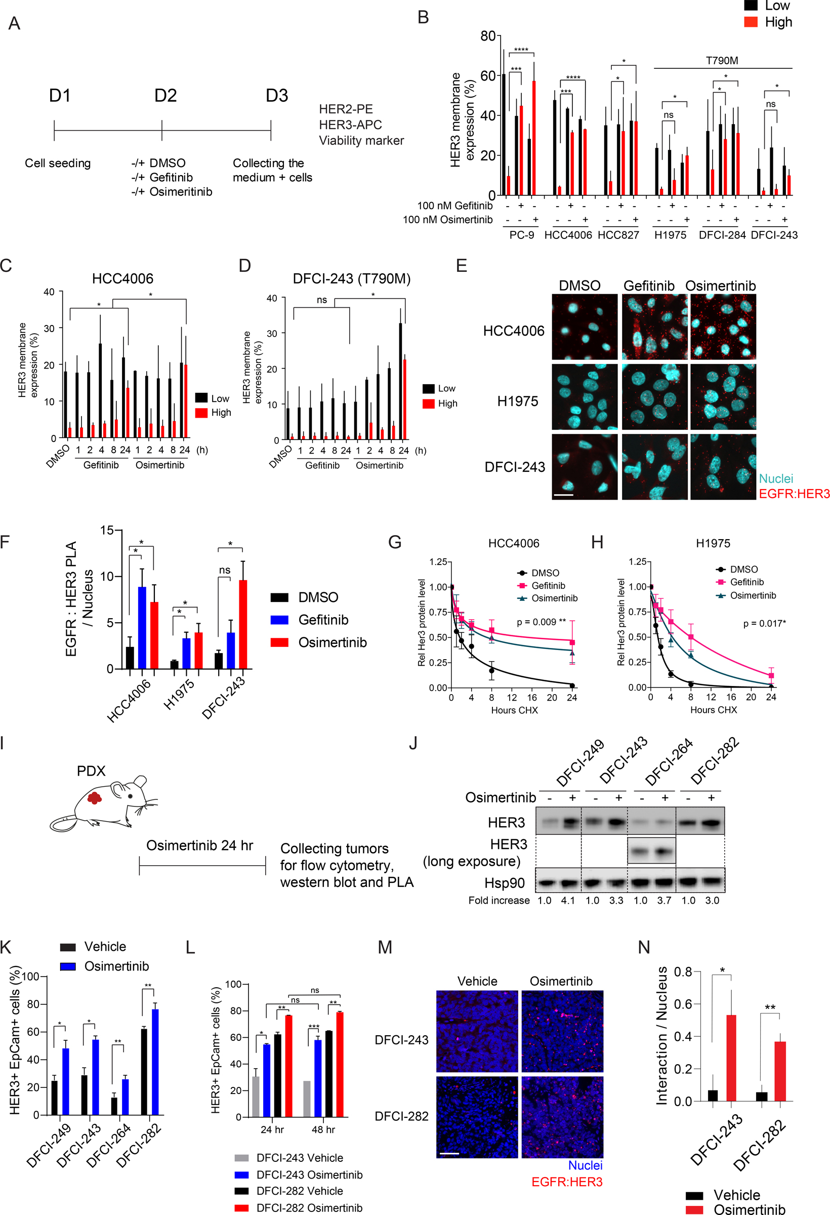
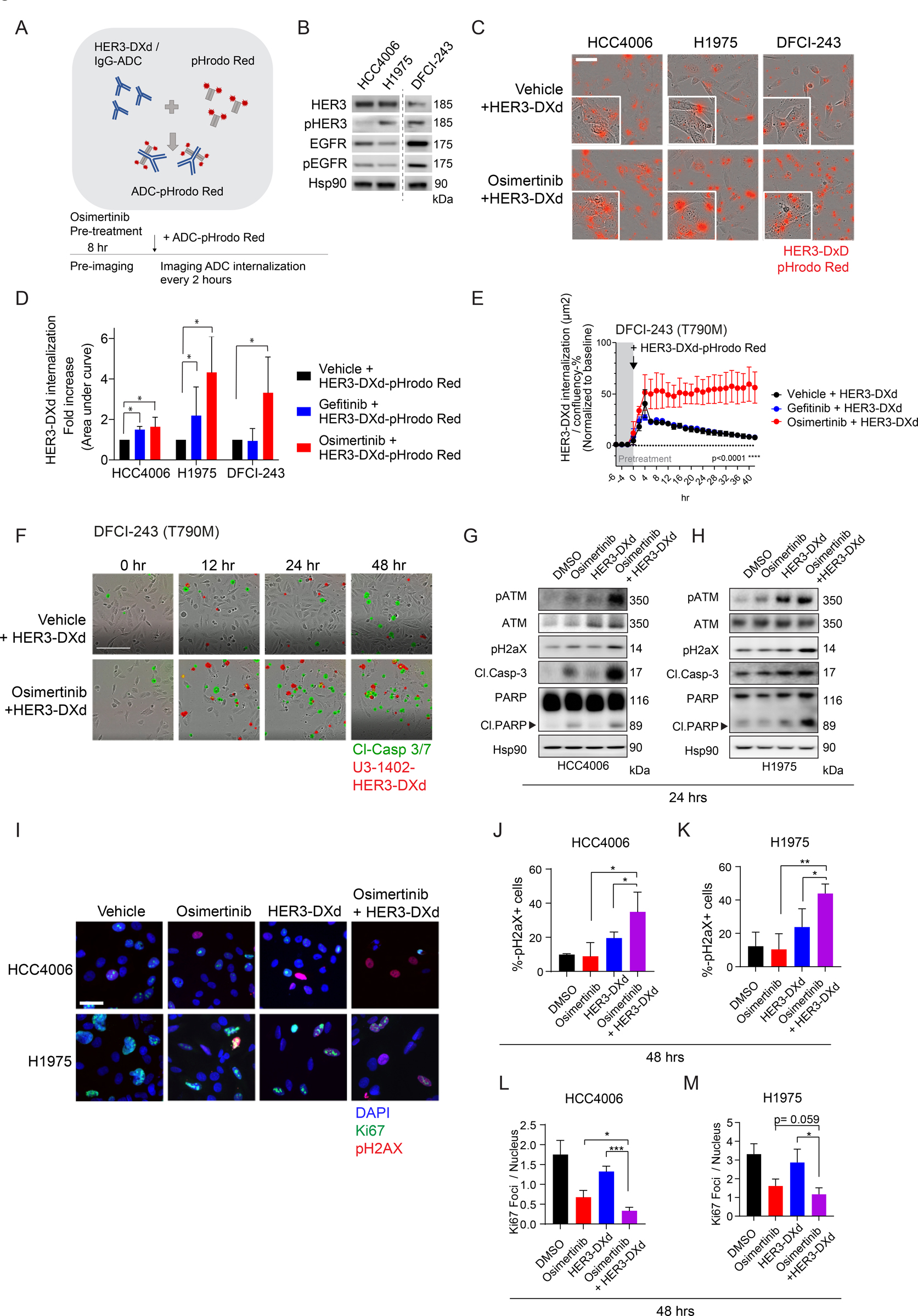
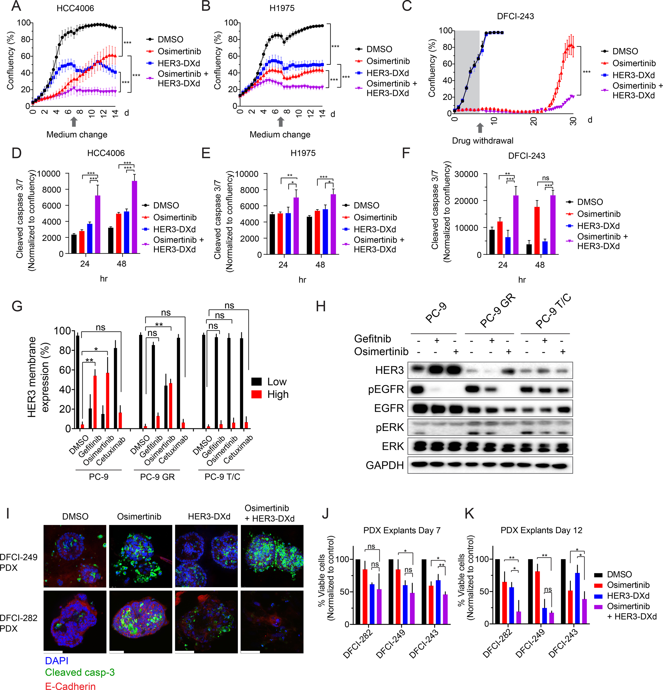
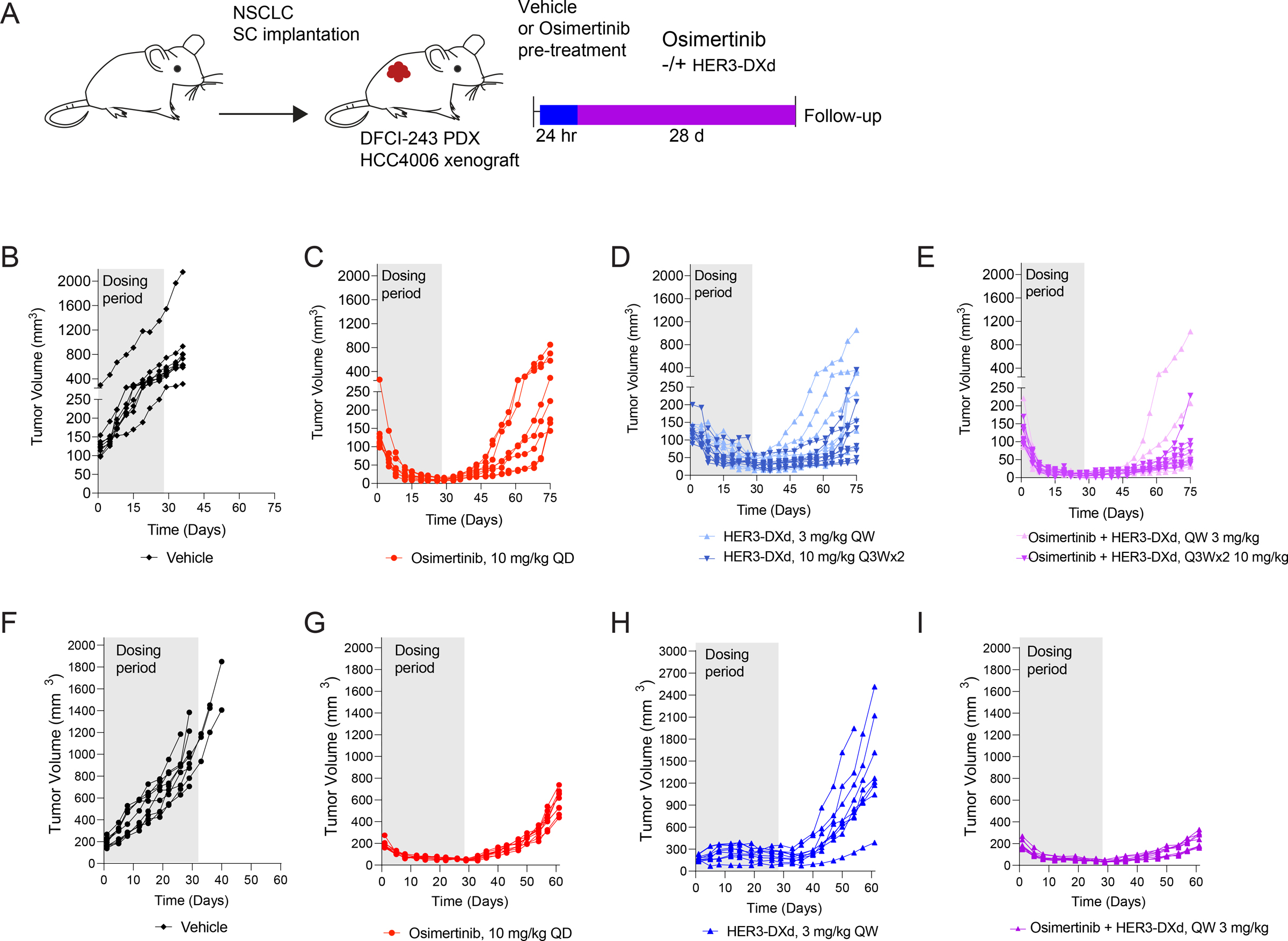
Epidermal growth factor receptor (EGFR) tyrosine kinase inhibitors (TKI) are the standard-of-care treatment for EGFR-mutant non-small cell lung cancers (NSCLC). However, most patients develop acquired drug resistance to EGFR TKIs. HER3 is a unique pseudokinase member of the ERBB family that functions by dimerizing with other ERBB family members (EGFR and HER2) and is frequently overexpressed in EGFR-mutant NSCLC. Although EGFR TKI resistance mechanisms do not lead to alterations in HER3, we hypothesized that targeting HER3 might improve efficacy of EGFR TKI. HER3-DXd is an antibody-drug conjugate (ADC) comprised of HER3-targeting antibody linked to a topoisomerase I inhibitor currently in clinical development. In this study, we evaluated the efficacy of HER3-DXd across a series of EGFR inhibitor-resistant, patient-derived xenografts and observed it to be broadly effective in HER3-expressing cancers. We further developed a preclinical strategy to enhance the efficacy of HER3-DXd through osimertinib pretreatment, which increased membrane expression of HER3 and led to enhanced internalization and efficacy of HER3-DXd. The combination of osimertinib and HER3-DXd may be an effective treatment approach and should be evaluated in future clinical trials in EGFR-mutant NSCLC patients. SIGNIFICANCE: EGFR inhibition leads to increased HER3 membrane expression and promotes HER3-DXd ADC internalization and efficacy, supporting the clinical development of the EGFR inhibitor/HER3-DXd combination in EGFR-mutant lung cancer.See related commentary by Lim et al., p. 18.


Figures

Figure 1:
Figure 1:. Single agent activity of HER3-DXd in EGFR inhibitor resistant patient-derived xenografts.
(A) HER2 and HER3 expression across EGFR inhibitor resistant patient-derived xenograft models. HER2 and HER3 were stained with immunohistochemistry and quantified using H score. The models used for HER3-DXd efficacy studies are bolded and marked with an asterisk. (B)-(E) Immunohistochemistry stainings for HER3 and single agent efficacy of HER3-DXd in PDX models with variable baseline HER3 expression. 10 mg/kg HER3-DXd or IgG control, qw. Scale bar is 50 μm. Student’s t test (unpaired), SEM. (F) Waterfall plot for all PDX models treated with HER3-DXd. The graph shows maximal tumor volume change induced by HER3-DXd treatment. (G) In vivo- grafted PC-9 T790M/C797S cells are resistant to osimertinib but are still sensitive for HER3-DXd. Mice were treated with either 25 mg/kg osimertinib qd., or 10 mg/kg HER3-DXd, qw. ANOVA, SEM. **** p<0.0001, *** p < 0.001, ** p < 0.01, ns = not significant.
Figure 2:
Figure 2:. EGFR inhibitors increase the membrane and total expression of HER3 in cell lines and patient-derived xenografts.
(A) Schematic for the experimental design. EGFR mutant non-small cell lung cancer lines were treated with gefitinib or osimertinib for 24 hr after which they were stained for the viability marker, HER2, and HER3. (B) HER3 membrane expression in cell lines after EGFR inhibitor treatment. HER3 positive cells were categorized into low and high expression groups. N >3 biological replicates. Student’s t test (unpaired), SD. (C-D) Time series for the HER3 membrane expression after EGFR inhibitor treatment in HCC4006 and DFCI243 cell lines. N = 3 biological replicates. Student’s t test (unpaired), SD. (E) EGFR inhibitors increase HER3:EGFR interaction measured by proximity-ligation assay (PLA). The cells were treated with 100 nM gefitinib or osimertinib for 24 hr before the assay. Scale bar is 20 μm. (F) Quantification of the HER3:EGFR PLA. N = 4 biological replicates. Student’s t test (unpaired), SD. (G-H) EGFR inhibitors stabilize HER3 in protein level. Cells were treated with 100 nM gefitnib or osimertinib for 24 hr before adding 30 μg/ml cycloheximide (CHX). Graph shows the average of quantifications of western blots from 3 biological replicates. ANOVA (one way), SD. (I) Treatment scheme for PDX tumors. Tumor-bearing mice were treated once with vehicle or 25 mg/kg osimertinib for 24 hrs, after which the tumors were collected for analysis. (J) Osimertinib pre-treatment increases total HER3 protein levels. Quantification of the bands is shown below the blot. Long exposure = longer blot exposure time. Hsp90 is the loading control. (K) Osimertinib pre-treatment increases the amount of viable HER3+ EpCam+ cells in PDX tumors. EpCam is an epithelial cell marker. N >3 / group. Student’s t test (unpaired), SD. (L) 48 hr treatment does not further increase HER3 membrane expression. Tumor-bearing mice were treated either for 24 hr or 48 hr with vehicle or 25 mg/kg osimertinib before HER3:EGFR interaction in vivo. (N) Quantification of the PLA assay. N >3 mice / group, 6–8 fields of views / tumor. Student’s t test (unpaired). Scale bar is 200 μm. **** p<0.0001, *** p < 0.001, ** p < 0.01, ns = not significant.
Figure 3:
Figure 3:. EGFR inhibitor- induced HER3 expression increases the internalization and efficacy of HER3-DXd in vitro.
(A) Schematic for the ADC internalization assay. HER3-DXd (or control IgG-ADC) was conjugated with pH-sensitive pHrodo Red fluorescent probes. After conjugation the labeled antibodies were added to the cells pre-treated either with vehicle or 100 nM osimertinib for 8 hr, and fluorescence was measured over time using Incucyte live cell imager. (B) Western blot showing the baseline expression of (p)HER3 and (p)EGFR in the used cell lines. Hsp90 is a loading control. Lanes are from the same blot; dashed line indicates where the blot was cut. (C) Representative images of HER3-DXd intake in all three cell lines. Scale bar is 60 μm. (D) Area under curve for HER3-DXd internalization in 3 different cell lines. Each cell line was normalized to the parental control cell line. ANOVA, N= 4 biological replicates, SD. (E) Quantification of the HER3-DXd internalization in DFCI-243 cells. N= 3 biological replicates, SD. (F) Representative pictures from the ADC internalization assay in DFCI-243 cells over time. HER3-DXd internalization is shown in red, green is cleaved caspase 3. Scale bar is 200 μm. (G)-(H) Western blots showing the DNA damage and apoptosis response followed by single agent or combination treatment with 50 nM osimertinib and 10 μg/ml HER3-DXd. Hsp90 is the loading control. (I) Immunofluorescence staining for pH2aX and Ki67 after 24 hr osimertinib (50 nM for HCC4006 and H1975, 10nM for DFCI-243) pre-treatment followed by 24 hrs of 10 μg/ml HER3-DXd. Scale bar is 25 μm. (J)-(M) Quantification for pH2aX and Ki67 immunofluorescence. N = 3 biological replicates. **** p<0.0001, *** p < 0.001, ** p < 0.01, ns = not significant.
Figure 4:
Figure 4:. Osimertinib pre-treatment increases the HER3-DXd efficacy in cell lines and in ex vivo patient-derived xenografts.
(A)-(B) Growth of HCC4006 and H1975 cells treated with DMSO, 50 nM osimertinib, 10 μg/ml HER3-DXd, or combination of osimertinib and HER3-DXd. Arrows indicate when medium and drugs were changed to the cells. ANOVA, SD. (C) Regrowth of DFCI-243 cells treated with 10 nM osimertinib, 10 μg/ml HER3-DXd, or combination of osimertinib and HER3-DXd. Cells were treated for 1 week after which the drug was withdrawn. ANOVA, SD. (D)-(F) Apoptosis quantification in cells treated like A-C. Values were normalized to cell confluency. ANOVA, SD. (G) Increased HER3 membrane expression by EGFR inhibition is target-dependent in PC-9 cells. HER3 membrane expression in PC-9, PC-9 gefitinib resistant cells (GR) and PC-9 osimertinib resistant (T/C: T790M/C797S) cells. Cells were treated with DMSO, 100 nM gefitnib, 100 nM osimertinib, or 10 ng/ml cetuximab for 24 hours. Students t test, SD. (H) Western blot showing the total HER3 levels and inhibition of EGFR/pERK signaling by on-target EGFR inhibition. GAPDH is a loading control. Cells were treated like in (G). (I) Example images from patient-derived xenograft fragment cultures stained for cleaved caspase-3 and epithelial marker E-Cadherin. Scale bar is 50 μm (J)-(K) Viability of PDX fragments on day 7 and day 12 (cell titer glo assay), normalized to baseline viability. ANOVA, SD.
Figure 5:
Figure 5:. Osimertinib pre-treatment increases the HER3-DXd efficacy ex vivo and in vivo patient-derived xenografts.
(A) Treatment scheme for DFCI-243 PDX. Tumor-bearing mice were first treated with vehicle or osimertinib (10 mg/kg) for 24 hr, after which HER3-DXd (3/10 mg/kg) was added. Mice were treated for 28 days,and followed up for tumor regrowth. (B)-(E) Individual tumor growths for all treated mice (DFCI-243 PDX). (F)-(I) Individual tumor growths for all treated mice (HCC4006 xenograft). Osimertinib vs U3–1402 p<0.0001 ****, Osimertinib vs U3–1402 + Osimertinib p<0.0002 ***, U3–1402 vs U3–1402 Osimertinib p<0.0001 ****, 2-way ANOVA. **** p<0.0001, *** p < 0.001, ** p < 0.01, ns = not significant.

Comment in

References

    1. Janne PA, Yang JC, Kim DW, Planchard D, Ohe Y, Ramalingam SS, et al. AZD9291 in EGFR inhibitor-resistant non-small-cell lung cancer. N Engl J Med 2015;372(18):1689–99 doi 10.1056/NEJMoa1411817. - DOI - PubMed
    1. Mok TS, Wu YL, Ahn MJ, Garassino MC, Kim HR, Ramalingam SS, et al. Osimertinib or Platinum-Pemetrexed in EGFR T790M-Positive Lung Cancer. N Engl J Med 2017;376(7):629–40 doi 10.1056/NEJMoa1612674. - DOI - PMC - PubMed
    1. Soria JC, Ohe Y, Vansteenkiste J, Reungwetwattana T, Chewaskulyong B, Lee KH, et al. Osimertinib in Untreated EGFR-Mutated Advanced Non-Small-Cell Lung Cancer. N Engl J Med 2018;378(2):113–25 doi 10.1056/NEJMoa1713137. - DOI - PubMed
    1. Oxnard GR, Hu Y, Mileham KF, Husain H, Costa DB, Tracy P, et al. Assessment of Resistance Mechanisms and Clinical Implications in Patients With EGFR T790M-Positive Lung Cancer and Acquired Resistance to Osimertinib. JAMA Oncol 2018;4(11):1527–34 doi 10.1001/jamaoncol.2018.2969. - DOI - PMC - PubMed
    1. Leonetti A, Sharma S, Minari R, Perego P, Giovannetti E, Tiseo M. Resistance mechanisms to osimertinib in EGFR-mutated non-small cell lung cancer. Br J Cancer 2019;121(9):725–37 doi 10.1038/s41416-019-0573-8. - DOI - PMC - PubMed

Publication types