. 2022 Jul;71(7):1399-1411.
doi: 10.1136/gutjnl-2020-323771. Epub 2021 Sep 21.

The human liver microenvironment shapes the homing and function of CD4+ T-cell populations

Affiliations

The human liver microenvironment shapes the homing and function of CD4+ T-cell populations

Benjamin G Wiggins et al. Gut. 2022 Jul.

Abstract

Objective: Tissue-resident memory T cells (TRM) are vital immune sentinels that provide protective immunity. While hepatic CD8+ TRM have been well described, little is known about the location, phenotype and function of CD4+ TRM.

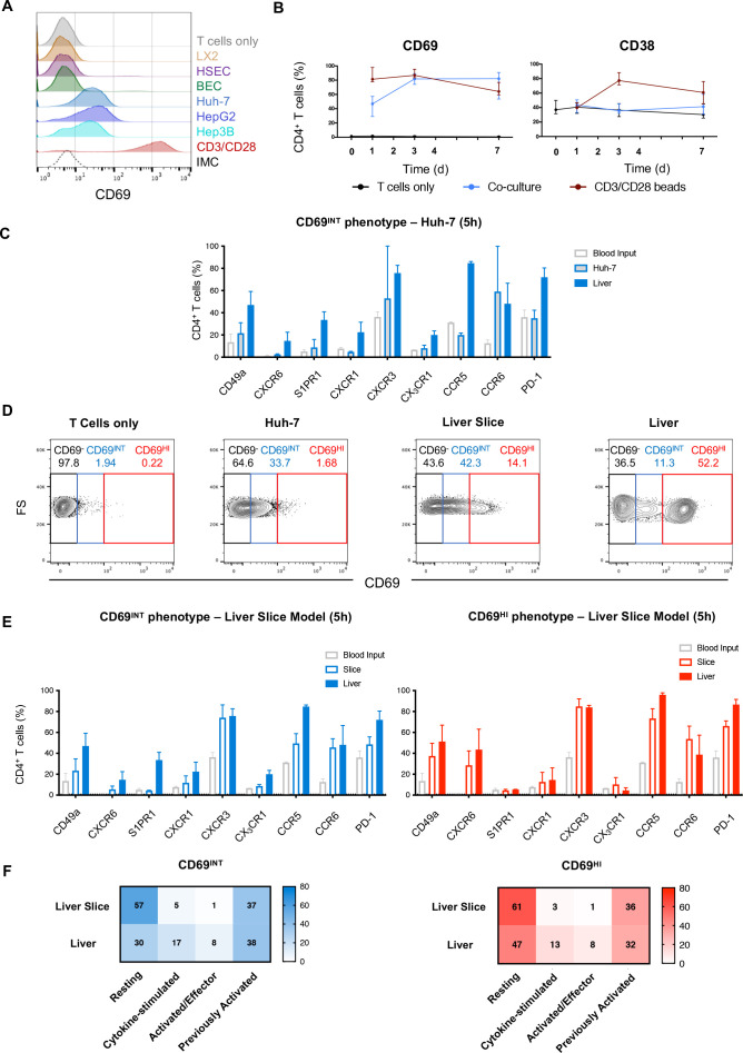
Design: We used multiparametric flow cytometry, histological assessment and novel human tissue coculture systems to interrogate the ex vivo phenotype, function and generation of the intrahepatic CD4+ T-cell compartment. We also used leukocytes isolated from human leukocyte antigen (HLA)-disparate liver allografts to assess long-term retention.

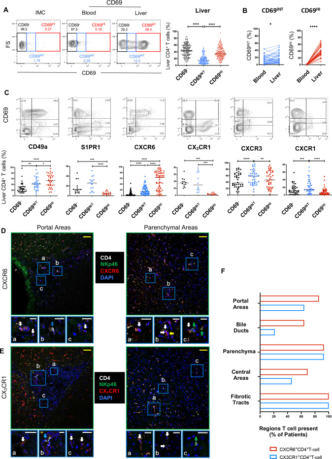
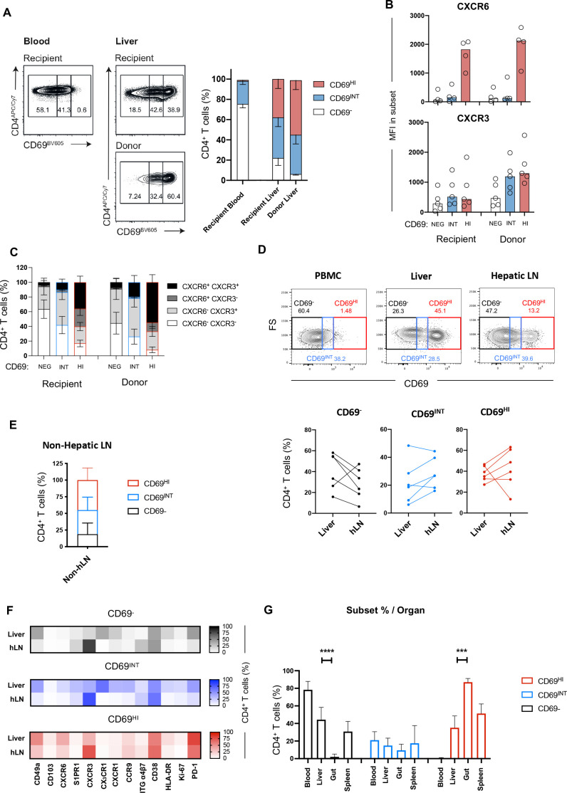
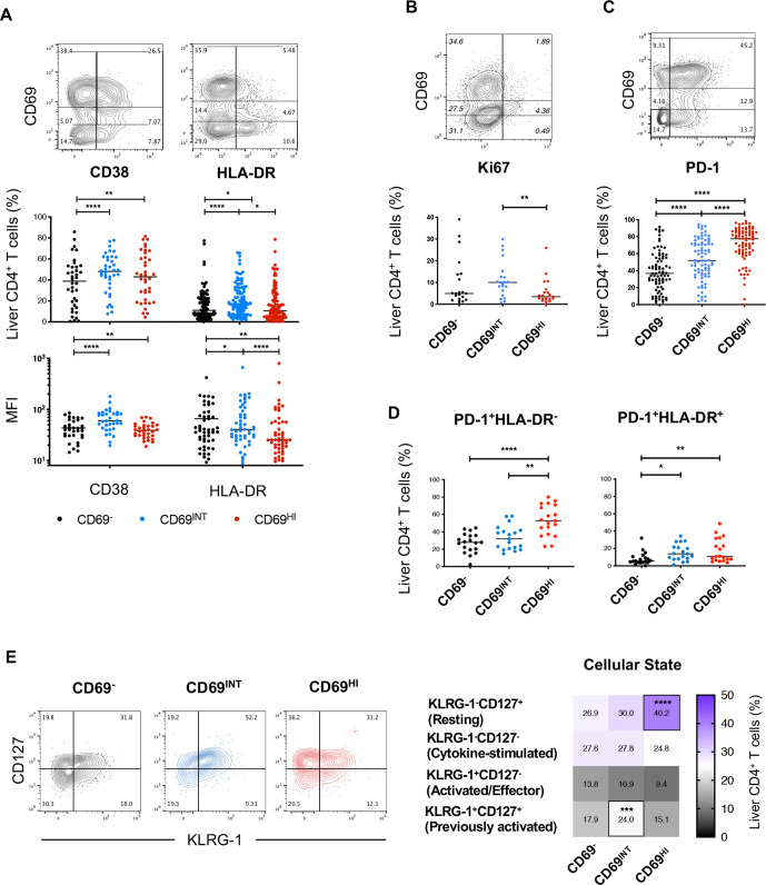
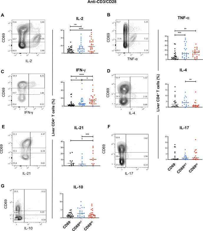
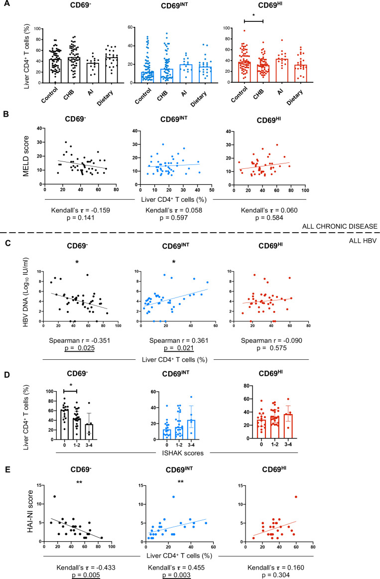
Results: Hepatic CD4+ T cells were delineated into three distinct populations based on CD69 expression: CD69-, CD69INT and CD69HI. CD69HICD4+ cells were identified as tissue-resident CD4+ T cells on the basis of their exclusion from the circulation, phenotypical profile (CXCR6+CD49a+S1PR1-PD-1+) and long-term persistence within the pool of donor-derived leukcoocytes in HLA-disparate liver allografts. CD69HICD4+ T cells produced robust type 1 polyfunctional cytokine responses on stimulation. Conversely, CD69INTCD4+ T cells represented a more heterogenous population containing cells with a more activated phenotype, a distinct chemokine receptor profile (CX3CR1+CXCR3+CXCR1+) and a bias towards interleukin-4 production. While CD69INTCD4+ T cells could be found in the circulation and lymph nodes, these cells also formed part of the long-term resident pool, persisting in HLA-mismatched allografts. Notably, frequencies of CD69INTCD4+ T cells correlated with necroinflammatory scores in chronic hepatitis B infection. Finally, we demonstrated that interaction with hepatic epithelia was sufficient to generate CD69INTCD4+ T cells, while additional signals from the liver microenvironment were required to generate liver-resident CD69HICD4+ T cells.

Conclusions: High and intermediate CD69 expressions mark human hepatic CD4+ TRM and a novel functionally distinct recirculating population, respectively, both shaped by the liver microenvironment to achieve diverse immunosurveillance.

Keywords: T lymphocytes; cellular immunity; hepatitis B; immunology; liver immunology.


Conflict of interest statement

Competing interests: BW collaborated with and received funding from Bioniz. LJP sat on advisory boards/provided consultancy for Gilead Sciences and SQZ Biotech. KA was funded by a studentship with Dr Falk. MKM received research funding from Gilead Sciences, Hoffmann La Roche and Immunocore. MKM sat on advisory boards/provided consultancy for Gilead, Hoffmann La Roche, Immunocore, VIR, Galapagos NV, GSK, Abbvie and Freeline. ZS collaborated with Bioniz and AstraZeneca and consulted for Boehringer Ingelheim. All other authors declare no conflict of interest.

Figures

Figure 1
Figure 1
CD69 expression distinguishes three intrahepatic CD4+ T-cell populations with differential homing potentials. (A) Gating strategy showing CD69, CD69INT and CD69HI populations. Representative flow cytometry plot for CD4+ T-cell distribution in blood and liver, and summary data showing % CD4+ T cells in IHL from two independent centres (n=162). Isotype-matched controls were used to set CD69 gates. (B) % CD69-expressing T-cell populations in paired blood and liver (n=39). (C) Expression of key homing and retention markers on CD69-expressing CD4+ T cells (% of total CD4+ T cells). Images depicting localisation of CXCR6+ CD4+ T cells (D) or CX3CR1+ CD4+ T cells (E) in portal and parenchymal areas of human livers (representative of n=14 livers (5 control, 4 patients with HBV and 5 patients with PBC)). Sections stained for CD4, NKp46, DAPI and chemokine receptor indicated. Cells of interest expressed both the chemokine receptor and CD4 and lacked NK cell marker NKp46. Areas of interest (A–C) shown at higher magnification below each main image. White arrows: cell of interest, green arrows: NKp46+ cell, yellow arrow: chemokine receptor+ CD4 cell, blue arrows: CD4+ NKp46- CXCR6- cells (D) or CD4+NKp46-CX3CR1- cells (E). Yellow scale bars: 50 µm, white scale bars: 20 µm. (F) Cumulative scoring of the presence of each cell of interest within different liver regions. Cells of interest were scored as present in specific areas if at least three cells were present within each region. Plot shows the % of each region that contained cells of interest (n=14, as above; fibrotic tracts in non-control livers only, n=9). Cells were classed as present in portal regions and central regions if they were identified within 50 µm of their respective vasculature. Association with bile ducts was scored if cells were making direct contact. Statistical comparisons by Freidman tests with Dunn’s multiple tests (A, C); Wilcoxon matched-pair, signed-rank tests (B). p < 0.05 (*), < 0.01 (**), < 0.001 (***), < 0.0001 (****) FS, forward scatter; IHL, intrahepatic lymphocyte; IMC, isotype-matched control; NK, natural killer; PBC, primary biliary cholangitis.
Figure 2
Figure 2
High CD69 expression marks a CD4+ T-cell population capable of long-term residence within the liver. (A) HLA-mismatched allograft sampling allows assessment of resident T cells. Donor-derived T cells are distinguished from recipient-derived T cells through HLA staining. Example distributions of CD69, CD69INT and CD69HI cells in recipient and donor pools of liver and blood samples, and combined data across five patient samples. (B) MFI of CXCR6 and CXCR3 expressions in the three populations in donor and recipient pools. (C) Breakdown of CXCR3/CXCR6 coexpression patterns in different donor and recipient subpopulations (n=4). (D) Staining and combined data showing population distribution from liver (n=6), hepatic LNs (n=6) and non-hepatic (mesenteric) LNs (n=6). Example plots show hLN, PBMC and liver as gating controls. (E) Subset breakdown in distal non-hLNs. (F) Heatmap of % marker expression in CD69 (top), CD69INT (middle) and CD69HI (bottom) from matched liver and hLN samples. CD103, n=6; CD49a, n=5; CXCR6 and HLA-DR, n=4; CXCR3, CXCR1, PD-1 and CD38, n=3; S1PR1, CCR9, integrin α4β7 and Ki-67, n=2; CX3CR1, n=1. (G) Frequency of CD69, CD69INT and CD69HI CD4+ T cells in blood (n=103), liver (n=118), gut (n=6) and spleen (n=4) samples. Statistical comparisons on paired populations by Wilcoxon matched-pair, signed-rank tests (A–D, F), and Kruskal-Wallis tests with duns post hoc tests on liver, gut and spleen samples within each CD4+ T-cell subset (G). HI, high; hLN, hepatic lymph node; INT, intermediate; LN, lymph node; MFI, Median fluorescence intensity, NEG, negative.
Figure 3
Figure 3
CD69HICD4+ TRM demonstrate a restrained, resting phenotype, while CD69INTCD4+ T cells exhibit features of activation. (A) % and MFI expression of CD38 and HLA-DR. % expression of (B) Ki-67 and (C) PD-1 expressions among the three CD4+ T-cell populations. (D) Subset representation among PD-1+HLA-DR and PD-1+HLA-DR+ designations (n=19). (E) Analysis of the four differentiation/cellular states based on KLRG-1 and CD127 expressions (n=41). Heatmap shows % expression of each designation. Freidman’s tests with Dunn’s multiple tests were used for statistical analysis (A–E) p < 0.05 (*), < 0.01 (**), < 0.001 (***), < 0.0001 (****). MFI, mean fluorescence intensity.
Figure 4
Figure 4
Liver CD69HICD4+ and CD69INTCD4+ T cells are skewed towards TH1 and TH2 functional profiles, respectively. Each sample was stained with CD69 prior to stimulation to exclude effects of altered CD69 levels due to cellular activation. Representative flow plots and combined data of % expression of six prototypical TH cytokines after 5 hours of stimulation of IHL with anti-CD3/CD28: (A) IL-2 (n=27), (B) TNF-α (n=24), (C) IFN-γ (n=24), (D) IL-4 (n=18), (E) IL-21 (n=11), (F) IL-17 (n=16), (G) IL-10 (n=22). See online supplemental table 4 for disease breakdowns. Freidman’s tests with Dunn’s multiple tests were used for statistical analysis (A–F). IFN-γ, interferon gamma; IHL, intrahepatic lymphocyte; IL, interleukin; TNF-α, tumour necrosis factor alpha.
Figure 5
Figure 5
Increased CD69INTCD4+ T-cell frequencies correlate with necroinflammation in CHB. (A) Representation of each population in control livers (n=62 (11 donor explant transplant rejections, 5 healthy tissue biopsies, 36 colorectal cancer margin liver explants, 8 HCC margin liver explants and 2 cyst-free areas of PLD explants)), patients with chronic HBV (CHB, n=54), autoimmune liver disease (n=15 (6 PBC, 8 PSC and 1 AIH)), and dietary liver disease (n=24 (16 ALD and 8 NASH)). (B) Correlation analysis of patient MELD scores versus % of each of the three subsets for all donors with end-stage liver disease from centre A. (C) HBV DNA, Ishak scoring (D) and HAI-NI scoring (E) plotted against % of each T-cell population in the HBV cohort. Correlation and p values reported for each plot. Statistical testing used: Kruskal-Wallis tests with Dunn’s multiple post hoc tests (A, C), Kendall’s tau rank correlation tests (B, E), Spearman’s rank order correlation (C). AI, autoimmune; AIH, autoimmune hepatitis; ALD, alcoholic liver disease; CHB, chronic HBV infection; HAI-NI, Histology Activity Index-Necroinflammatory; HCC, hepatocellular carcinoma; NASH, non-alcoholic steatohepatitis; PBC, primary biliary cholangitis; PLD, polycystic liver disease; PSC, primary sclerosing cholangitis.
Figure 6
Figure 6
CD69INTCD4+ and CD69HI CD4+ T cells are differentially induced by the liver microenvironment. (A) % CD69 expression on PBMC-derived CD4+ T cells cultured for 16 hours with primary HSEC, primary BEC; hepatic stellate cell line LX-2; hepatocyte cell lines HuH-7, HepG2 or Hep3B; with anti-CD3/CD28; or alone. Histogram displays representative CD69 expression levels in each condition. (B) % CD69 and CD38 expressions on blood CD4+ T cells over a 7-day culture period with HuH-7 (n=8–10/timepoint). (C) Comparison of key phenotypical markers in Huh-7-generated CD69INT cells from PBMC following 5-hour culture, matched patient IHL CD69INT cells and blood T cells alone (n=2). (D) Representative flow plots showing degree of CD69INT and CD69HI generation within PBMC after 5 hours of culture: alone, with Huh-7 cells, with precision-cut donor-matched liver slices; or from directly isolated IHLs from matched human liver. (E) Comparison of CD69INT cells (left) and CD69HI cells (right) generated from donor-matched PBMCs in a precision-cut liver slice model, with matched donor-derived liver populations, and input blood CD4+ T cells alone (n=2). (F) Activation/differentiation statuses of CD69INT and CD69HI cells in the different conditions as assessed by KLRG-1/CD127 costaining patterns (as in figure 3E). Colour intensity and displayed numbers represent median % in each KLRG-1/CD127 designation. BEC, biliary epithelial cell; FS, forward scatter; HL, intrahepatic lymphocyte; HSEC, hepatic sinusoidal endothelial cell; IMC, isotype-matched control.

References

    1. Clark RA. Resident memory T cells in human health and disease. Sci Transl Med 2015;7:269rv1. 10.1126/scitranslmed.3010641 - DOI - PMC - PubMed
    1. Schenkel JM, Fraser KA, Vezys V, et al. . Sensing and alarm function of resident memory CD8⁺ T cells. Nat Immunol 2013;14:509–13. 10.1038/ni.2568 - DOI - PMC - PubMed
    1. Beura LK, Fares-Frederickson NJ, Steinert EM, et al. . CD4+ resident memory T cells dominate immunosurveillance and orchestrate local recall responses. J Exp Med 2019;216:1214–29. 10.1084/jem.20181365 - DOI - PMC - PubMed
    1. Park CO, Kupper TS. The emerging role of resident memory T cells in protective immunity and inflammatory disease. Nat Med 2015;21:688–97. 10.1038/nm.3883 - DOI - PMC - PubMed
    1. Pallett LJ, Davies J, Colbeck EJ, et al. . IL-2 high tissue-resident T cells in the human liver: Sentinels for hepatotropic infection. J Exp Med 2017;214:1567–80. 10.1084/jem.20162115 - DOI - PMC - PubMed

Publication types