. 2021 Sep 28;118(39):e2104490118.
doi: 10.1073/pnas.2104490118.

Assembly and organization of the N-terminal region of mucin MUC5AC: Indications for structural and functional distinction from MUC5B

Affiliations

Assembly and organization of the N-terminal region of mucin MUC5AC: Indications for structural and functional distinction from MUC5B

Jerome Carpenter et al. Proc Natl Acad Sci U S A. .

Abstract

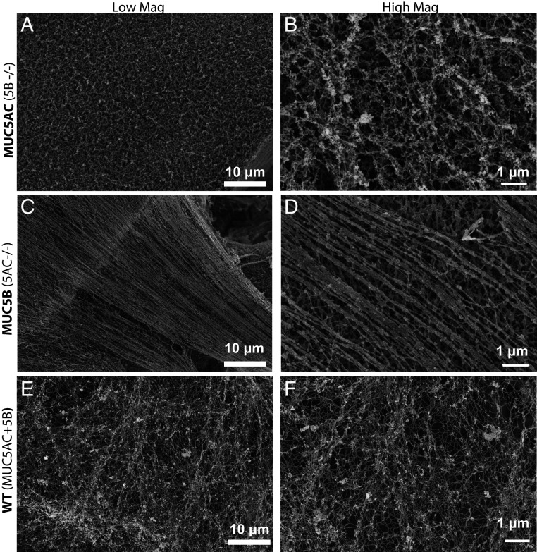
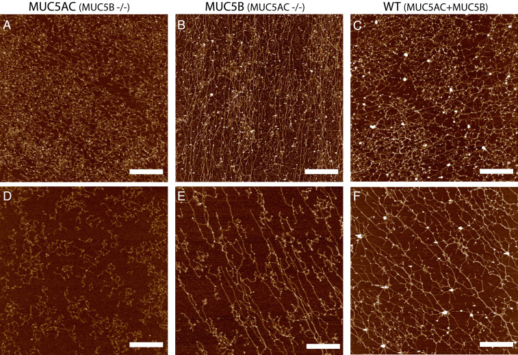
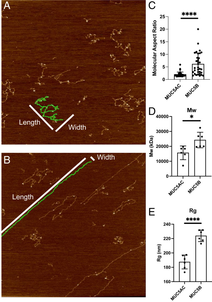
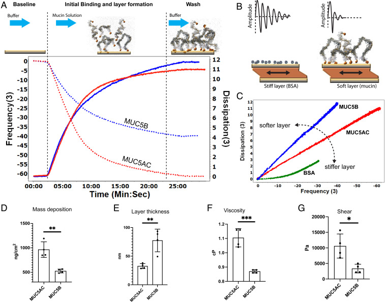
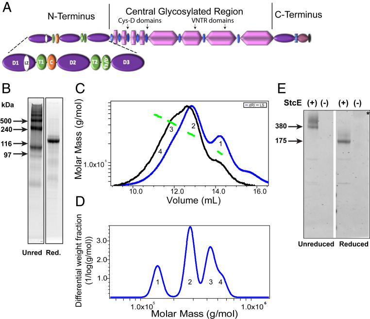
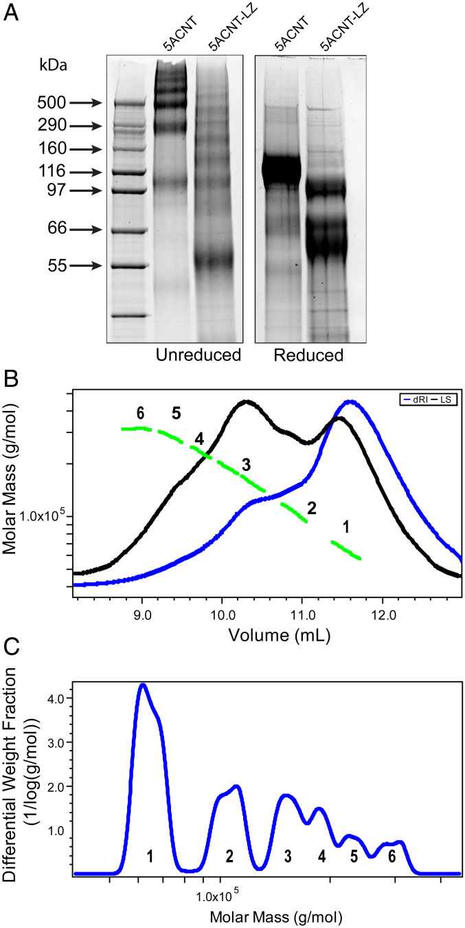
Elevated levels of MUC5AC, one of the major gel-forming mucins in the lungs, are closely associated with chronic obstructive lung diseases such as chronic bronchitis and asthma. It is not known, however, how the structure and/or gel-making properties of MUC5AC contribute to innate lung defense in health and drive the formation of stagnant mucus in disease. To understand this, here we studied the biophysical properties and macromolecular assembly of MUC5AC compared to MUC5B. To study each native mucin, we used Calu3 monomucin cultures that produced MUC5AC or MUC5B. To understand the macromolecular assembly of MUC5AC through N-terminal oligomerization, we expressed a recombinant whole N-terminal domain (5ACNT). Scanning electron microscopy and atomic force microscopy imaging indicated that the two mucins formed distinct networks on epithelial and experimental surfaces; MUC5B formed linear, infrequently branched multimers, whereas MUC5AC formed tightly organized networks with a high degree of branching. Quartz crystal microbalance-dissipation monitoring experiments indicated that MUC5AC bound significantly more to hydrophobic surfaces and was stiffer and more viscoelastic as compared to MUC5B. Light scattering analysis determined that 5ACNT primarily forms disulfide-linked covalent dimers and higher-order oligomers (i.e., trimers and tetramers). Selective proteolytic digestion of the central glycosylated region of the full-length molecule confirmed that MUC5AC forms dimers and higher-order oligomers through its N terminus. Collectively, the distinct N-terminal organization of MUC5AC may explain the more adhesive and unique viscoelastic properties of branched, highly networked MUC5AC gels. These properties may generate insight into why/how MUC5AC forms a static, "tethered" mucus layer in chronic muco-obstructive lung diseases.

Keywords: MUC5AC; airways; lung; mucin; mucus gel.


Conflict of interest statement

The authors declare no competing interest.

Figures

Fig. 1.
Fig. 1.
SEM images of the apical surfaces of Calu3 monomucin cultures indicate that MUC5AC and MUC5B create distinct supramolecular frameworks: Calu3 cultures were grown on 12-well transwells for 10 d and subjected to SEM analysis on a Zeiss Supra 25 field emission SEM at 5 kV. The MUC5AC monomucin culture (A and B) shows a mucus gel that is tighter, more branched/cross-linked, and has no indication of linear threads/bundles formation, while the MUC5B monomucin culture mucus gel (C and D) is dominated by linear, less-branched, hair-like mucin strands. E and F show the surfaces of WT Calu3 cells, which produce both MUC5AC and MUC5B and have structural elements of both gels produced by monomucin cultures.
Fig. 2.
Fig. 2.
Isolated MUC5AC and MUC5B from monomucin cultures exhibit distinct macromolecular organization on surfaces. AFM images of mucins MUC5AC (A), MUC5B (B), and WT secretions (C) at a high concentration (∼100 μg/mL) and also imaged at a lower concentration ∼30 μg/mL (DF). MUC5AC mucin polymers (A, D) form more compact structures with fewer (if any) long linear structures, while MUC5B (B, E) networks form polymers with a higher frequency of linear straight structures. WT Calu3 secretions (C, F), which contain both MUC5AC and MUC5B mucins at a 3:1 ratio exhibit a distinct mucin network that combines elements of both monomucin networks. (Scale bar, 1 μm.)
Fig. 3.
Fig. 3.
Distinct macromolecular properties of MUC5AC and MUC5B mucins on surfaces: Aspect ratio analysis was performed on images of dilute MUC5AC (A) and MUC5B (B) preparations using AR16.10.211 software (Oxford Instruments). Selected images had less than 4% mucin coverage to enable the analysis of isolated macromolecules. Images were first flattened (Flatten 0) and then segmented by masking the image at 350 picometers, dilating the mask, and then running the analyze particles job to segment particles excluding particles with diameters less than 50 nm. The aspect ratio (length/width, with length being the longer dimension) of particles was recorded across several images for both the MUC5AC sample and the MUC5B sample. (C) MUC5AC macromolecules have a low aspect ratio, while MUC5B macromolecules have both high (dominant) and sometimes low aspect ratio macromolecules. SEC-MALLS analysis shows that MUC5B has both a higher average (D) MW and (E) radius of gyration. *P = 0.05, ****P < 0.0001.
Fig. 4.
Fig. 4.
QCM-D experiments indicate that MUC5AC forms stiffer, denser, and more viscoelastic layers compared to MUC5B. (A) A cartoon depicting the time course of a QCM-D experiment with representative data for measurements with MUC5AC and MUC5B shown underneath. At the same concentration (100 μg/mL), the frequency shift of the F3 overtones (dotted lines) and the dissipation shift for D3 (solid lines) were monitored while MUC5AC (red) and MUC5B (blue) were being deposited in the gold QCM-D chip. (B) The cartoon shows that for a material that is tightly coupled to the crystal (e.g. bovine serum albumin, BSA) there is a gradual decay in the amplitude. For a “softer” layer that has more interaction with the buffer (such as mucin), the damping occurs much faster. (C) A typical plot for dissipation versus frequency shifts (ΔD/ΔF) during the absorption of MUC5AC (red) and MUC5B (blue). MUC5AC forms a stiffer, denser layer. The slope of a BSA layer, which forms a solid and nondissipating layer, is shown for comparison (green). (D) The Sauerbrey model calculated an absorbed MUC5AC layer of 964 ± SD 159 ng ⋅ cm−2 and MUC5B layer of 526 ± SD 39 ng ⋅ cm−2, n = 4. (E–G) Voight modeling determined that (E) the MUC5AC layer was much thinner/denser (32.6 nm ± SD 4.4, n = 4) compared to the MUC5B layer (77.4 nm ± SD 19.7, n = 4, P = 0.0045) and that (F) the relative viscosity (1.105 cP ± SD 0.06, n = 4) and (G) shear elasticity (10,600 Pascal ± SD 3,800, n = 4) of MUC5AC layers were significantly higher compared to the relative viscosity (0.87 cP ± SD 0.01, n = 4) and shear elasticity (3,300 P ± SD 1,400, n = 4) (P = 0.02) of the MUC5B layer. *P ≤ 0.05, **P ≤ 0.01, ***P ≤ 0.005 (a more detailed legend can be found in SI Appendix, Fig. S6).
Fig. 5.
Fig. 5.
5ACNT forms dimers, trimers, and higher-order oligomers: (A) Cartoon showing the 5ACNT construct in relation to the entire molecule. The construct contains the D1, D2, D′, and D3 domains of the N-terminal region. (B) SYPRO Ruby staining of SDS-PAGE of purified protein shows that the unreduced (Unred) 5ACNT construct produced monomers, dimers, trimers, and higher-order oligomers. A second gel shows that upon reduction (Red.), these all collapse into a single band. (C) This panel shows traces of the purified 5ACNT protein on a Superose 6 column monitored by the tREX refractometer (blue) and Heleos II multiangle laser light scattering (black). The molar masses, as obtained from the MALLS, are plotted across the 5ACNT distribution (green). (D) Molar mass distribution of MUC5AC N-terminal multimers (1 = monomer, 2 = dimer, 3 = trimer, and 4 = tetramer). (E) A Western blot stained with MUC5AC antibodies against the D2 and D3 domains reveals that enzymatically treating whole-length MUC5AC with StcE, which cleaves the molecule within the O-linked glycosylation region, thus leaving the terminals intact, shows that the native MUC5AC N-terminal domains are organized into dimers, trimers, and higher-order oligomers. These bands collapse upon reduction. * = Untreated, reduced native MUC5AC (MW around 2.5 MDa) hardly enters the gel. VNTR, Variable number of tandem repeats.
Fig. 6.
Fig. 6.
MUC5AC N-terminal domain with a mutated LZ (5ACNT-LZ) displays a different multimerization profile: (A) SYPRO Ruby staining of SDS-PAGE of purified protein products shows that 5ACNT-LZ maintains the ability to form higher-order oligomers, bands ranging from 55 to 500 kDa, despite no longer having the D3 domain present. For mass spectrometry analyses of the bands, see SI Appendix, Fig. S5. When reduced, the 5ACNT-LZ shows three monomeric bands, as compared to 5ACNT, which produced only one band after reduction. (B) SEC-MALLS analysis of the 5ACNT-LZ zipper–expressed protein shows traces of the purified 5ACNT-LZ protein on a Superose 6 column monitored by the tREX refractometer (blue) and Heleos II multiangle laser light scattering (black). The molar masses, obtained from MALLS, are shown across the 5ACNT-LZ distribution (green). (C) Molar mass distribution of 5ACNT-LZ multimers. Increasing molecular weight peaks are represented with numbers (1 to 6).

References

    1. Johansson M. E., et al., The inner of the two Muc2 mucin-dependent mucus layers in colon is devoid of bacteria. Proc. Natl. Acad. Sci. U.S.A. 105, 15064–15069 (2008). - PMC - PubMed
    1. Nordman H., et al., Gastric MUC5AC and MUC6 are large oligomeric mucins that differ in size, glycosylation and tissue distribution. Biochem. J. 364, 191–200 (2002). - PMC - PubMed
    1. Veerman E. C., et al., Distinct localization of MUC5B glycoforms in the human salivary glands. Glycobiology 13, 363–366 (2003). - PubMed
    1. Kesimer M., et al., Airway mucin concentration as a marker of chronic bronchitis. N. Engl. J. Med. 377, 911–922 (2017). - PMC - PubMed
    1. Kesimer M., et al., Tracheobronchial air-liquid interface cell culture: A model for innate mucosal defense of the upper airways? Am. J. Physiol. Lung Cell. Mol. Physiol. 296, L92–L100 (2009). - PMC - PubMed

Publication types

LinkOut - more resources