. 2021 Sep 21;4(1):1109.
doi: 10.1038/s42003-021-02630-z.

Prenylcysteine oxidase 1, an emerging player in atherosclerosis

Affiliations

Prenylcysteine oxidase 1, an emerging player in atherosclerosis

C Banfi et al. Commun Biol. .

Abstract

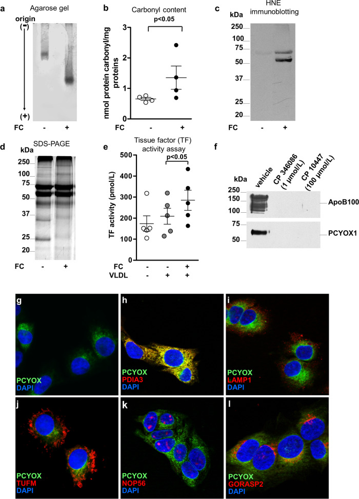
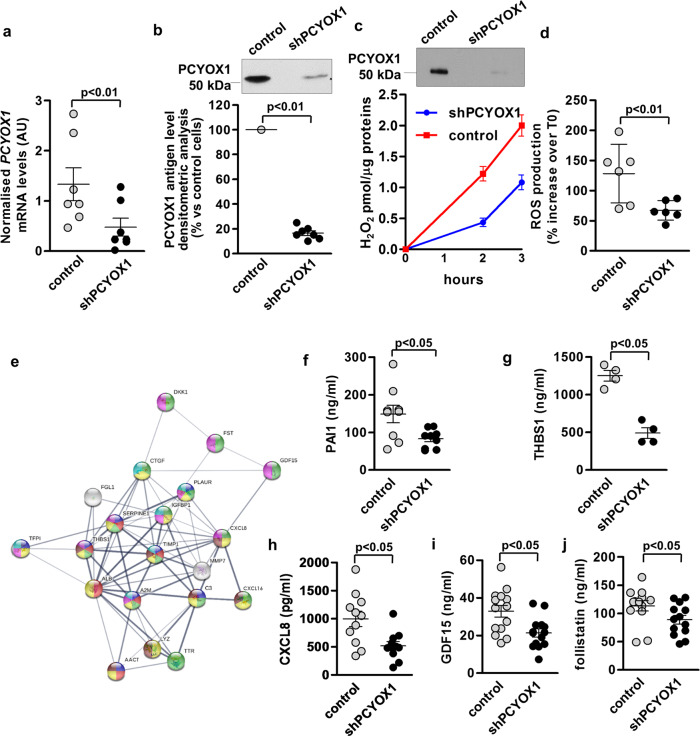
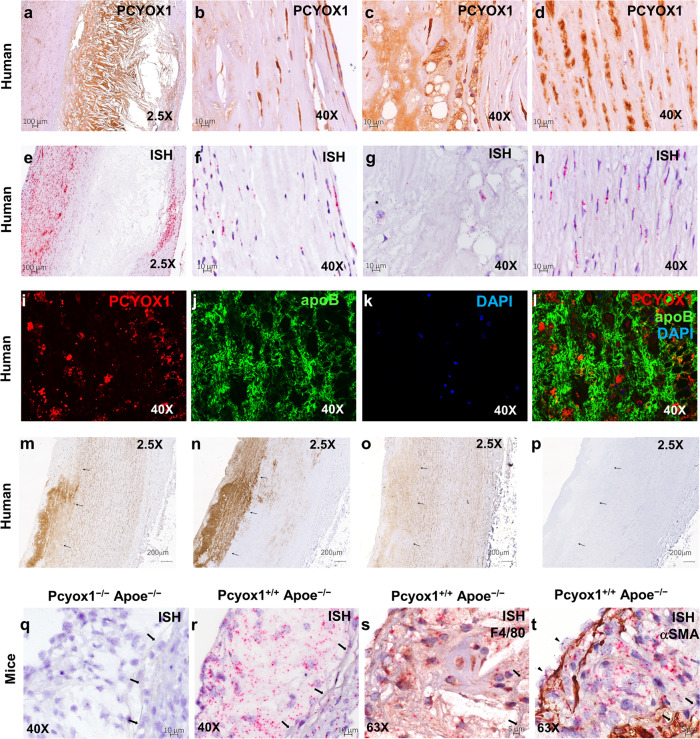
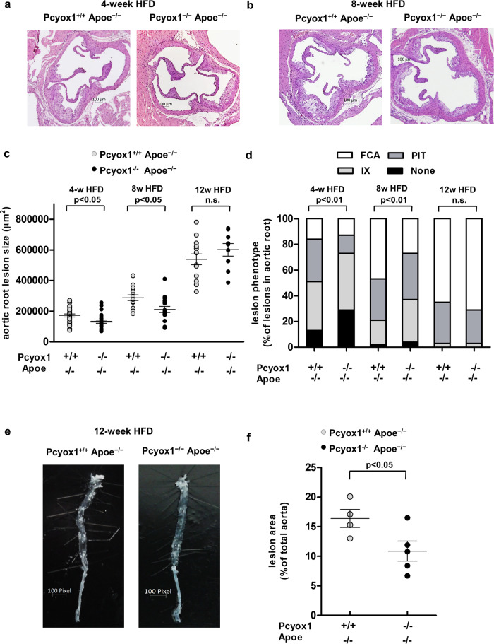
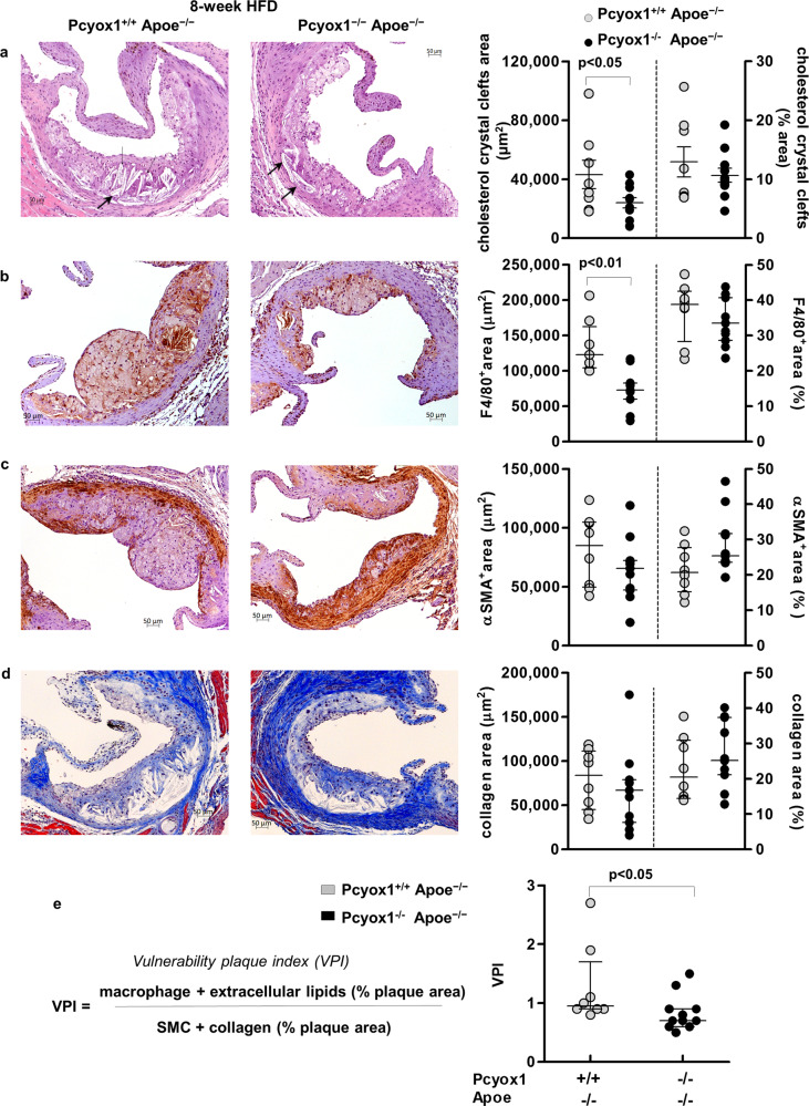
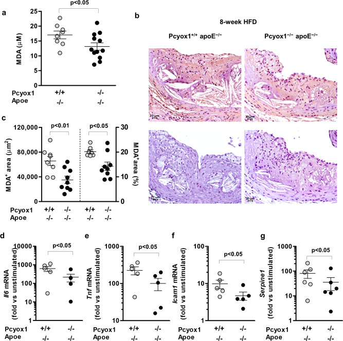
The research into the pathophysiology of atherosclerosis has considerably increased our understanding of the disease complexity, but still many questions remain unanswered, both mechanistically and pharmacologically. Here, we provided evidence that the pro-oxidant enzyme Prenylcysteine Oxidase 1 (PCYOX1), in the human atherosclerotic lesions, is both synthesized locally and transported within the subintimal space by proatherogenic lipoproteins accumulating in the arterial wall during atherogenesis. Further, Pcyox1 deficiency in Apoe-/- mice retards atheroprogression, is associated with decreased features of lesion vulnerability and lower levels of lipid peroxidation, reduces plasma lipid levels and inflammation. PCYOX1 silencing in vitro affects the cellular proteome by influencing multiple functions related to inflammation, oxidative stress, and platelet adhesion. Collectively, these findings identify the pro-oxidant enzyme PCYOX1 as an emerging player in atherogenesis and, therefore, understanding the biology and mechanisms of all functions of this unique enzyme is likely to provide additional therapeutic opportunities in addressing atherosclerosis.


Conflict of interest statement

We disclose the following European Patent application, under examination, EP15817414.4 entitled “Prenylcysteine oxidase 1 inhibitors for the prevention and/or treatment of oxidative stress-related degenerative diseases and prenylcysteine oxidase 1 as diagnostic marker”, applicant Centro Cardiologico Monzino IRCCS, inventors C.B., M.B., R.B, and S.B., with no financial competing interest. The remaining authors declare no competing interest.

Figures

Fig. 1
Fig. 1. PCYOX1 bound to human atherogenic apoB100-containing lipoproteins generates H2O2, which in turn induces lipoprotein oxidation in vitro, and associates to nascent apoB100-containing lipoproteins in the endoplasmic reticulum.
Agarose gel electrophoresis (a), carbonyl content (b), 4-hydroxy-2-nonenal (HNE) immunoblotting (c), and SDS-polyacrylamide gel (d) of human VLDL incubated with FC (200 µmol/L) for 72 h. e Tissue factor (TF) activity in HAEC incubated with FC-treated VLDL. p < 0.05 vs vehicle-treated VLDL, by Student’s t test and ANOVA, respectively. Data in b, e are presented as circle plot, with each circle representing an individual sample and bars showing the mean value ± SEM of 4 and 5 independent experiments, respectively. f PCYOX1 and apoB100 immunoreactivity of apoB100-containing lipoproteins isolated from HepG2 cells incubated with vehicle or MTP inhibitors, CP346086 and CP10447. gl Immunofluorescence of PCYOX1 (green) (g) in HepG2 cells and co-staining (red) with antibodies against PDIA3 (h), LAMP1 (i), TUFM (j), NOP56 (k), and GORASP2 (l). Nuclear reference DAPI in blue, objective ×40.
Fig. 2
Fig. 2. PCYOX1 silencing affects the cellular proteome.
PCYOX1 mRNA normalized to the housekeeping gene 18S rRNA (a, n = 7) and protein (b, n = 7) in PCYOX1-silenced HepG2 cells (shPCYOX1). p < 0.01 vs control cells by Student’s t test. c PCYOX1 immunoblotting (upper panel, n = 7) and H2O2 production (lower panel, n = 3) of apoB100-containing lipoproteins isolated from control or shPCYOX1 cell conditioned media. Data are expressed as mean value ± SEM of H2O2 pmol/µg proteins over time. d ROS production in PCYOX1-silenced HepG2 cells (n = 6). p < 0.01 vs control cells by Student’s t test. e GO analysis of secreted proteins downregulated by PCYOX1 silencing highlighting enriched biological processes: blue, negative regulation of peptidase activity; red, platelet degranulation; green, regulation of signal transduction; yellow, response to stress; violet, negative regulation of response to stimulus; brown, inflammatory response; light blue, response to wounding. fj Levels of PAI-1, THBS1, CXCL8, GDF15, and Follistatin measured by ELISA. n > 4. p < 0.05 vs control cells by Student’s t test. Data are presented as circle plot, with each circle representing an individual sample and bars showing the mean value ± SEM.
Fig. 3
Fig. 3. PCYOX1 is abundant in human and Apoe−/− mice atherosclerotic lesions.
Sections of human atherosclerotic lesions were subjected to IHC (ad) or ISH (fh) analysis for the detection of PCYOX1 protein and mRNA, respectively, and counterstained with hematoxylin. (a; objective ×2.5; bd; objective ×40). e Representative image of the positive control probe (housekeeping gene, Ubiquitin C), demonstrating mRNA integrity (objective ×2.5). fh PCYOX1 mRNA staining (red punctate dots, objective ×40). b, f luminal side; c, g lipid-rich regions; d, h tunica media. il PCYOX1 and apoB immunofluorescence in human atherosclerotic lesions counterstained with DAPI (confocal images, objective ×40). Shown are representative images from one donor. mp Serial sections immunostained for PCYOX1 (m), apoB (n), and farnesyl groups deriving from isoprenylated proteins (o) or in the absence of primary antibody (negative control, p). Arrows indicate the internal elastic lamina. Shown are representative images from another donor. Negative controls are shown in Supplementary Fig. 2. qt Aortic root sections from Pcyox1+/+/Apoe−/− and Pcyox1−/−/Apoe−/− mice fed a HFD for 8 weeks subjected to ISH for the detection of Pcyox1 mRNA alone (q, r objective ×40) or in combination with immunostaining for cell-type-specific protein markers (objective ×63), F4/80 for macrophages (s), and α-SMA for SMC (t). Tissue sections were counterstained with hematoxylin. Arrows in qs indicate internal elastic lamina. Arrowheads in t indicate the expression of PCYOX1 in luminal ECs.
Fig. 4
Fig. 4. Progression of atherosclerosis is retarded in Pcyox1−/−/Apoe−/− mice.
Representative images of H&E staining of the aortic root from Pcyox1+/+/Apoe−/− and Pcyox1−/−/Apoe−/− mice fed a HFD for 4 (a) or 8 (b) weeks. c Quantification of atherosclerotic lesion size. Data are presented as circle plot, with each circle representing an individual mouse and bars showing the mean value ± SEM. d Characterization of lesion phenotype according to the atherosclerosis stages: IX intimal xanthoma, PIT pathological intimal thickening, FCA fibrous cap atheroma. Statistical significance was calculated by Fisher exact test. e, f Representative images and quantification of atherosclerotic areas in the whole aorta by en face analysis. The bright white material is unstained atherosclerotic lesions. Statistical significance calculated by Student’s t test. Data are presented as circle plot, with each circle representing an individual mouse and bars showing the mean value ± SEM.
Fig. 5
Fig. 5. Pcyox1 deficiency is associated with reduced lesion vulnerability.
Representative images and quantification of: a cholesterol crystal clefts, indicated by arrows (H&E staining); b macrophages (F4/80 staining); c α-smooth muscle cells (α-SMA staining); d collagen (Masson’s Trichrome staining) in atherosclerotic lesions from aortic roots of Pcyox1+/+/Apoe−/− (n = 8) and Pcyox1−/−/Apoe−/− (n = 11) mice fed a HFD for 8 weeks. Shown on the right are the results of the morphometric analysis for the different stainings, expressed either as absolute positive areas within the intima or as percentage of plaque that stained for each component. e Calculation of vulnerability plaque index. Data are presented as circle plot, with each circle representing an individual sample and bars showing the mean value ± SEM (a) or median with interquartile range (be). Statistical significance was calculated by Student’s t test (a) or Mann–Whitney test (be).
Fig. 6
Fig. 6. Pcyox1 deficiency is associated with lower levels of lipid peroxidation and decreased LPS-induced inflammatory response in murine peritoneal macrophages.
MDA in plasma (a) and in the aortic root lesions (b, c) from Pcyox1+/+/Apoe−/− (n = 8) and Pcyox1−/−/Apoe−/− (n = 11) mice fed a HFD for 8 weeks. b Representative images of MDA immunostaining (upper panels) and negative control sections incubated with no primary antibody (lower panels); c results of the morphometric analysis of MDA-stained sections, expressed either as absolute positive areas within the intima or as the percentage of total lesion area. Data are presented as circle plot, with each circle representing an individual mouse and bars showing the mean value ± SEM. p < 0.05 or p < 0.01 by Student’s t test. dg mRNA levels normalized to the housekeeping gene 18S rRNA of Il6 (d), Tnf (e), Icam1 (f), and Serpine1 (g) in macrophages isolated from Pcyox1+/+/Apoe−/− and Pcyox1−//Apoe−/− mice and treated with LPS 5 ng/mL for 2 h. Data are expressed as fold increase induced by LPS with respect to unstimulated cells and presented as circle plot, with each circle representing an individual mouse and bars showing the mean value ± SEM (n = 5). p < 0.05 by Student’s t test.

Similar articles

Cited by

References

    1. Jernberg T, et al. Cardiovascular risk in post-myocardial infarction patients: nationwide real world data demonstrate the importance of a long-term perspective. Eur. Heart J. 2015;36:1163–1170. doi: 10.1093/eurheartj/ehu505. - DOI - PubMed
    1. Benjamin EJ, et al. Heart disease and stroke statistics-2019 update: a report from the American Heart Association. Circulation. 2019;139:e56–e528. doi: 10.1161/CIR.0000000000000659. - DOI - PubMed
    1. Libby P, et al. Atherosclerosis. Nat. Rev. Dis. Prim. 2019;5:56. doi: 10.1038/s41572-019-0106-z. - DOI - PubMed
    1. Libby P, Bornfeldt KE, Tall AR. Atherosclerosis: successes, surprises, and future challenges. Circ. Res. 2016;118:531–534. doi: 10.1161/CIRCRESAHA.116.308334. - DOI - PMC - PubMed
    1. Musunuru K, Kathiresan S. Surprises from genetic analyses of lipid risk factors for atherosclerosis. Circ. Res. 2016;118:579–585. doi: 10.1161/CIRCRESAHA.115.306398. - DOI - PMC - PubMed

Publication types