Infective endocarditis caused by Enterobacteriaceae: phenotypic and molecular characterization of Escherichia coli and Klebsiella pneumoniae in Rio de Janeiro, Brazil
- PMID: 34549374
- PMCID: PMC8578509
- DOI: 10.1007/s42770-021-00528-w
Infective endocarditis caused by Enterobacteriaceae: phenotypic and molecular characterization of Escherichia coli and Klebsiella pneumoniae in Rio de Janeiro, Brazil
Abstract
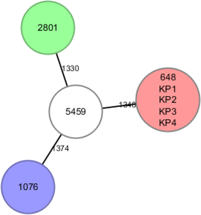
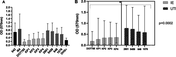
The etiological agent for infective endocarditis (IE), a life-threatening disease, is usually gram-positive bacteria. However, gram-negative bacteria can rarely cause IE and 4% of cases are associated with morbidity and mortality. This study aimed to characterize Escherichia coli and Klebsiella pneumoniae isolates from the blood of patients with IE. The characteristics of blood isolates were compared with those of urinary isolates from patients with urinary tract infections (UTIs). The results of this study revealed that K. pneumoniae isolates from patients with IE were phylogenetically related to those from patients with UTI. Additionally, the resistance phenotype, resistance gene, virulence gene, and plasmid profiles were similar between the blood and urinary isolates. The isolates belonging to the sequence types (STs) 76, 36, 101 (K. pneumoniae), and 69 (E. coli) are reported to be associated with drug resistance. The Enterobacteriaceae isolates from patients with IE did not produce extended-spectrum β-lactamase or carbapenemase. Additionally, this study investigated the virulence phenotype, biofilm formation ability, and the ability to adhere to the epithelial cells in vitro of the isolates. The isolates from patients with IE exhibited weaker biofilm formation ability than the urinary isolates. All isolates from patients with IE could adhere to the renal epithelial cells. However, three isolates from patients with UTIs could not adhere to the epithelial cells. The closely related K. pneumoniae isolates (648, KP1, KP2, KP3, and KP4) could not form biofilms or adhere to the epithelial cells. In summary, the molecular analysis revealed that the genetic characteristics of IE-causing K. pneumoniae and E. coli were similar to those of UTI-causing isolates. These isolates belonged to the STs that are considered treatable. Genetically similar isolates did not exhibit the same virulence phenotype. Thus, these non-hypervirulent clones must be monitored as they can cause complex infections in susceptible hosts.
Keywords: Adhesion; Biofilm; Brazil; Endocarditis; Enterobacteriaceae; Next-generation sequencing.
© 2021. The Author(s).
Conflict of interest statement
The authors declare no competing interests.
Figures
References
-
- Habib G, Lancellotti P, Antunes MJ, Bongiorni MG, Casalta JP, et al. 2015 ESC guidelines for the management of infective endocarditis: the task force for the management of infective endocarditis of the European Society of Cardiology (ESC) Eur Heart J. 2015;36:3075–3128. doi: 10.1093/eurheartj/ehv319. - DOI - PubMed
-
- Falcone M, Tiseo G, Durante-Mangoni E, Ravasio V, Barbaro F. Risk factors and outcomes of endocarditis due to non-HACEK gram-negative bacilli: data from the prospective multicenter Italian endocarditis study cohort. Antimicrob Agents Chemother. 2018;62:e02208–e2217. doi: 10.1128/AAC.02208-17. - DOI - PMC - PubMed
MeSH terms
Substances
Grants and funding
LinkOut - more resources
Full Text Sources
Medical