. 2021 Oct;69(10):633-643.
doi: 10.1369/00221554211046777. Epub 2021 Sep 22.

Picrosirius Red Staining: Revisiting Its Application to the Qualitative and Quantitative Assessment of Collagen Type I and Type III in Tendon

Affiliations

Picrosirius Red Staining: Revisiting Its Application to the Qualitative and Quantitative Assessment of Collagen Type I and Type III in Tendon

Consuelo M López De Padilla et al. J Histochem Cytochem. 2021 Oct.

Abstract

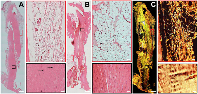
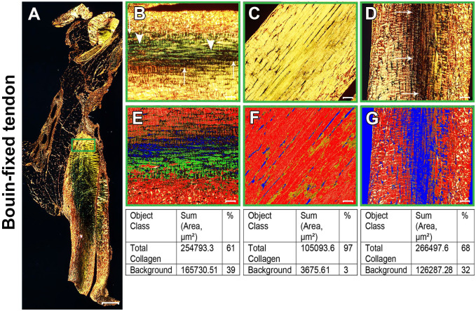
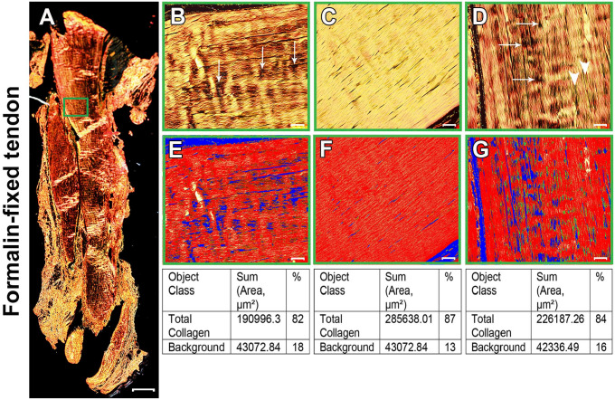
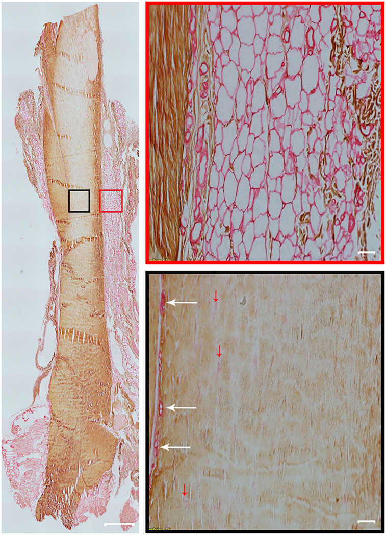
Collagen has a major role in the structural organization of tendons. Picrosirius red (PSR) staining viewed under polarized light microscopy is the standard method to evaluate the organization of collagen fibers in tissues. It is also used to distinguish between type I and type III collagen in tissue sections. However, accurate analysis and interpretation of PSR images are challenging because of technical factors and historical misconceptions. The aim of this study was to clarify whether collagen types I and III can be distinguished by PSR staining in rat Achilles tendons, using double immunohistochemistry as the positive control. Our findings showed that PSR staining viewed with polarized light microscopy was suitable for qualitative and quantitative assessment of total collagen but was not able to distinguish collagen types. We found it critical to use a polarizing microscope equipped with a rotating stage; tendon section orientation at 45° with respect to crossed polarizers was optimal for the qualitative and quantitative assessment of collagen organization. Immunohistochemistry was superior to PSR staining for detection of collagen type III. We also compared formalin and Bouin solution as fixatives. Both produced similar birefringence, but formalin-fixed tendons provided higher quality histological detail with both hematoxylin-eosin and immunostaining.

Keywords: Achilles tendon; Sirius red; collagen type; immunostaining; microscopy; polarization.


Conflict of interest statement

Competing Interests: The author(s) declared no potential conflicts of interest with respect to the research, authorship, and/or publication of this article.

Figures

Figure 1.
Figure 1.
Histological structure of healthy Achilles tendon. Representative serial longitudinal sections of Achilles tendons, with two fields of view of paratenon (red boxes) and a region of the mid tendon section (black boxes). (A) Formalin-fixed, hematoxylin–eosin (H&E)-stained section shows uniformly organized parallel collagen bundles and elongated tenocyte cell nuclei (black arrows). The paratenon is seen as loose connective tissue surrounding the tendon. (B) Bouin-fixed, H&E-stained tendon shows collagen fibers appearing intensely eosinophilic with nuclei stained darkly. In addition, the shrinkage effect of picric acid in Bouin solution causes artifactual changes in the normal appearance of tendon. (C) Formalin-fixed, picrosirius red staining on a sequential tissue section from the same tendon as panel A shows thick, bright yellowish-orange fibers. Another tendon area with greenish collagen fibers (white arrowhead) also contains some fibers that are at extinction (white arrows). The paratenon is seen with both thin fibers and thick fibers of yellowish-orange birefringence by polarizing microscopy. Tendon overview images were obtained with a 4× magnification lens (scale bar: 500 µm). Higher magnification images within the boxes were obtained with a 20× magnification lens (scale bar: 50 µm).
Figure 2.
Figure 2.
Bouin-fixed normal Achilles tendon stained with picrosirius red (PSR). (A) Polarized view of representative entire longitudinal Achilles tendon section. (B–D) Magnifications of approximately the same region as the green box in panel A at three different orientations: 0°(B), 45°(C), and 90°(D). At 0°(B), collagen fibers showed a mixture of birefringence with yellowish-orange and greenish-yellow (arrowheads). As the microscope stage was rotated, the birefringence of collagen changed gradually. At 45° (C), the tendon appeared uniformly bright yellow. At 90°(D), birefringence changed to yellowish-orange with more collagen fibers at extinction (arrows in B and D). (E–G) PSR images acquired in the same regions of interest as panels B–D were quantitatively compared for the percentage of total collagen using the Count & Measure module of the cellSens software. Blue indicates background; the sum of red and yellow is total collagen, represented as red in the quantification panel. Tendon overview image (A) was obtained with a 4× magnification lens (scale bar: 500 µm). Higher magnification images within the boxes (B–F) were obtained with a 20× magnification lens (scale bar: 50 µm).
Figure 3.
Figure 3.
Formalin-fixed normal Achilles tendon stained with picrosirius red (PSR). (A) Polarized view of representative entire longitudinal Achilles tendon section. (B–D) Magnifications of approximately the same region as the green box in panel A at three different orientations: 0°(B), 45°(C), and 90°(D). Collagen fibers showed different birefringence colors depending on the tendon orientation. Overall, collagen was displayed predominantly as a parallel arrangement of thick, yellowish-orange birefringent fibers. At 45°(C), the collagen fibers appeared uniformly bright yellow. At 0°(B) and 90°(D), a striped pattern was seen, with some fibers at extinction (arrows). White arrowheads in D show some greenish collagen fibers colocalized with thick, yellowish-orange fibers. (E–G) PSR images acquired in the same regions of interest as panels B–D were quantitatively compared for the percentage of total collagen using the Count & Measure module of the cellSens software. Blue indicates background; the sum of red and yellow is total collagen, represented as red in the quantitation panel. Tendon overview image (A) was obtained with a 4× magnification lens (scale bar: 500 µm). Higher magnification images within the boxes (B–F) were obtained with a 20× magnification lens (scale bar: 50 µm).
Figure 4.
Figure 4.
Collagen type I and type III expression in rat Achilles tendon. A representative entire longitudinal Achilles tendon section is double immunolabeled for collagen type I and collagen type III. Positive immunolabelling for collagen type I is brown (diaminobenzidine chromogen) and for collagen type III is red (permanent red chromogen). Two high-magnification fields of view are shown of paratenon (red box) and the mid tendon section (black box). Collagen type I is present in tendon. Collagen type III is found mostly in the paratenon (red box) and blood vessels (black box, white arrows). However, a small amount of collagen type III is also found in the tendon core and indicated with red arrows (black box). Tendon overview image was obtained with a 4× magnification lens (scale bar: 500 µm). Higher magnification images within boxes obtained with a 20× magnification lens (scale bar: 50 µm).

References

    1. Kirkendall DT, Garrett WE. Function and biomechanics of tendons. Scand J Med Sci Sports. 1997;7(2):62–6. - PubMed
    1. Wang JH. Mechanobiology of tendon. J Biomech. 2006; 39(9):1563–82. - PubMed
    1. Somaiah C, Kumar A, Mawrie D, Sharma A, Patil SD, Bhattacharyya J, Swaminathan R, Jaganathan BG. Collagen promotes higher adhesion, survival and proliferation of mesenchymal stem cells. PLoS ONE. 2015; 10(12):e0145068. - PMC - PubMed
    1. Merkel JR, DiPaolo BR, Hallock GG, Rice DC. Type I and type III collagen content of healing wounds in fetal and adult rats. Proc Soc Exp Biol Med. 1988;187(4):493–7. - PubMed
    1. Volk SW, Wang Y, Mauldin EA, Liechty KW, Adams SL. Diminished type III collagen promotes myofibroblast differentiation and increases scar deposition in cutaneous wound healing. Cells Tissues Organs. 2011;194(1):25–37. - PMC - PubMed

Publication types