. 2021 Oct 27;6(5):e0064121.
doi: 10.1128/mSphere.00641-21. Epub 2021 Sep 22.

The Vancomycin Resistance-Associated Regulatory System VraSR Modulates Biofilm Formation of Staphylococcus epidermidis in an ica-Dependent Manner

Affiliations

The Vancomycin Resistance-Associated Regulatory System VraSR Modulates Biofilm Formation of Staphylococcus epidermidis in an ica-Dependent Manner

Youcong Wu et al. mSphere. .

Abstract

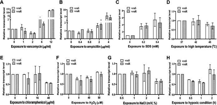
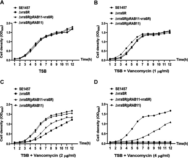
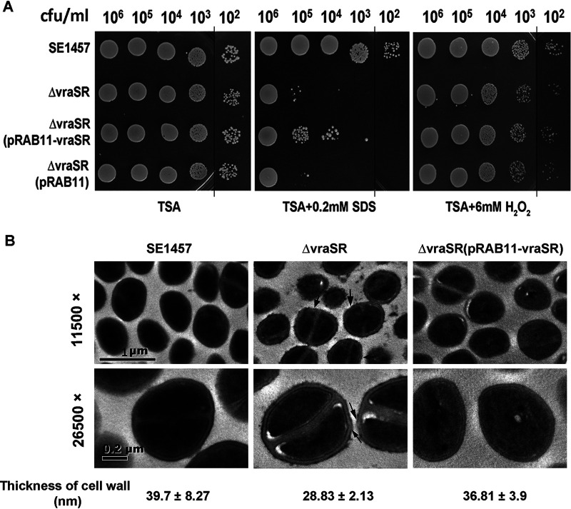
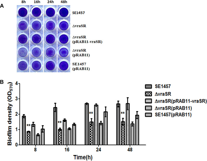
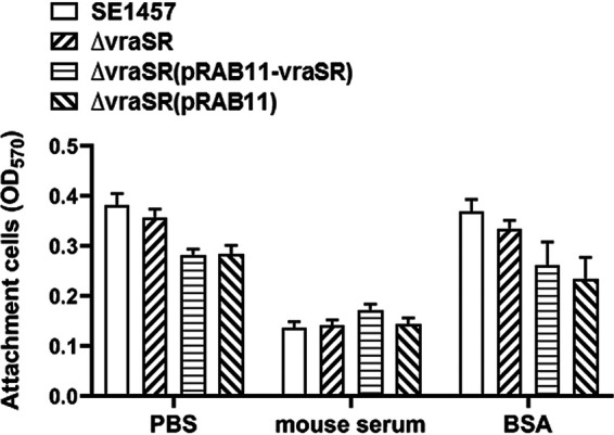
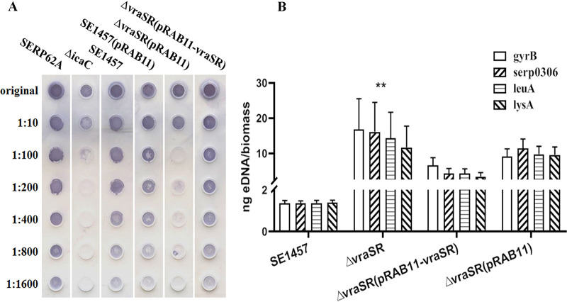
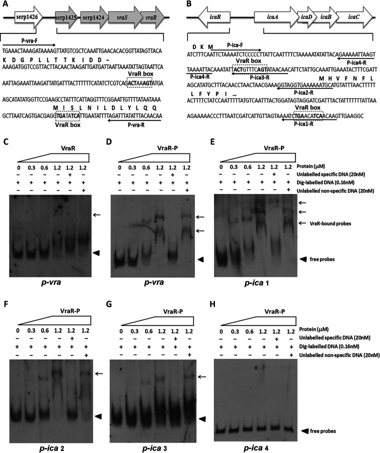
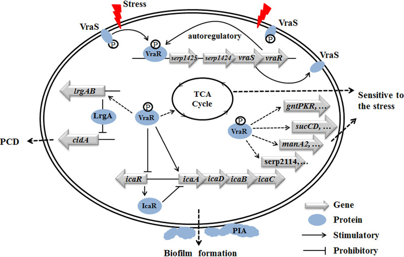
The two-component system VraSR responds to the cell wall-active antibiotic stress in Staphylococcus epidermidis. To study its regulatory function in biofilm formation, a vraSR deletion mutant (ΔvraSR) was constructed using S. epidermidis strain 1457 (SE1457) as the parent strain. Compared to SE1457, the ΔvraSR mutant showed impaired biofilm formation both in vitro and in vivo with a higher ratio of dead cells within the biofilm. Consistently, the ΔvraSR mutant produced much less polysaccharide intercellular adhesin (PIA). The ΔvraSR mutant also showed increased susceptibility to the cell wall inhibitor and SDS, and its cell wall observed under a transmission electron microscope (TEM) appeared to be thinner and interrupted, which is in accordance with higher susceptibility to the stress. Complementation of vraSR in the ΔvraSR mutant restored the biofilm formation and the cell wall thickness to wild-type levels. Transcriptome sequencing (RNA-Seq) showed that the vraSR deletion affected the transcription levels of 73 genes, including genes involved in biofilm formation, bacterial programmed cell death (CidA-LrgAB system), glycolysis/gluconeogenesis, the pentose phosphate pathway (PPP), and the tricarboxylic acid (TCA) cycle, etc. The results of RNA-Seq were confirmed by quantitative real-time reverse transcription-PCR (qRT-PCR). In the ΔvraSR mutant, the expression of icaA and lrgAB was downregulated and the expression of icaR and cidA was upregulated, in comparison to that of SE1457. The transcriptional levels of antibiotic-resistant genes (pbp2, serp1412, murAA, etc.) had no significant changes. An electrophoretic mobility shift assay further revealed that phosphorylated VraR bound to the promoter regions of the ica operon, as well as its own promoter region. This study demonstrates that in S. epidermidis, VraSR is an autoregulator and directly regulates biofilm formation in an ica-dependent manner. Upon cell wall stress, it indirectly regulates cell death and drug resistance in association with alterations to multiple metabolism pathways. IMPORTANCE S. epidermidis is a leading cause of hospital-acquired catheter-related infections, and its pathogenicity depends mostly on its ability to form biofilms on implants. The biofilm formation is a complex procedure that involves multiple regulating factors. Here, we show that a vancomycin resistance-associated two-component regulatory system, VraSR, plays an important role in modulating S. epidermidis biofilm formation and tolerance to stress. We demonstrate that S. epidermidis VraSR is an autoregulated system that selectively responds to stress targeting cell wall synthesis. Besides, phosphorylated VraR can bind to the promoter region of the ica operon and directly regulates polysaccharide intercellular adhesin production and biofilm formation in S. epidermidis. Furthermore, VraSR may indirectly modulate bacterial cell death and extracellular DNA (eDNA) release in biofilms through the CidA-LrgAB system. This work provides a new molecular insight into the mechanisms of VraSR-mediated modulation of the biofilm formation and cell death of S. epidermidis.

Keywords: Staphylococcus epidermidis; VraSR; biofilm formation; cell death; two-component regulatory system.


Figures

FIG 1
FIG 1
Transcriptional levels of vraS and vraR in SE1457 under diverse stresses. After culturing for 4 h, staphylococci were treated with different concentrations of vancomycin, ampicillin, chloramphenicol, H2O2, NaCl, or SDS for 30 min of incubation. Under microaerobic or heat stress, cultures were transferred into a 50-ml syringe (sealed entirely, with no bubbles inside) or shifted from 37°C to 42°C or 45°C, respectively. Bacterial cells were collected, and total RNA was extracted. The relative expression levels of vraS and vraR were analyzed by qRT-PCR in comparison to the transcription level of gyrB (housekeeping gene). Data are represented as means ± standard deviations (SD) of results from three independent experiment.
FIG 2
FIG 2
Growth curves of SE1457 isogenic vraSR deletion mutants with or without vancomycin. Overnight cultures of SE1457, ΔvraSR, ΔvraSR(pRAB11-vraSR), and ΔvraSR(pRAB11) strains were diluted (1:200) in fresh TSB medium with (B, C, and D) or without (A) vancomycin and inoculated into a flask (1:10 culture-to-flask volume ratio) at 37°C with shaking. The cultures were measured hourly at an OD600 for 12 h. The curve represents the results of one of the three independent experiments.
FIG 3
FIG 3
Tolerance of the ΔvraSR mutant to SDS and H2O2. (A) Overnight cultures of S. epidermidis strains were inoculated (1:200) in fresh TSB medium and grown to logarithmic phase (6 h; OD600, 2.5) at 37°C. The cultures were serially diluted (1:10), and the aliquot (5 μl) was spotted onto the TSA plate containing 6 mM H2O2 or 0.2 mM SDS and then incubated at 37°C for 24 h. The colonies on the TSA were photographed. (B) Bacterial morphology of the ΔvraSR mutant observed by TEM. The ultrastructure of the log-phase bacteria was observed using TEM. The thickness of the cell wall was measured using Image-Pro Plus 6.0 software and is expressed as the mean ± SD. Arrows indicate the roughness or interruption of the cell wall in the ΔvraSR mutant.
FIG 4
FIG 4
Biofilm formation by the ΔvraSR mutant on microtiter plates. Overnight cultures of the S. epidermidis strains were diluted (1:200) with fresh TSB and inoculated into 96-well polystyrene plates in triplicate. After static incubation for 8, 16, 24, and 48 h, biofilms were stained with crystal violet and detected at an OD570. The experiments were repeated at least 3 times, and the data represent means ± SD. **, P < 0.01 (ΔvraSR mutant versus SE1457).
FIG 5
FIG 5
Biofilms of the ΔvraSR mutant observed by CLSM. The 24-h biofilms grown on a cover glass in a cell culture dish were visualized using Live/Dead viability staining under a CLSM. Three-dimensional (3-D) structural images (zoom 1, ×63 magnification) were reconstructed, and the thickness of the biofilms was measured using Imaris software. The viable and dead cells were stained in green (SYTO9) and red (PI), respectively. The total amount of fluorescence from the bottom to the top layer of the biofilm was quantified using ImageJ software (zoom 3, ×63 magnification). The PI/total fluorescence value indicates the proportion of dead cells within the biofilm. The figures represent one of three independent experiments.
FIG 6
FIG 6
Biofilm formation in vivo of the ΔvraSR mutant observed under SEM. The New Zealand rabbit model of local S. epidermidis biofilm infection was used. The incisions were subcutaneously made on the back of the animal. Sterile polyethylene disks were implanted, and then overnight cultures (108 CFU) resuspended in 1 ml of TSB were inoculated into the cavities. (A) After 72 h, the disks covered by biofilms were removed, fixed with 2.5% glutaraldehyde, and observed using SEM. As a control, 24-h biofilms cultured in a 6-well plate in vitro were observed under SEM. (B) The biofilms formed on the disks were scraped, and the viable bacteria were determined by CFU counting. The data are from one of three independent experiments. **, P < 0.01 [ΔvraSR mutant versus SE1457, ΔvraSR(pRAB11-vraSR) versus ΔvraSR mutant].
FIG 7
FIG 7
Initial adherence capacity in vitro of the ΔvraSR mutant. Overnight cultures were diluted (1:200) into fresh TSB medium, and bacteria grown to the log phase (OD600, 1.0) were pipetted into the microplate coated with mouse serum or BSA. After 2 h of incubation at 37°C, the plates were washed with PBS and then stained with crystal violet. The initial adherence capacity of the S. epidermidis strains was measured at OD570. The results (means ± SD) are from three independent experiments.
FIG 8
FIG 8
Extracellular matrix biosynthesis of the ΔvraSR mutant. (A) PIA biosynthesis was semiquantified by dot blot assay with WGA. The biofilms cultured for 24 h were scraped off and suspended in EDTA. Serial dilutions of PIA extracts were spotted onto nitrocellulose membranes, subsequently incubated with the HRP-conjugated WGA, and then visualized using chromogenic detection. (B) eDNA was quantified by qPCRs of four chromosomal loci (gyrB, serp0306, leuA, and lysA). The OD600 of unwashed 24-h biofilms was measured to normalize biofilm biomass, and then eDNA was isolated from the biofilms using phenol-chloroform-isoamyl alcohol extraction and ethanol precipitation. The results are shown as the amount of eDNA per biofilm biomass (means ± SD) from three independent experiments.
FIG 9
FIG 9
EMSA analysis of S. epidermidis VraR with the putative promoter regions. His-tagged VraR was purified and phosphorylated (VraR-P) by incubation with 50 mM acetyl phosphate. The putative promoter regions of vraSR, ica, pbp2, sgtB, and murAA genes were PCR amplified. DNA probes were labeled with digoxigenin (Dig). Electrophoretic mobility shift assays (EMSAs) were performed by incubating labeled probes with increasing amounts of VraR-P (range, 0.3 to 1.2 μM). For each blot, lane 1 contained a no-protein control and lanes 5 and 6 contained a 125-fold excess of the unlabeled specific probe (competitor control) and unlabeled nonspecific probe (DNA fragment within the rpsJ coding region), respectively. Reaction mixtures were incubated for 20 min at 25°C, separated in a nondenaturing polyacrylamide gel (6%), and then blotted onto a nylon membrane. After incubation with anti-digoxigenin antibody, CSPD chemiluminescent reagent was added. Triangles indicate the positions of free probes; arrows indicate the positions of the VraR-DNA complex.
FIG 10
FIG 10
Proposed model of VraSR regulation in S. epidermidis. VraS represents the membrane-associated sensor kinase that becomes activated and autophosphorylated (indicated by a circled “P”) upon the cell wall/membrane damage (indicated by a red flash). The VraS-P phosphorylates VraR to VraR-P, which acts as a response regulator that directly regulates its own vraSR operon, as well as the icaADBC operon (solid lines). At the same time, VraR-P acts as a repressor for icaR, which encodes the repressor of the ica operon. Genes that are indirectly positively regulated are indicated by dotted lines.

Similar articles

Cited by

References

    1. O'Connor AM, McManus BA, Kinnevey PM, Brennan GI, Fleming TE, Cashin PJ, O'Sullivan M, Polyzois I, Coleman DC. 2018. Significant enrichment and diversity of the staphylococcal arginine catabolic mobile element ACME in Staphylococcus epidermidis isolates from subgingival peri-implantitis sites and periodontal pockets. Front Microbiol 9:1558. doi:10.3389/fmicb.2018.01558. - DOI - PMC - PubMed
    1. Igbinosa EO, Beshiru A, Akporehe LU, Ogofure AG. 2016. Detection of methicillin-resistant staphylococci isolated from food producing animals: a public health implication. Vet Sci 3:14. doi:10.3390/vetsci3030014. - DOI - PMC - PubMed
    1. Fey PD, Olson ME. 2010. Current concepts in biofilm formation of Staphylococcus epidermidis. Future Microbiol 5:917–933. doi:10.2217/fmb.10.56. - DOI - PMC - PubMed
    1. Otto M. 2017. Staphylococcus epidermidis: a major player in bacterial sepsis. Future Microbiol 12:1031–1033. doi:10.2217/fmb-2017-0143. - DOI - PMC - PubMed
    1. Mann EE, Rice KC, Boles BR, Endres JL, Ranjit D, Chandramohan L, Tsang LH, Smeltzer MS, Horswill AR, Bayles KW. 2009. Modulation of eDNA release and degradation affects Staphylococcus aureus biofilm maturation. PLoS One 4:e5822. doi:10.1371/journal.pone.0005822. - DOI - PMC - PubMed

Publication types

Substances

LinkOut - more resources