Review
. 2022 Jan 20;34(1):146-173.
doi: 10.1093/plcell/koab235.

Molecular mechanisms of endomembrane trafficking in plants

Affiliations
Review

Molecular mechanisms of endomembrane trafficking in plants

Fernando Aniento et al. Plant Cell. .

Abstract

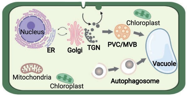
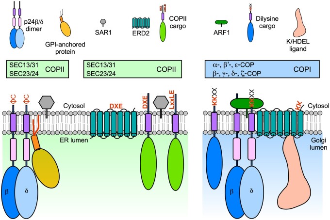
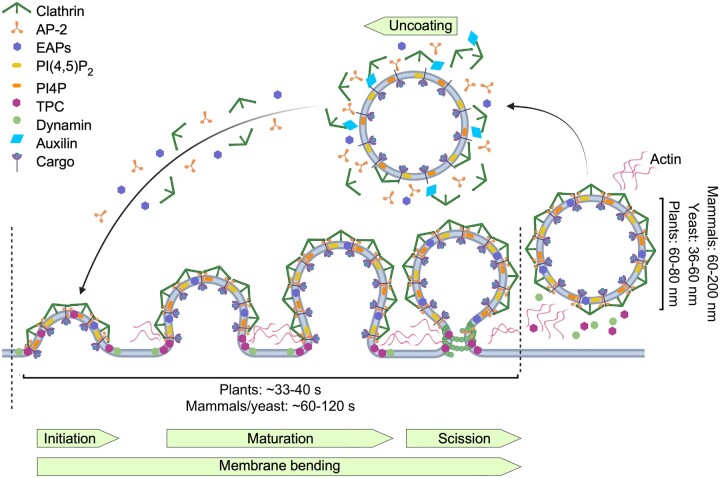
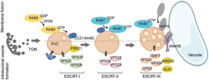
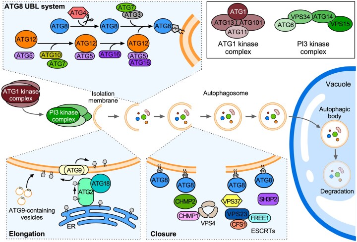
Endomembrane trafficking is essential for all eukaryotic cells. The best-characterized membrane trafficking organelles include the endoplasmic reticulum (ER), Golgi apparatus, early and recycling endosomes, multivesicular body, or late endosome, lysosome/vacuole, and plasma membrane. Although historically plants have given rise to cell biology, our understanding of membrane trafficking has mainly been shaped by the much more studied mammalian and yeast models. Whereas organelles and major protein families that regulate endomembrane trafficking are largely conserved across all eukaryotes, exciting variations are emerging from advances in plant cell biology research. In this review, we summarize the current state of knowledge on plant endomembrane trafficking, with a focus on four distinct trafficking pathways: ER-to-Golgi transport, endocytosis, trans-Golgi network-to-vacuole transport, and autophagy. We acknowledge the conservation and commonalities in the trafficking machinery across species, with emphasis on diversity and plant-specific features. Understanding the function of organelles and the trafficking machinery currently nonexistent in well-known model organisms will provide great opportunities to acquire new insights into the fundamental cellular process of membrane trafficking.


Figures

Figure 1
Figure 1
Endomembrane trafficking in plants. Major protein trafficking pathways in the endomembrane system include the biosynthetic routes for secreted proteins and those destined to the vacuole, involving vesicle trafficking and compartment maturation between the ER, Golgi apparatus, TGN, and the prevacuolar compartment (PVC)/MVB. The endocytic pathway removes PM proteins and delivers them to the TGN, where they are sorted to be either recycled back to the PM or intended for degradation in the vacuole. The endocytic and biosynthetic routes converge at the TGN, a main hub for protein sorting. Autophagosomes are formed de novo in the cytoplasm and enclose cellular components, including organelles. Fusion with the vacuole results in autophagosome cargo degradation.
Figure 2
Figure 2
Protein sorting at the ER-Golgi interface is mediated by Coat Protein I (COPI) and COPII proteins. ER export is mediated by COPII proteins that recognize sorting signals in cargo proteins via the SEC24 subunit. ER export signals include DXE and LxxLE motifs or a C-terminal diaromatic (ΦC) motif in p24 proteins. ER export of proteins with a bulky luminal portion [p24 proteins and glycosylphosphatidylinositol (GPI)-anchored proteins] may involve specific COPII vesicles (probably including specific COPII isoforms), but this remains to be demonstrated in plants. p24 proteins are required for ER export of GPI-anchored proteins in mammals, yeast, and plants. Sorting of the K/HDEL receptor ERD2 within COPII vesicles may involve a DXE motif exposed by a conformational change occurring at the neutral pH of the ER. Retrograde Golgi-to-ER transport is mediated by COPI proteins that recognize sorting signals in cargo proteins (including a dilysine KK motif in p24 proteins and other cargoes). Sorting of ERD2 within COPI vesicles may implicate a KK motif exposed by a conformational change occurring at the acidic pH of the Golgi apparatus.
Figure 3
Figure 3
Clathrin-mediated endocytosis (CME) in plants. CME is a multistep process involving initiation and stabilization of clathrin-coated pits (CCPs), maturation and membrane bending, followed by dynamin-catalyzed scission and uncoating. Clathrin-coated vesicles (CCVs) are wider in mammalian cells than in yeast and plants, but plant CCVs form faster than in yeast, where CCVs are pinched off from a narrow tubular invagination. In plants, internalized CCVs display a delayed uncoating, whereas actin is not essential for CME. Adaptor and accessory proteins involved in plant CME are shown. AP-2, AP-2; TPC, TPLATE complex; EAPs, endocytic accessory proteins; PIP, phosphatidylinositol phosphate.
Figure 4
Figure 4
Post-Golgi trafficking to the vacuole. Trafficking to the vacuole involves the maturation of prevacuolar compartments (PVCs) in preparation for fusion. The recruitment of fusiogenic proteins starts with the activation of the GTPase RAB5 and is followed by RAB7 activation and HOPS recruitment. The SNARE complex mediates the final membrane fusion step. PVC maturation also involves the formation of intraluminal vesicles carrying ubiquitinated cargo proteins for vacuolar degradation. This process initiates with the recruitment of ubiquitinated cargo by TOL proteins and the activity of the plant-specific component FREE1, followed by the sequential action of the ESCRT-I to III complexes, resulting in the inward bending of the membrane to form internal vesicles.
Figure 5
Figure 5
Overview of autophagosome biogenesis. Autophagy-inducing conditions activate signaling pathways with the ATG1 kinase (ATG1K) complex (consisting of ATG1, ATG11, ATG13, and ATG101) as a common target to initiate autophagosome biogenesis. The ATG1 complex phosphorylates the PI3 kinase (PI3K) complex (consisting of ATG6, ATG14, VPS15, and VPS34) that drives the nucleation of the isolation membrane, also termed the phagophore. Phosphatidylinositol 3-phosphate (PI3P) production leads to the recruitment of the PI3P-binding proteins ATG18 and ATG2. ATG9-containing vesicles serve as membrane seeds for the phagophore, whose development is maintained by the lipid transfer activity of ATG2, while the lipid scramblase activity of ATG9 ensures the even distribution of transferred phospholipids between the inner and outer phagophore membranes. During phagophore elongation, ATG8 is covalently bound to phosphatidylethanolamine (PE) via the ubiquitin-like (UBL) protein conjugation system, composed of an E1 (ATG7), E2 (ATG3), and E3 (ATG5-ATG12-ATG16 complex), allowing its insertion into the growing phagophore. Phagophore closure is mediated by ESCRT complexes: the ESCRT-I subunit VPS37 can recruit the ESCRT-III subunits CHMP1/CHMP2 and the ATPase VPS4, while VPS23, another ESCRT-I subunit, interacts with the FYVE domain-containing proteins CFS1 and FREE1, enabling the recruitment of SH3P2 proteins. Upon closure, autophagosomes are delivered to the vacuole. The inner membrane-bound autophagosome structure, known as an autophagic body, is released into the vacuolar lumen and is degraded by resident hydrolases, allowing recycling of its constituents.

Comment in

  • Back to the roots: A focus on plant cell biology.
    Weijers D, Bezanilla M, Jiang L, Roeder AHK, Williams M. Weijers D, et al. Plant Cell. 2022 Jan 20;34(1):1-3. doi: 10.1093/plcell/koab278. Plant Cell. 2022. PMID: 34755878 Free PMC article. No abstract available.

References

    1. Acheampong AK, Shanks C, Cheng C-Y, Schaller GE, Dagdas Y, Kieber JJ (2020) EXO70D isoforms mediate selective autophagic degradation of type-A ARR proteins to regulate cytokinin sensitivity. Proc Natl Acad Sci USA 117: 27034–27043 - PMC - PubMed
    1. Adamowski M, Narasimhan M, Kania U, Glanc M, De Jaeger G, Friml J (2018) A functional study of AUXILIN-LIKE1 and 2, two putative clathrin uncoating factors in Arabidopsis. Plant Cell 30: 700–716 - PMC - PubMed
    1. Adolf F, Rhiel M, Hessling B, Gao Q, Hellwig A, Béthune J, Wieland FT (2019) Proteomic profiling of mammalian COPII and COPI vesicles. Cell Rep 26: 250–265 - PubMed
    1. Ahn H-K, Kang YW, Lim HM, Hwang I, Pai H-S (2015) Physiological functions of the COPI complex in higher plants. Mol Cells 38: 866–875 - PMC - PubMed
    1. Ahn G, Kim H, Kim DH, Hanh H, Yoon Y, Singaram I, Wijesinghe KJ, Johnson KA, Zhuang X, Liang Z, et al. (2017) SH3 Domain-containing protein 2 plays a crucial role at the step of membrane tubulation during cell plate formation. Plant Cell 29: 1388–1405 - PMC - PubMed

Publication types

LinkOut - more resources