Randomized Controlled Trial
. 2021 Dec;32(12):1411-1424.
doi: 10.1111/clr.13841. Epub 2021 Oct 13.

Vertical and horizontal ridge augmentation using customized CAD/CAM titanium mesh with versus without resorbable membranes. A randomized clinical trial

Affiliations
Randomized Controlled Trial

Vertical and horizontal ridge augmentation using customized CAD/CAM titanium mesh with versus without resorbable membranes. A randomized clinical trial

Alessandro Cucchi et al. Clin Oral Implants Res. 2021 Dec.

Abstract

Objectives: The aim was to evaluate the role of resorbable membranes applied over customized titanium meshes related to soft tissue healing and bone regeneration after vertical/horizontal bone augmentation.

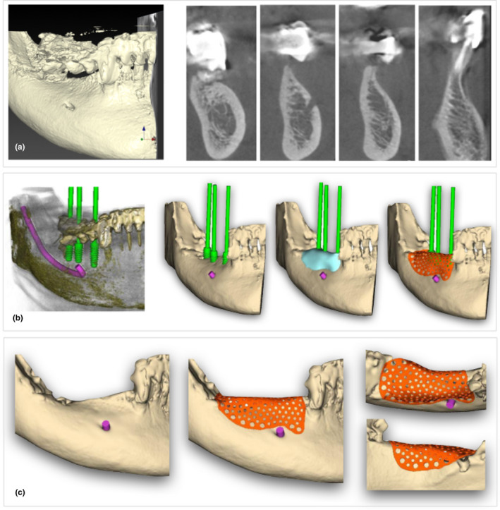
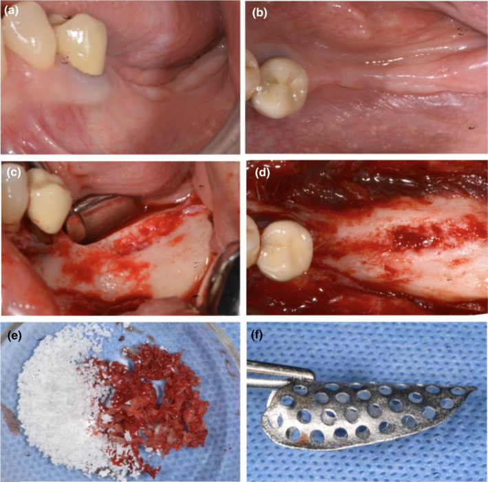
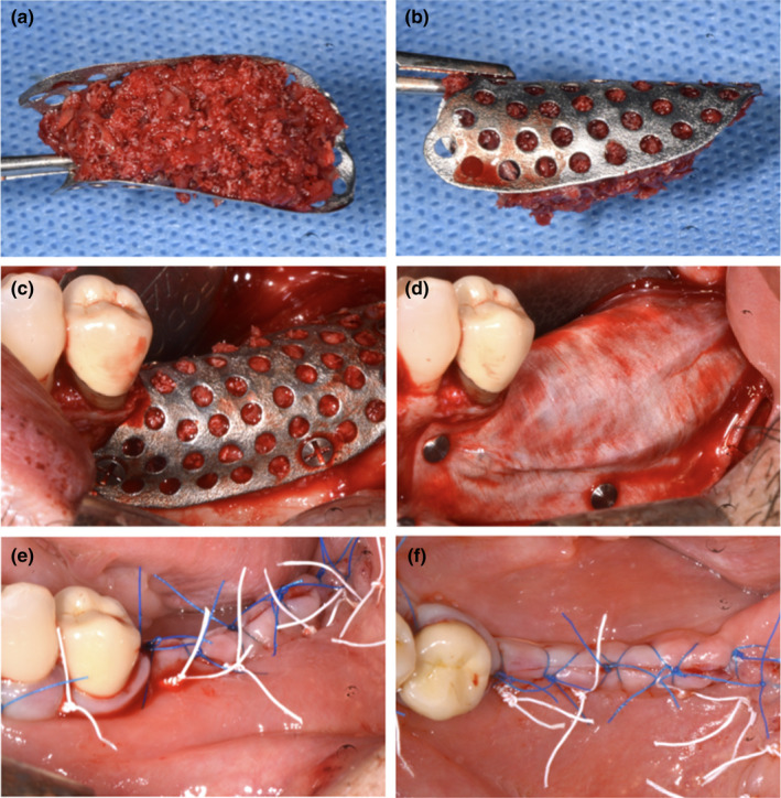
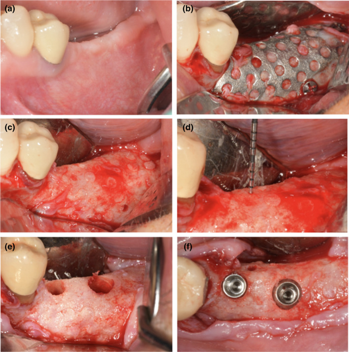
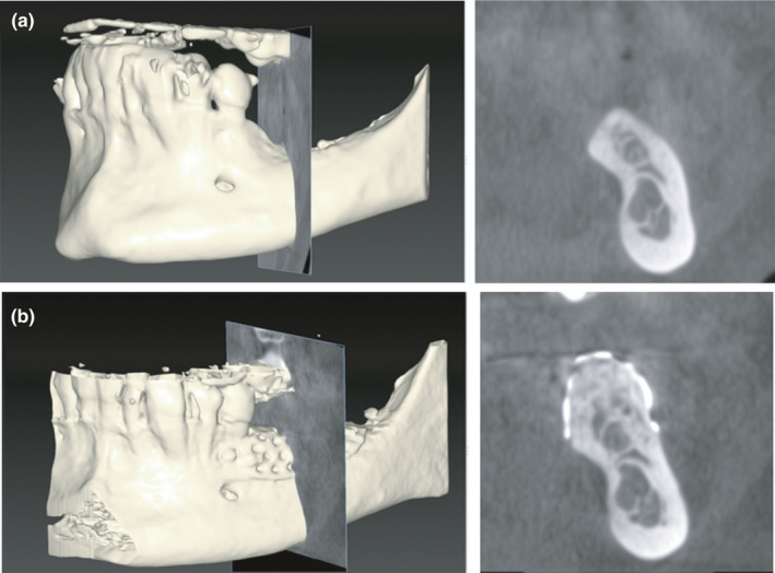
Materials and methods: Thirty patients with partial edentulism of the maxilla/mandible, with vertical/horizontal reabsorption of the alveolar bone, and needing implant-supported restorations, were randomly divided into two groups: Group A was treated using only custom-made meshes (Mesh-) and Group B using custom-made meshes with cross-linked collagen membranes (Mesh+). Data collection included surgical/technical and healing complications, "pseudo-periosteum" thickness, bone density, planned bone volume (PBV), regenerated bone volume (RBV), regeneration rate (RR), vertical bone gain (VBG), and implant survival in regenerated areas. Statistical analysis was performed between the two study groups using a significance level of α = .05.

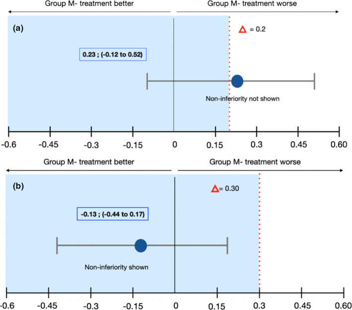
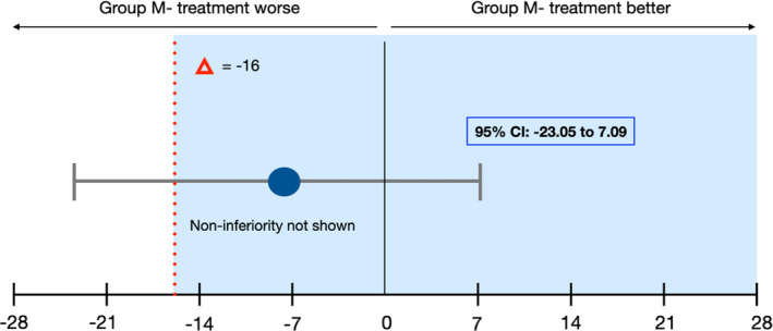
Results: Regarding the healing complications, the noninferiority analysis proved to be inconclusive, despite the better results of group Mesh+ (13%) compared to group Mesh- (33%): estimated value -1.13 CI-95% from -0.44 to 0.17. Superiority approach confirmed the absence of significant differences (p = .39). RBV was 803.27 mm3 and 843.13 mm3 , respectively, and higher RR was observed in group Mesh+ (82.3%) compared to Mesh- (74.3%), although this value did not reach a statistical significance (p = .44). All 30 patients completed the study, receiving 71 implants; 68 out of them were clinically stable and in function.

Conclusion: The results showed that customized meshes alone do not appear to be inferior to customized meshes covered by cross-linked collagen membranes in terms of healing complication rates and regeneration rates, although superior results were observed in group Mesh+compared to group Mesh- for all variables.

Keywords: alveolar ridge augmentation; collagen membrane; healing complication; osseointegrated implants; titanium mesh.


Figures

FIGURE 1
FIGURE 1
(a) A dedicated software for.dicom files allowed to have the 3D rendering and the ortho‐radial slices for digital planning; (b) digital work‐flow included the following steps: creation of.stl model, implant‐prosthetic planning, bone augmentation planning, and mesh customization; and (c) approval of the customized mesh after buccal, crestal, and lingual evaluation [Colour figure can be viewed at wileyonlinelibrary.com]
FIGURE 2
FIGURE 2
(a and b) Preoperative evaluation of soft tissue defect—lateral and occlusal views; (c and d) intraoperative evaluation of bone defect—lateral and occlusal views; (e) mixture 50:50 of autogenous bone and high porosity xenograft; (f) presentation of customized mesh [Colour figure can be viewed at wileyonlinelibrary.com]
FIGURE 3
FIGURE 3
(a and b) Customized mesh filled using bone graft—lateral and occlusal views; (c) fixation of the customized mesh using self‐tapping 5‐mm titanium mini‐screws; (d) application of a cross‐linked collagen membrane over the mesh using titanium tacks; (e and f) primary closure of surgical flaps using horizontal maîtresse sutures (white) and single interrupted sutures (blue)—lateral and occlusal views [Colour figure can be viewed at wileyonlinelibrary.com]
FIGURE 4
FIGURE 4
(a) Preoperative evaluation of soft tissue defect 6 months after surgery; (b) re‐entry surgery after flap elevation and mesh identification; (c and d) intraoperative evaluation of bone augmentation, bone density, and pseudoperiosteum; (e) preparation of implant sites using drilling burs; (f) placement of two implants in the molar region [Colour figure can be viewed at wileyonlinelibrary.com]
FIGURE 5
FIGURE 5
(a and b) Cone beam computed tomography (CBCT) before and after bone augmentation in correspondence with maximum vertical defect and calculation of vertical bone gain (VBG)
FIGURE 6
FIGURE 6
Healing and Surgical/technical Complications: Noninferiority Analysis. Error bars indicated one‐sided 95% confidence intervals of the difference in the healing complication mean values between the M‐ and M+ groups (Mesh‐ minus Mesh+). The red broken line delineating the difference in the score shows the noninferiority margin (delta); tinted area indicates the zone of noninferiority. (a) Healing Complications: The CI includes Δ and zero and the data do not prove noninferiority of Mesh‐ group compared with Mesh+group. Although there is no statistically significant difference between the two treatments, Mesh‐ group tends to be worse than Mesh+group in terms of healing complications. (b) Surgical/technical Complications: The CI does not include Δ and the data prove noninferiority of Mesh‐ group compared with Mesh+group. Although there is no statistically significant difference between the two treatments, Mesh‐ group tends to be superior to Mesh+group in terms of surgical/technical complications [Colour figure can be viewed at wileyonlinelibrary.com]
FIGURE 7
FIGURE 7
Regeneration Rates (RR): Noninferiority Analysis. Error bars indicated one‐sided 95% confidence intervals of the difference in the healing complication mean values between the M‐ and M+ groups (M‐ minus M+). The red broken line delineating the difference in the score (Δ = −16) shows the noninferiority margin (delta); tinted area indicates the zone of noninferiority. The CI includes Δ and zero and the data do not prove noninferiority of group M‐ compared with group M+ [Colour figure can be viewed at wileyonlinelibrary.com]

Similar articles

Cited by

References

    1. Aghaloo, T. L. , Misch, C. , Lin, G. H. , Iacono, V. J. , & Wang, H. L. Bone augmentation of the edentulous maxilla for implant placement: A systematic review. The International Journal of Oral & Maxillofacial Implants, 31, s19–s30. 10.11607/jomi.16suppl.g1 - DOI - PubMed
    1. Alayan, J. , & Ivanovski, S. (2018). A prospective controlled trial comparing xenograft/autogenous bone and collagen‐stabilized xenograft for maxillary sinus augmentation‐complications, patient‐reported outcomes and volumetric analysis. Clinical Oral Implants Research, 29(2), 248–262. 10.1111/clr.13107 - DOI - PubMed
    1. Assenza, B. , Piattelli, M. , Scarano, A. , Lezzi, G. , Petrone, G. , & Piattelli, A. (2001). Localized ridge augmentation using titanium micromesh. Journal of Oral Implantology, 27(6), 287–292. 10.1563/1548-1336(2001)027<0287:LRAUTM>2.3.CO;2 - DOI - PubMed
    1. Bornstein, M. M. , Al‐Nawas, B. , Kuchler, U. , & Tahmaseb, A. (2014). Consensus statements and recommended clinical procedures regarding contemporary surgical and radiographic techniques in implant dentistry. The International Journal of Oral & Maxillofacial Implants, 29S, 78–82. 10.11607/jomi.2013.g1 - DOI - PubMed
    1. Boyne, P. J. , Cole, M. D. , Stringer, D. , & Shafqat, J. P. (1985). A technique for osseous restoration of deficient edentulous maxillary ridges. Journal of Oral and Maxillofacial Surgery, 43, 87–91. 10.1016/0278-2391(85)90054-0 - DOI - PubMed

Publication types