. 2021 Nov;41(11):2756-2769.
doi: 10.1161/ATVBAHA.121.316867. Epub 2021 Sep 23.

Receptor and Molecular Mechanism of AGGF1 Signaling in Endothelial Cell Functions and Angiogenesis

Affiliations

Receptor and Molecular Mechanism of AGGF1 Signaling in Endothelial Cell Functions and Angiogenesis

Jingjing Wang et al. Arterioscler Thromb Vasc Biol. 2021 Nov.

Abstract

Objective: Angiogenic factor AGGF1 (angiogenic factor with G-patch and FHA [Forkhead-associated] domain 1) promotes angiogenesis as potently as VEGFA (vascular endothelial growth factor A) and regulates endothelial cell (EC) proliferation, migration, specification of multipotent hemangioblasts and venous ECs, hematopoiesis, and vascular development and causes vascular disease Klippel-Trenaunay syndrome when mutated. However, the receptor for AGGF1 and the underlying molecular mechanisms remain to be defined.

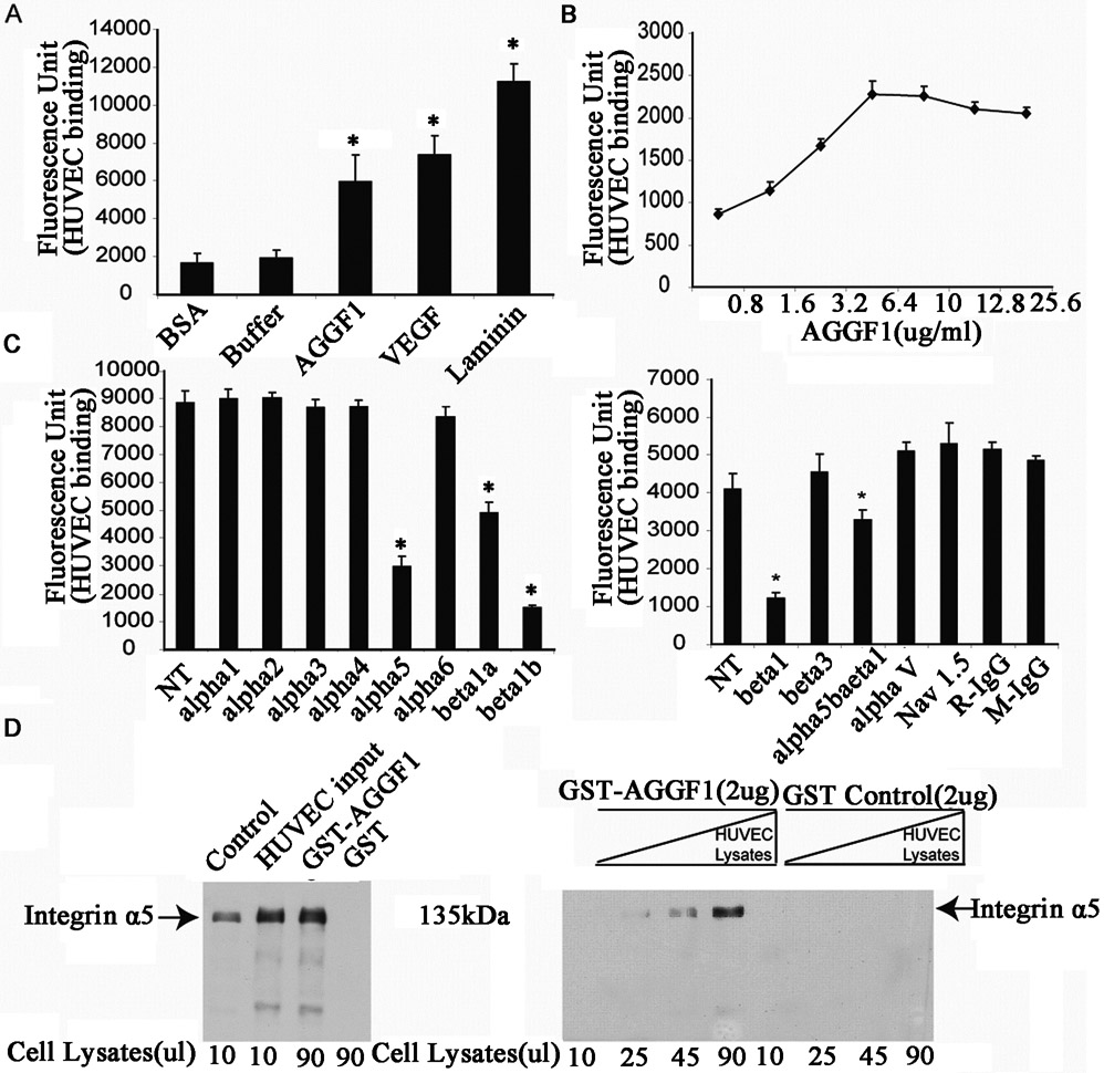
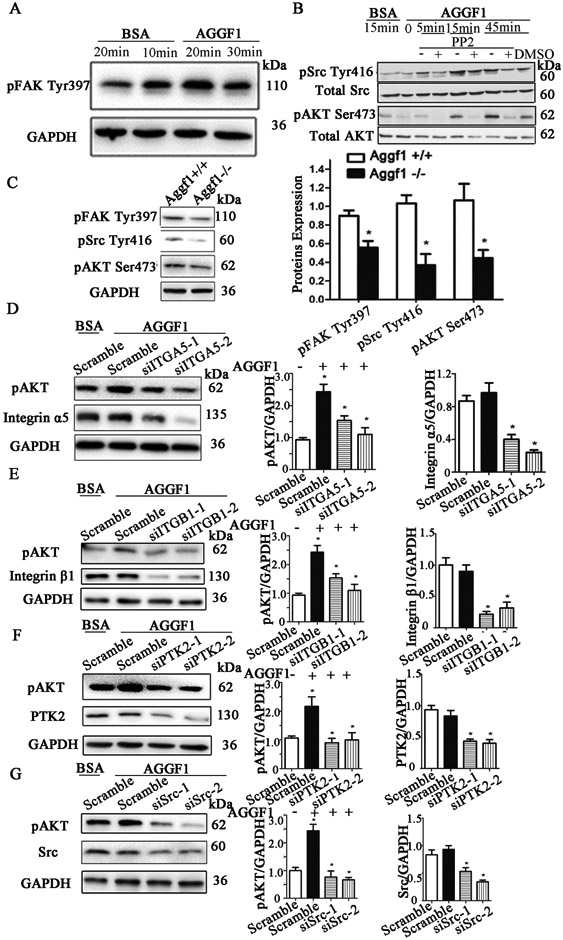
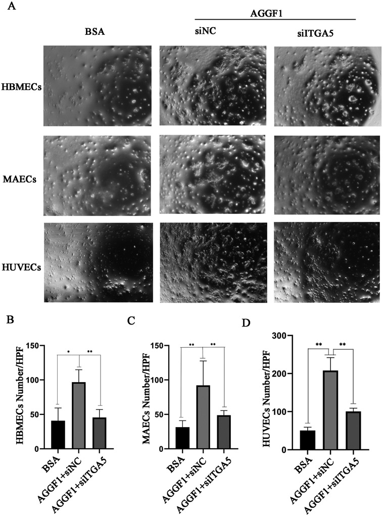
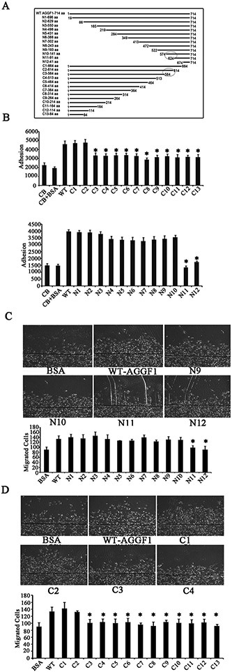
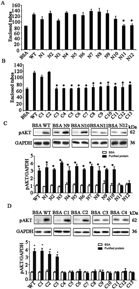
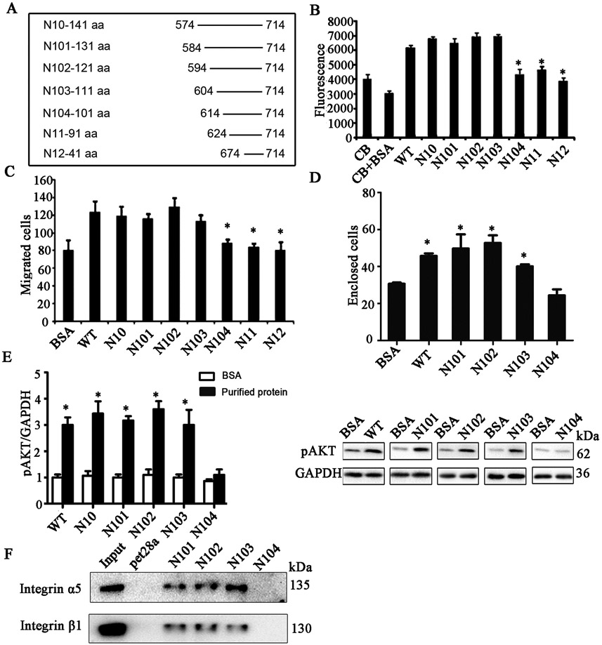
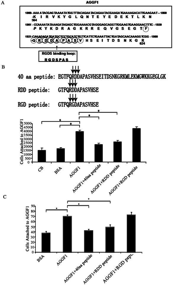
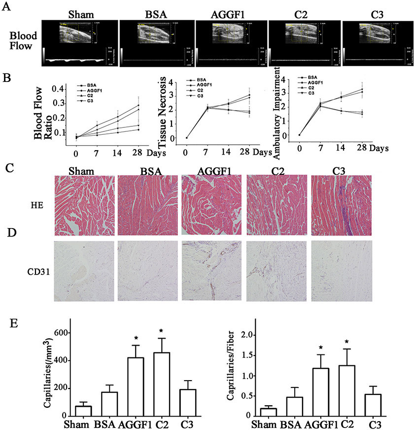
Approach and results: Using functional blocking studies with neutralizing antibodies, we identified [alpha]5[beta]1 as the receptor for AGGF1 on ECs. AGGF1 interacts with [alpha]5[beta]1 and activates FAK (focal adhesion kinase), Src (proto-oncogene tyrosine-protein kinase), and AKT (protein kinase B). Functional analysis of 12 serial N-terminal deletions and 13 C-terminal deletions by every 50 amino acids mapped the angiogenic domain of AGGF1 to a domain between amino acids 604-613 (FQRDDAPAS). The angiogenic domain is required for EC adhesion and migration, capillary tube formation, and AKT activation. The deletion of the angiogenic domain eliminated the effects of AGGF1 on therapeutic angiogenesis and increased blood flow in a mouse model for peripheral artery disease. A 40-mer or 15-mer peptide containing the angiogenic domain blocks AGGF1 function, however, a 15-mer peptide containing a single amino acid mutation from -RDD- to -RGD- (a classical RGD integrin-binding motif) failed to block AGGF1 function.

Conclusions: We have identified integrin [alpha]5[beta]1 as an EC receptor for AGGF1 and a novel AGGF1-mediated signaling pathway of [alpha]5[beta]1-FAK-Src-AKT for angiogenesis. Our results identify an FQRDDAPAS angiogenic domain of AGGF1 crucial for its interaction with [alpha]5[beta]1 and signaling.

Keywords: amino acids; endothelial cells; hemangioblasts; integrins; peripheral artery disease.


Figures

Figure 1.
Figure 1.. Identification of integrin α5β1 as the first receptor for angiogenic factor AGGF1.
(A) Cell adhesion assays showing binding of HUVECs (150 μl of 2x10⁵ cells/ml) to AGGF1-coated wells as compared to VEGFA and laminin (100 μl x 5 μg/ml). BSA and coating buffer (100 μl) were used as negative control. *P<0.05, n=4/group (one-way ANOVA with Dunnett post hoc tests). (B) Dose response curve of HUVEC adhesion to AGGF1 (100 μl x 0.8–25.6 μg/ml). Data points are presented as mean ± SEM from quadruple wells. *P<0.05, n=4/group. The half maximum binding of HUVECs was achieved at the AGGF1 dose of 2.27 μg/ml (equal to 100 μl x 2.27 μg/ml = 2.80 pmole of AGGF1). In the cell adhesion system, the estimated concentration of AGGF1 was 2.80 pmole in 150 μl of volume = 18.7 nM. (C) Neutralizing antibodies against integrin α5, β1, and α5β1 disrupt HUVEC adhesion to AGGF1. 150 μl of HUVECs were pre-incubated with functional blocking antibodies against various integrin subunits or control antibodies, and plated into microtiter wells coated with AGGF1 (100 μl x 6.40 μg/ml) for cell adhesion assays. An anti-α5 antibody, two different anti-β1 antibodies (P5D2 as beta1a; TDM2 as beta1b), and an anti-α5β1 showed a blocking effect. An anti-cardiac sodium channel antibody (Nav1.5) and normal rabbit (R-IgG) and mouse IgG (M-IgG) were used as negative controls. NT refers to HUVECs without any antibody treatment. Data are shown as mean ± SEM from quadruple wells. *P≤0.05, n=4/group (one-way ANOVA with Dunnett post hoc tests). (D) AGGF1 interacts with integrin α5 in GST pull-down assays. GST-AGGF1 or control GST was incubated with varying amounts of HUVEC cell lysates, followed by precipitation with glutathione-sepharose 4B beads. The precipitates were used for western blot analysis with an anti-α5 antibody.
Figure 2.
Figure 2.. AGGF1 activates a novel angiogenic signaling pathway involving α5β1-FAK-Src-AKT.
(A) Western blot analysis showing an increased phosphorylation level of FAK in HUVECs stimulated with AGGF1. (B) Western blot analysis showing an increased phosphorylation level of Src or AKT in HUVECs stimulated with AGGF1. PP2, a Src specific inhibitor (30 μM), prevented the induction of AKT activation by AGGF1. (C) Aggf1 haploinsufficiency reduces the phosphorylation levels of FAK, Src and AKT. Microvascular endothelial cells from heterozygous Aggf1Geo/+ KO mice vs. wild type Aggf1+/+ mice were lysed and used for western blot analysis with antibodies against phosphor-FAK, phosphor-Src or phosphor-AKT antibodies. GAPDH was used as loading control. (D-F) Western blot analysis showing the effects of siRNAs for ITGA5 (D), ITGB1 (E), PTK2 (F), and Src (G) on the phosphorylation level of AKT in HUVECs stimulated with AGGF1 (5 μg/ml). Representative Western blotting images are shown on the left, and the quantified data are shown on the right as mean ± SEM. *P≤0.05, n=3/group (a Student’s t test).
Figure 3.
Figure 3.. Three different types of endothelial cells adhere to the AGGF1 protein through integrin α5.
(A) Representative images from cell adhesion assays for HBMEC, MAECs and HUVECs in wells coated with 5 μg/ml of purified recombinant AGGF1. BSA was used as a negative control. NC, negative scramble siRNA; siITGA5, siRNA specific for ITGA5. (B) siITGA5 markedly reduces adhesion of HBMECs to AGGF1. (C) siITGA5 markedly reduces adhesion of MAECs to AGGF1. (D) siITGA5 markedly reduces adhesion of HUVECs to AGGF1. Quantitative data were shown as mean±SEM, and analyzed using a Student’s t test (*P<0.05, **P<0.01, n=6/group).
Figure 4.
Figure 4.. Identification of a functional angiogenic domain of AGGF1 involved in cell adhesion and migration between amino acid 574 and 614.
(A) A diagram showing the full length AGGF1 (WT), 12 serial N-terminal deletions and 13 C-terminal deletion mutants. (B) HUVECs adhesion assays with HUVECs treated with WT AGGF1 and serial AGGF1 deletion mutants. Coating buffer (CB) with and without BSA was used as negative controls. (C) HUVEC migration assays with HUVECs treated with WT AGGF1 (5 μg/ml) and serial N-terminal AGGF1 deletion mutants (equal moles as WT). BSA was used as negative control. Representative photomicrographs of endothelial cell migration are shown on the right. (D) HUVEC migration assays with HUVECs treated with WT AGGF1 (5 μg/ml) and serial C-terminal AGGF1 deletion mutants (equal moles as WT). All data are shown as mean ± SEM. *P≤0.05, n=4/group (one-way ANOVA with Dunnett post hoc tests).
Figure 5.
Figure 5.. The 40 amino acid functional angiogenic domain of AGGF1 between amino acid 574 and 614 is involved in AGGF1-induced capillary endothelial tube formation and AKT activation in HUVECs.
(A) Endothelial tube formation assays with HUVECs treated with WT AGGF1 (5 μg/ml) and serial N-terminal AGGF1 deletion mutants (equal moles as WT). BSA was used as negative control. (B) Endothelial tube formation assays with HUVECs treated with WT AGGF1 (5 μg/ml) and serial N-terminal AGGF1 deletion mutants (equal moles as WT). (C) AKT activation in HUVECs treated with WT AGGF1 (5 μg/ml) and serial N-terminal AGGF1 deletion mutants (equal moles as WT). BSA was used as negative control. Representative western blotting images are shown at the top. (D) AKT activation in HUVECs treated with WT AGGF1 (5 μg/ml) and serial N-terminal AGGF1 deletion mutants (equal moles as WT). Representative western blotting images are shown at the top. All data are shown as mean ± SEM. *P≤0.05, n=3-4/group (one-way ANOVA with Dunnett post hoc tests).
Figure 6.
Figure 6.. Fine mapping of the functional angiogenic domain of AGGF1 to a 10-amino acid domain between amino acid 604 and 614.
(A) A diagram showing the full length AGGF1 (WT) and N-terminal deletion mutants N-10, N101, N102, N103, N104, N11 and N12. (B) Endothelial cell adhesion assays with HUVECs treated with WT AGGF1 and 7 N-terminal deletion mutants of N10, N101, N102, N103, N104, N11 and N12. Coating buffer (CB) with and without BSA was used as negative controls. (C) Endothelial cell migration assays with HUVECs treated with WT AGGF1 and 7 N-terminal deletion mutants of N10, N101, N102, N103, N104, N11 and N12. (D) Endothelial tube formation assays with HUVECs treated with WT AGGF1 and N-terminal deletion mutants of N101, N102, N103, and N104. (E) Western blot analysis for pAKT (Ser473) in HUVECs treated with WT AGGF1, and 7 N-terminal deletion mutants of N10, N101, N102, N103, N104, N11 and N12. Representative western blotting images are shown on the right. (F) Interaction between AGGF1 mutants N101, N102 and N103 with the Angiogenic Domain, but not N104 without the Angiogenic Domain. His pull-down was performed with His-tagged N101, N102, N103 or N104 and HUVEC lysates and analyzed using western blot analysis with an anti-α5 or anti-β1 antibody. pet28a, His tag alone as negative control. All data are shown as mean ± SEM. *P≤0.05, n=3-4/group (one-way ANOVA with Dunnett post hoc tests).
Figure 7.
Figure 7.. Effects of three peptides derived from AGGF1 functional angiogenic domain on HUVEC adhesion and capillary tube formation.
(A) A diagram showing the location and amino acid sequences of the functional Angiogenic Domain of AGGF1. The RDDAPAS motif of AGGF1 is similar, but not identical, to the RGDSPAS integrin binding loop of fibronectin. (B) HUVEC-AGGF1 adhesion assays with three different AGGF1 peptides. HUVECs were pre-incubated with a peptide (0.12 mM) before being added to the wells coated with AGGF1. Coating buffer (CB) with and without BSA was used as negative controls. (C) Endothelial tube formation assays with HUVECs treated with the AGGF1 protein in combination with or without three different peptides. HUVECs were pre-incubated with a peptide (0.12 mM) before being added to the wells containing solidified matrigel with WT AGGF1. Matrigel with BSA was used as negative control (left panel). The data are shown as mean ± SEM. *P≤0.05, n=4/group (one-way ANOVA with Dunnett post hoc tests).
Figure 8.
Figure 8.. Analysis of WT AGGF1 and mutant C2 with the functional angiogenic domain, and mutant C3 without the domain in male mice.
(A) The treatment effect of WT and mutant AGGF1 proteins for PAD in a hindlimb ischemia mouse model. Representative Doppler ultrasound images are shown for male mice treated with WT AGGF1, mutant C2, and mutant C3. Sham mice and BSA were used as negative controls. The structure of mutants C2 and C3 can be found in Figure 4A. (B) The ratios of blood flow in the ischemic leg over that in the non-ischemic limb, tissue necrosis scores and ambulatory impairment scores are shown for different time points in days. The blood flow was measured by high resolution micro-ultrasound (left panel). Effects of different treatments on tissue necrosis are shown in the middle panel. Effects of different treatments on ambulatory impairment are shown in the right panel. Histological examinations of muscle tissue. (C) Representative H&E staining images of sections of ischemic hindlimb muscles. (D) Representative immunostaining images of sections of ischemic hindlimb muscles stained with an anti-CD31 antibody. (E) Quantification of density of CD-31-positive vessels per mm2 in ischemic muscles 28 days after different treatments (left panel). The number of CD-31-positive vessels per muscular fiber is plotted for different treatments for the time point of 28 days (right panel). All data are shown as mean ± SEM. *P≤0.05, n=13/group (one-way ANOVA with Dunnett post hoc tests).

Comment in

References

    1. Timur AA, Driscoll DJ, Wang Q. Biomedicine and diseases: the Klippel-Trenaunay syndrome, vascular anomalies and vascular morphogenesis. Cell Mol Life Sci. 2005;62:1434–1447. - PMC - PubMed
    1. Auluck A, Suhas S, Pai KM. Klippel-Trenaunay syndrome. Oral Dis. 2005;11:255–258. - PubMed
    1. Berry SA, Peterson C, Mize W, Bloom K, Zachary C, Blasco P, Hunter D. Klippel-Trenaunay syndrome. Am J Med Genet. 1998;79:319–326. - PubMed
    1. Aggarwal K, Jain VK, Gupta S, Aggarwal HK, Sen J, Goyal V. Klippel-Trenaunay syndrome with a life-threatening thromboembolic event. J Dermatol. 2003;30:236–240. - PubMed
    1. Jacob AG, Driscoll DJ, Shaughnessy WJ, Stanson AW, Clay RP, Gloviczki P. Klippel-Trenaunay syndrome: spectrum and management. Mayo Clin Proc. 1998;73:28–36. - PubMed

Publication types

MeSH terms