Review
. 2021 Dec;41(12):3157-3170.
doi: 10.1177/0271678X211046992. Epub 2021 Sep 22.

Mitochondrial quality control in acute ischemic stroke

Affiliations
Review

Mitochondrial quality control in acute ischemic stroke

Hong An et al. J Cereb Blood Flow Metab. 2021 Dec.

Abstract

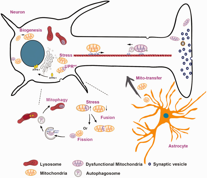
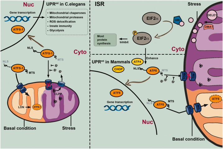
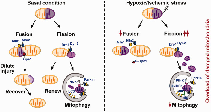
Mitochondria play a central role in the pathophysiological processes of acute ischemic stroke. Disruption of the cerebral blood flow during acute ischemic stroke interrupts oxygen and glucose delivery, leading to the dysfunction of mitochondrial oxidative phosphorylation and cellular bioenergetic stress. Cells can respond to such stress by activating mitochondrial quality control mechanisms, including the mitochondrial unfolded protein response, mitochondrial fission and fusion, mitophagy, mitochondrial biogenesis, and intercellular mitochondrial transfer. Collectively, these adaptive response strategies contribute to retaining the integrity and function of the mitochondrial network, thereby helping to recover the homeostasis of the neurovascular unit. In this review, we focus on mitochondrial quality control mechanisms occurring in acute ischemic stroke. A better understanding of how these regulatory pathways work in maintaining mitochondrial homeostasis will provide a rationale for developing innovative neuroprotectants when these mechanisms fail in acute ischemic stroke.

Keywords: Acute ischemic stroke; intercellular mitochondrial transfer; mitochondrial biogenesis; mitochondrial dynamics; mitochondrial quality control; mitochondrial unfolded protein response; mitophagy.


Conflict of interest statement

Declaration of conflicting interests: The author(s) declared no potential conflicts of interest with respect to the research, authorship, and/or publication of this article.

Figures

Figure 1.
Figure 1.
Mitochondrial quality control and intercellular mitochondrial transfer. When exposed to stressors, mitochondria can initiate a set of quality control mechanisms, including the mitochondrial unfolded protein response (UPRmt), mitochondrial fission and fusion, mitophagy, and mitochondrial biogenesis. In addition, mitochondria can be transferred from adjacent astrocytes to injured neurons as a compensatory internal adaptive response, which is a novel type of mitochondrial quality control mechanism.
Figure 2.
Figure 2.
Mechanisms of the mitochondrial unfolded protein response (UPRmt) in Caenorhabditis elegans and mammalian cells. The UPRmt is an adaptive transcriptional response that can be activated by various mitochondrial stressors. In C. elegans, the key regulator of UPRmt is the activation transcription factor associated with stress 1 (ATFS-1), which is continuously transported into mitochondria via mitochondrial targeting sequence (MTS) guidance and cleaved by the Lon protease under physiological conditions. However, the ATFS-1 mitochondria targeting can be suppressed by the accumulation of aberrant peptides in the cytosol, which are unfolded or misfolded proteins that are cleaved by the caseinolytic protease P (CLPP) and extruded into the cytosol via the transporter HAF-1 under stress conditions. Stabilized ATFS-1 translocates through guidance of the nuclear localization sequence (NLS) into the nucleus from where it promotes gene transcription related to mitochondrial chaperones and proteases, detoxification of reactive oxygen species (ROS), innate immunity, and glycolysis. Together, these protective responses facilitate the recovery of mitochondrial homeostasis. UPRmt in mammalian cells may be regulated by basic leucine zipper (bZip) protein homologs including the C/EBP homologous protein (CHOP), activating transcription factor 4 (ATF4), and activating transcription factor 5 (ATF5), which are transcribed under the control of the integrated stress response (ISR) mechanism mediated by phosphorylated eukaryotic initiation factor-2 alpha subunit (eIF2α). The mitochondrial metallopeptidase OMA1 cleaves the DELE1 protein into short peptides, contributing to heme-regulated inhibitor (HRI) activation, eIF2α phosphorylation, and a general inhibition of gene transcription, while promoting transcription of genes encoding bZip proteins.
Figure 3.
Figure 3.
Mechanisms of mitochondrial dynamics and mitophagy under basal conditions and hypoxic/ischemic stress. Typically, fission and fusion machineries balance injured or aged mitochondria by dislodging damaged contents via dynamin-related protein (Drp)1- and dynamin (Dyn)2-mediated fission or merging with other healthy mitochondria via mitofusin (Mfn)1-, Mfn2-, or optic atrophy (Opa)1-mediated fusion. Mitochondria that fail to recover homeostasis will undergo mitophagy in a Parkin-dependent or Parkin-independent (FUNDC1 or NIX/Bnip3L) manner. However, the balance in mitochondrial dynamics shifts to fission under conditions of hypoxic/ischemic stress. Combined with an attenuated mitophagy capacity, these mechanisms collectively lead to the excessive accumulation of damaged mitochondria and irreversible cellular injury.

References

    1. Lees KR, Bluhmki E, von Kummer R, ECASS, ATLANTIS, NINDS and EPITHET rt-PA Study Group et al.. Time to treatment with intravenous alteplase and outcome in stroke: an updated pooled analysis of ECASS, ATLANTIS, NINDS, and EPITHET trials. Lancet 2010; 375: 1695–1703. - PubMed
    1. Goyal M, Menon BK, van Zwam WH, et al.. Endovascular thrombectomy after large-vessel ischaemic stroke: a Meta-analysis of individual patient data from five randomised trials. Lancet 2016; 387: 1723–1731. - PubMed
    1. Nogueira RG, Jadhav AP, Haussen DC, et al.. Thrombectomy 6 to 24 hours after stroke with a mismatch between deficit and infarct. N Engl J Med 2018; 378: 11–21. - PubMed
    1. Albers GW, Marks MP, Kemp S, et al.. Thrombectomy for stroke at 6 to 16 hours with selection by perfusion imaging. N Engl J Med 2018; 378: 708–718. - PMC - PubMed
    1. Savitz SI, Baron JC, Yenari MA, et al.. Reconsidering neuroprotection in the reperfusion era. Stroke 2017; 48: 3413–3419. - PubMed

Publication types