. 2021 Sep 22;19(1):400.
doi: 10.1186/s12967-021-03052-5.

Long non-coding RNA NORAD promotes pancreatic cancer stem cell proliferation and self-renewal by blocking microRNA-202-5p-mediated ANP32E inhibition

Affiliations

Long non-coding RNA NORAD promotes pancreatic cancer stem cell proliferation and self-renewal by blocking microRNA-202-5p-mediated ANP32E inhibition

Yu-Shui Ma et al. J Transl Med. .

Abstract

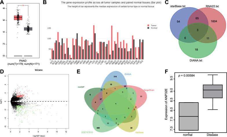
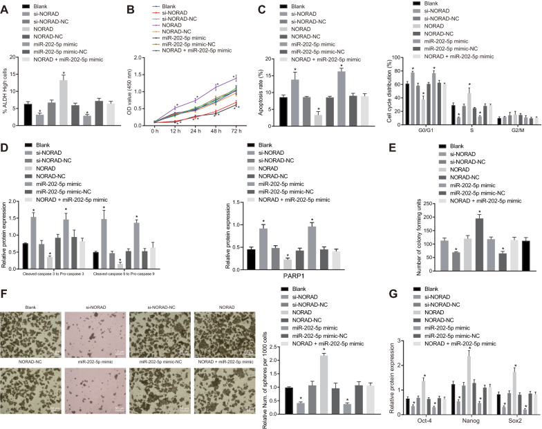
Background: Cancer stem cells (CSCs) are key regulators in the processes of tumor initiation, progression, and recurrence. The mechanism that maintains their stemness remains enigmatic, although the role of several long noncoding RNAs (lncRNAs) has been highlighted in the pancreatic cancer stem cells (PCSCs). In this study, we first established that PCSCs overexpressing lncRNA NORAD, and then investigated the effects of NORAD on the maintenance of PCSC stemness.

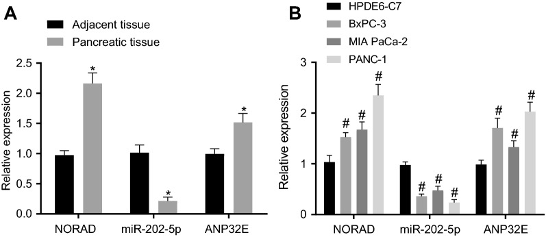
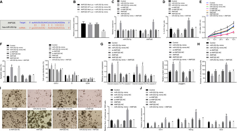
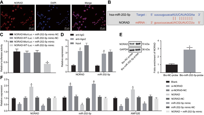
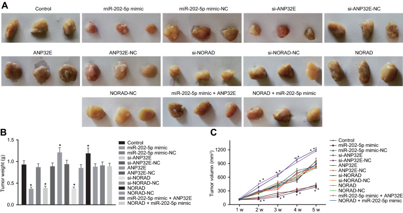
Methods: Expression of lncRNA NORAD, miR-202-5p and ANP32E in PC tissues and cell lines was quantified after RNA isolation. Dual-luciferase reporter assay, RNA pull-down and RIP assays were performed to verify the interactions among NORAD, miR-202-5p and ANP32E. We then carried out gain- and loss-of function of miR-202-5p, ANP32E and NORAD in PANC-1 cell line, followed by measurement of the aldehyde dehydrogenase activity, cell viability, apoptosis, cell cycle distribution, colony formation, self-renewal ability and tumorigenicity of PC cells.

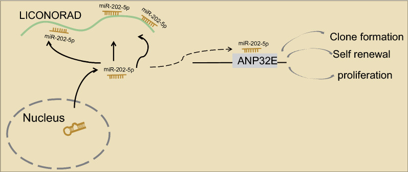
Results: LncRNA NORAD and ANP32E were upregulated in PC tissues and cells, whereas the miR-202-5p level was down-regulated. LncRNA NORAD competitively bound to miR-202-5p, and promoted the expression of the miR-202-5p target gene ANP32E thereby promoting PC cell viability, proliferation, and self-renewal ability in vitro, as well as facilitating tumorigenesis of PCSCs in vivo.

Conclusion: Overall, lncRNA NORAD upregulates ANP32E expression by competitively binding to miR-202-5, which accelerates the proliferation and self-renewal of PCSCs.

Keywords: ANP32E; Long non-coding RNA NORAD; Pancreatic cancer; Pancreatic cancer stem cells; Proliferation; Self-renewal; microRNA-202-5p.


Conflict of interest statement

The authors declare no conflict of interest.

Figures

Fig. 1
Fig. 1
LncRNA, miRNA and mRNA expression profiles in PC. A Expression of NORAD in PC where the X axis represents the grouping and the Y axis represents the NORAD expression; B Expression of NORAD in all patient tumor samples and paired normal tissues (black represents normal tissues, and red represents tumor samples); C Prediction of downstream miRNAs of NORAD (three circles in the figure represent the prediction results of the three databases, respectively, and the middle part represents their intersection); D Volcano plot of expression of differentially expressed genes in PC-related datasets, where the X axis denotes differential log10 p value and the Y axis denotes log FoldChange. Each point in the plot represents a gene, where red dots represent upregulated genes while green dots represent down-regulated genes; E Prediction of target genes of miR-202-5p (the three circles in the figure represent the prediction results of the three databases respectively, and the middle part represents their intersection); F Expression of ANP32E in GSE107610 (the X axis represents the tumor samples and normal tissues and the Y axis represents the level of ANP32E)
Fig. 2
Fig. 2
NORAD and ANP32E are highly expressed and miR-202-5p is poorly expressed in PC tissues and cells. A Expression of NORAD, ANP32E and miR-202-5p in PC tissues and adjacent normal tissues (n = 28); B Expression of NORAD, ANP32E and miR-202-5p in HPDE6-C7, BxPC-3, MIAPaCa-2, and PANC-1 cell lines; * p < 0.05 vs. the adjacent normal tissues; # p < 0.05 vs. HPDE6-C7 cell line; Measurement data were expressed as mean ± standard derivation; data between PC tissues and adjacent normal tissues were compared by paired t-test, and measurement data among multiple groups were analyzed by one-way analysis of variance with Tukey’s post hoc test. The experiment was repeated three times
Fig. 3
Fig. 3
miR-202-5p inhibits the viability, proliferation, and self-renewal of PCSCs, and promotes cell apoptosis by suppressing ANP32E expression. A Potential target gene of miR-202-5p predicted by mirDIP, DIANA, TargetScan and starBase databases; B Targeting relationship between miR-202-5p and ANP32E measured by dual-luciferase reporter assay (* p < 0.05 vs. cells co-transfected with ANP32E-WT-Luc and miR-202-5p mimic-NC, ANP32E-MUT-Luc and miR-202-5p mimic, ANP32E-MUT-Luc and miR-202-5p mimic-NC). PCSCs were treated with ANP32E, si-ANP32E and/or miR-202-5p mimic. C miR-202-5p expression and the mRNA expression of ANP32E in PCSCs measured by RT-qPCR; D ALDH activity of PCSCs assessed by Aldefluor assay, where mock means a NC with the addition of DEAB (a specific inhibitor of ALDH enzyme); E Proliferation of PCSCs detected by MTT; F Apoptosis and cell cycle changes of PCSCs measured by flow cytometry; G Protein expression of PARP1 and the ratios of cleaved-caspase3 to pro-caspase3 and cleaved-caspase9 to pro-caspase9 in PCSCs detected by Western blot analysis; H Colony formation of PCSCs assessed by colony formation assay; I Self-renewal ability of PCSCs detected by sphere formation assay (200×); J Protein expression of Oct4, Nanog, Sox2 in PCSCs measured by Western blot analysis; * p < 0.05 vs. cells without treatment. Measurement data were expressed as mean ± standard derivation. Data among multiple groups were analyzed by one-way analysis of variance with Tukey’s post hoc test, and data comparison among multiple groups at different time points was conducted using two-way analysis of variance with Bonferroni post hoc test. The experiment was repeated three times
Fig. 4
Fig. 4
NORAD competitively binds to miR-202-5p and reduces the expression of miR-202-5p in PCSCs. A Distribution of NORAD in PC cells (400×); B Specific binding sites of NORAD and miR-202-5p; C Targeting relationship between miR-202-5p and NORAD measured by dual-luciferase reporter assay (* p < 0.05 vs. cells co-transfected with NORAD-WT-Luc and miR-202-5p mimic-NC, NORAD-MUT-Luc and miR-202-5p mimic, NORAD-MUT-Luc and miR-202-5p mimic-NC); D NORAD targeted miR-202-5p verified by RIP assay (* p < 0.05 vs. IgG pulldown samples); E NORAD targeted miR-202-5p verified by RNA pull-down assay (* p < 0.05 vs. Bio-NC probe labeled samples); F NORAD and miR-202-5p expression as well as the mRNA expression of ANP32E in PCSCs measured by RT-qPCR (* p < 0.05 vs. cells without treatment). Measurement data were expressed as mean ± standard derivation. Data between two groups were compared by unpaired t-test, and data among multiple groups were analyzed by one-way analysis of variance with Tukey’s post hoc test. The experiment was repeated three times
Fig. 5
Fig. 5
NORAD stimulates the viability, proliferation, and stemness of PCSCs via competitively suppressing miR-202-5p expression. PCSCs were treated with si-NORAD, NORAD and/or miR-202-5p mimic. A ALDH activity of PCSCs assessed by Aldefluor assay, where mock means a NC with the addition of DEAB (a specific inhibitor of ALDH enzyme); B Proliferation of PCSCs detected by MTT; C Apoptosis and cell cycle changes of PCSCs measured by flow cytometry; D Protein expression of ratios of cleaved-caspase3 to pro-caspase3, and of cleaved-caspase9 to pro-caspase9, and PARP1 in PCSCs detected by Western blot analysis; E Colony formation of PCSCs assessed by colony formation assay; F Self-renewal ability of PCSCs detected by sphere formation assay; G Protein expression of Oct4, Nanog and Sox2 in PCSCs measured by Western blot analysis; * p < 0.05 vs. cells without treatment. Measurement data were expressed as mean ± standard derivation. Data among multiple groups were analyzed by one-way analysis of variance with Tukey’s post hoc test, and data comparison among multiple groups at different time points was conducted using two-way analysis of variance with Tukey’s post hoc test with Bonferroni post hoc test. The experiment was repeated three times
Fig. 6
Fig. 6
NORAD facilitates the tumorigenicity of PCSCs in vivo by competitively inhibiting miR-202-5p and upregulating ANP32E. A Representative images showing xenografts in nude mice treated with miR-202-5p mimic, si-ANP32E, si-NORAD, ANP32E, NORAD, miR-202-5p mimic + ANP32E or NORAD + miR-202-5p mimic. B Tumor weight of mice treated with miR-202-5p mimic, si-ANP32E, si-NORAD, ANP32E, NORAD, miR-202-5p mimic + ANP32E or NORAD + miR-202-5p mimic. C Tumor volume of mice treated with miR-202-5p mimic, si-ANP32E, si-NORAD, ANP32E, NORAD, miR-202-5p mimic + ANP32E or NORAD + miR-202-5p mimic. * p < 0.05 vs. mice without treatment. The above results are all measurements and expressed as mean ± standard derivation. One-way analysis of variance with Tukey’s post hoc test was used for data comparison among multiple groups. Repeated measures analysis of variance was performed for data comparison at different time points with Bonferroni’s post hoc test (n = 12)
Fig. 7
Fig. 7
Schematic diagram of the proposed mechanism of the NORAD axis in PCSC tumorgenicity. NORAD upregulates the expression of ANP32E by competitively binding to miR-202-5p, thus accelerating the proliferation and self-renewal of PCSCs

References

    1. Kamisawa T, Wood LD, Itoi T, Takaori K. Pancreatic cancer. Lancet. 2016;388:73–85. - PubMed
    1. Halbrook CJ, Lyssiotis CA. Employing metabolism to improve the diagnosis and treatment of pancreatic cancer. Cancer Cell. 2017;31:5–19. - PubMed
    1. Wolfgang CL, Herman JM, Laheru DA, Klein AP, Erdek MA, Fishman EK, Hruban RH. Recent progress in pancreatic cancer. CA Cancer J Clin. 2013;63:318–348. - PMC - PubMed
    1. Challa S, Husain K, Kim R, Coppola D, Batra SK, Cheng JQ, Malafa MP. Targeting the IκB kinase enhancer and its feedback circuit in pancreatic cancer. Transl Oncol. 2020;13:481–489. - PMC - PubMed
    1. Guo Z, Wang X, Yang Y, Chen W, Zhang K, Teng B, Huang C, Zhao Q, Qiu Z. Hypoxic tumor-derived exosomal long noncoding RNA UCA1 promotes angiogenesis via miR-96-5p/AMOTL2 in pancreatic cancer. Mol Ther Nucleic Acids. 2020;22:179–195. - PMC - PubMed

Publication types