Review
. 2021 Oct;15(5):1031-1038.
doi: 10.14444/8131. Epub 2021 Sep 22.

Distal Junctional Failure Secondary to Nontraumatic Fracture of Lower Instrumented Vertebra: Our Experience and Review of Literature

Affiliations
Review

Distal Junctional Failure Secondary to Nontraumatic Fracture of Lower Instrumented Vertebra: Our Experience and Review of Literature

Akash Hosthota et al. Int J Spine Surg. 2021 Oct.

Abstract

Background: Junctional kyphosis (JK) is usually observed in long-level instrumented fusion surgeries. Various contributing factors are proposed, the pre-existing and postoperative spinal imbalance is considered as the single most important factor for the development of JK in adult spinal deformity surgeries. Distal JK (DJK) is seldom reported compared to proximal JK (PJK), and scarce literature exists.

Methods: We report 2 unique cases of distal junctional failure (DJF) with worsening of neurology, secondary to nontraumatic fracture of a lower instrumented vertebra operated for thoracic canal stenosis without deformity. The first case had acute worsening of the Neurology during follow up and on evaluation, the supine CT and MRI scan revealed well decompressed spinal canal, no implant migration to the canal, no screw loosening, or rod failure. Supine sitting radiographs demonstrated DJK with Fracture and the patient underwent extension of fusion till the pelvis with 3-rod construct and interbody fusion, because of the instability at the L1 level.The second case remained neurologically stable for a month and then had an acute onset of back pain, sensory deficit, and urine incontinence. The supine-sitting dynamic radiograph done demonstrated L1 fracture with DJK at D12-L1 levels. The patient was counseled for extension of fusion, which was deferred by the patient.

Results: Patients in our series, had an acute worsening of neurological deficit within a month of posterior spinal fixation. Their supine imaging was almost normal, and the diagnosis of DJK with L1 fracture instability was possible only on a supine-sitting dynamic radiograph. Various factors like obesity, TL kyphosis, osteoporosis, etc. can be the attributing factors for the development of DJK CONCLUSION: A high index of suspicion is required for diagnosing nontraumatic fracture in long-level fusion patients with acute neurological worsening. The supine-sitting dynamic radiograph is an important diagnostic tool for DJF in patients having difficulty standing erect.

Level of evidence: 4.

Clinical relevance: Application of sitting and supine dynamic radiographs to diagnose instability in patients unable to stand for flexion and extension radiographs.

Keywords: Distal junctional failure; Junctional kyphosis; dynamic radiograph.


Conflict of interest statement

Disclosures and COI: The authors declare no conflict of interest. No financial assistance or sponsorship was received in support of the study. Anonymity of patients has been completely maintained, and no clinical pictures of patients are shown. Written consent for publishing radiological images and clinical data has been obtained.

Figures

Figure 1
Figure 1
Figure 1: (A-E) Of Case 1 (A) Pre-operative sagittal MRI of whole spine showing multi-level canal stenosis; (B) Sagittal CT showing ossified ligamentum flavum and posterior longitudinal ligament; (C, D) Coronal CT image of whole spine (E) Axial CT at D6-D7 with maximum stenosis.
Figure 2
Figure 2
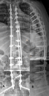
(A, B) Immediate post-operative X ray of case 1 after decompression and posterior instrumentation. (A) Antero posterior view, (B) Supine lateral view.
Figure 3
Figure 3
(A-C) MRI and X ray investigation after onset of neurological deterioration in case 1. (A) Sagittal MRI showing adequate decompression, (B) Supine AP X ray and (C) Supine lateral X ray of thoraco- lumbar spine.
Figure 4
Figure 4
(A-C) Retrospective analysis of the chance fracture. (A) Hairline fracture at posterior inferior corner of L1 vertebra, (B, C) Dynamic instability and failure at the LIV in sitting and supine position, (D) Intra operative image intensifier fracture displacement at L1 corner, secondary to prone position.
Figure 5
Figure 5
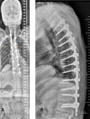
(A, B) Post-operative radiograph of extension of posterior instrumented fusion till pelvis, (A) AP view, (B) lateral view
Figure 6
Figure 6
Intra operative image (arrow) showing the discontinuity in the posterior elements, which was mobile with manipulation. (Case 1)
Figure 7
Figure 7
Pre-operative sagittal MRI of Case 2, showing canal stenosis at multi-level with cord changes maximum at D6-D7. (A) Coronal, (B, C) Sagittal view; (D) Axial view.
Figure 8
Figure 8
(A-B) Immediate supine post-operative radiograph of case 2 (A) AP view, (B) lateral view.
Figure 9
Figure 9
(A-D) MRI and X ray investigation after onset of neurological deterioration in case 2. (A) MRI showing adequate decompression, (B) AP view of thoraco- lumbar spine, (C) supine lateral view of spine showing change in the direction of LIV screw, so this was followed by, (D) sitting lateral view showing failure at LIV with kyphosis D12-L1.
Figure 10
Figure 10
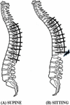
(A, B) demonstration of spine alignment (A) In supine position, anterior pelvic tilt with lumbar lordosis and fixed thoracic spine. (B) In sitting position, neutral pelvis with decrease in lumbar lordosis and no change in thoracic kyphosis due to instrumentation leading to stress at the LIV, resulting in failure.

References

    1. Denis F, Sun EC, Winter RB. Incidence and risk factors for proximal and distal junctional kyphosis following surgical treatment for Scheuermann kyphosis: minimum five-year follow-up. Spine (Phila Pa 1976) . 2009;34(20):E729–E734. doi: 10.1097/BRS.0b013e3181ae2ab2. - DOI - PubMed
    1. Lowe TG, Lenke L, Betz R, et al. Distal junctional kyphosis of adolescent idiopathic thoracic curves following anterior or posterior instrumented fusion: incidence, risk factors, and prevention. Spine (Phila Pa 1976) . 2006;31(3):299–302. doi: 10.1097/01.brs.0000197221.23109.fc. - DOI - PubMed
    1. Cho KJ, Lenke LG, Bridwell KH, Kamiya M, Sides B. Selection of the optimal distal fusion level in posterior instrumentation and fusion for thoracic hyperkyphosis: the sagittal stable vertebra concept. Spine (Phila Pa 1976) . 2009;34(8):765–770. doi: 10.1097/BRS.0b013e31819e28ed. - DOI - PubMed
    1. Cho KJ, Suk SI, Park SR, et al. Risk factors of sagittal decompensation after long posterior instrumentation and fusion for degenerative lumbar scoliosis. Spine (Phila Pa 1976) . 2010;35(17):1595–1601. doi: 10.1097/BRS.0b013e3181bdad89. - DOI - PubMed
    1. Hostin R, McCarthy I, O'Brien M, et al. Incidence, mode, and location of acute proximal junctional failures after surgical treatment of adult spinal deformity. Spine (Phila Pa 1976) . 2013;38(12):1008–1015. doi: 10.1097/BRS.0b013e318271319c. - DOI - PubMed