. 2021 Dec;20(12):2469-2482.
doi: 10.1158/1535-7163.MCT-20-0144. Epub 2021 Sep 22.

Targeting the IGF-Axis Potentiates Immunotherapy for Pancreatic Ductal Adenocarcinoma Liver Metastases by Altering the Immunosuppressive Microenvironment

Affiliations

Targeting the IGF-Axis Potentiates Immunotherapy for Pancreatic Ductal Adenocarcinoma Liver Metastases by Altering the Immunosuppressive Microenvironment

Masakazu Hashimoto et al. Mol Cancer Ther. 2021 Dec.

Abstract

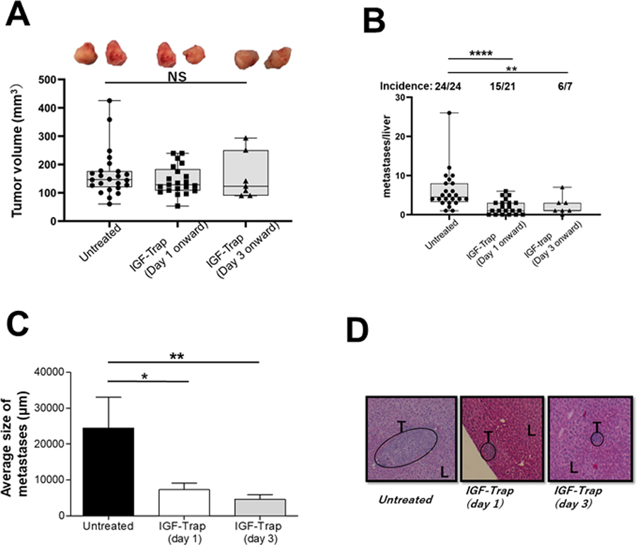
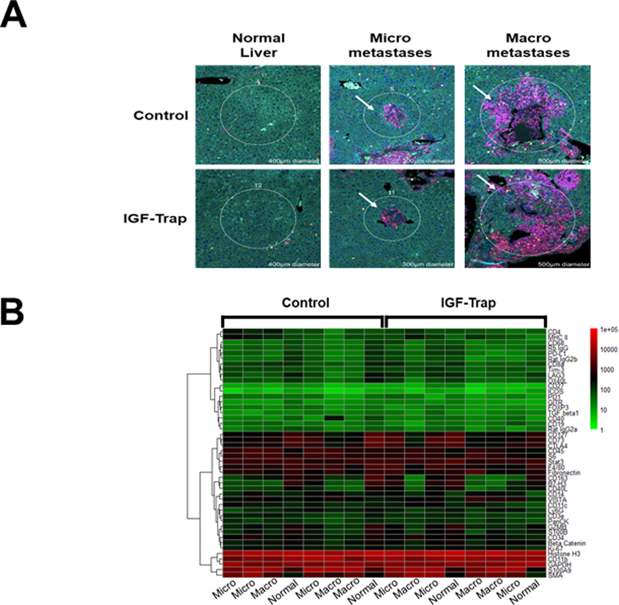
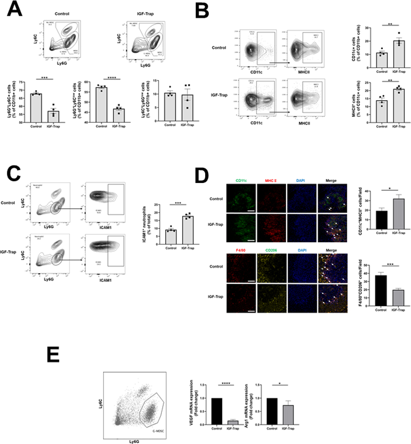
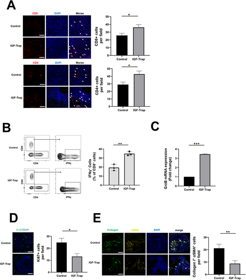
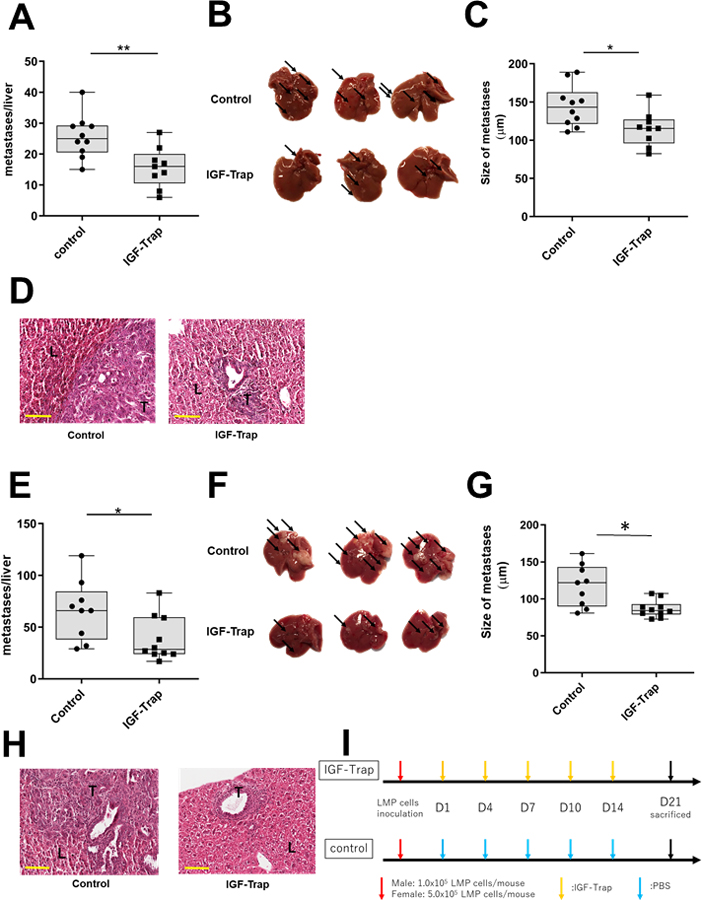
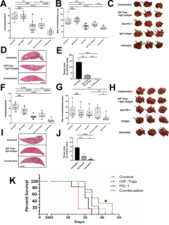
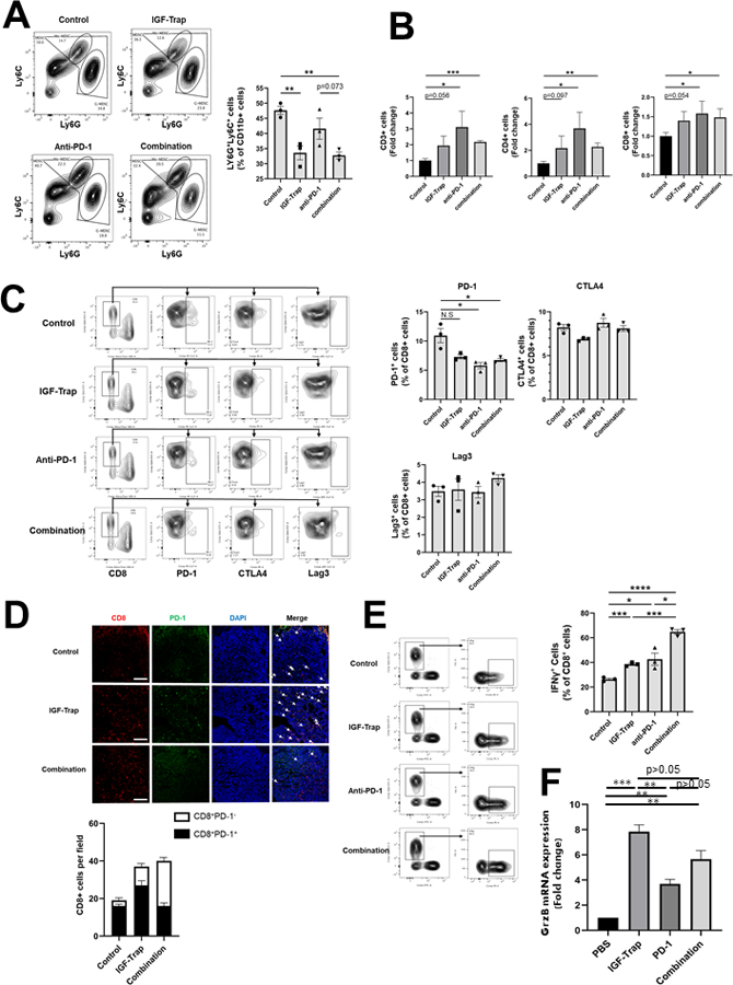
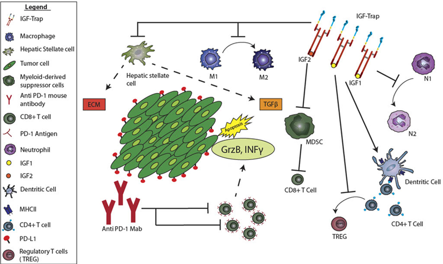
Pancreatic ductal adenocarcinoma (PDAC) is a highly aggressive malignancy, resistant to chemotherapy and associated with high incidence of liver metastases and poor prognosis. Using murine models of aggressive PDAC, we show here that in mice bearing hepatic metastases, treatment with the IGF-Trap, an inhibitor of type I insulin-like growth factor receptor (IGF-IR) signaling, profoundly altered the local, immunosuppressive tumor microenvironment in the liver, curtailing the recruitment of myeloid-derived suppressor cells, reversing innate immune cell polarization and inhibiting metastatic expansion. Significantly, we found that immunotherapy with anti-PD-1 antibodies also reduced the growth of experimental PDAC liver metastases, and this effect was enhanced when combined with IGF-Trap treatment, resulting in further potentiation of a T-cell response. Our results show that a combinatorial immunotherapy based on dual targeting of the prometastatic immune microenvironment of the liver via IGF blockade, on one hand, and reversing T-cell exhaustion on the other, can provide a significant therapeutic benefit in the management of PDAC metastases.


Conflict of interest statement

Competing interests: The authors declare no potential conflicts of interest.

Figures

Figure 1.
Figure 1.. The IGF-Trap preferentially inhibits the growth of liver metastases in an orthotopic PDAC model.
LMP cells (5x10⁵ in Matrigel) were implanted in the pancreas of immunocompetent, histocompatible B6/129F1 (B129) male mice. Treatment with 5mg/kg IGF-Trap 3.3 (or PBS) was initiated 1 (IGF-Trap (Day1)) or 3 (IGF-Trap (Day 3)) days later and continued on alternate days for a total of 5 injections per mouse. Animals were euthanized 3 weeks post tumor implantation, local pancreatic tumors measured and visible metastases on the surface of the liver enumerated. Results are based on pooled data from 4 independent experiments in which each treatment group consisted of 5–7 mice. Shown in (A) are the numbers of metastases per liver and (on top) the incidence of hepatic metastases per group. Shown in (B) are the average sizes of the metastases expressed as means (±SD) in each group and in (C) representative H&E stained PPFE sections of livers from each of the treatment groups. Shown in (D) are the volumes of individual local pancreatic tumors of the same mice calculated using the formula 1/2(length x width2) with 2 representative tumors from each of the treatment groups shown on top. Box and whiskers graphs: the box extends from the 25th to 75th percentiles, the middle line denotes de median and the whiskers extends from the minimum to the maximum value. *p ≤ 0.05; **P < 0.01; ****P < 0.0001; NS-not significant.
Figure 2.
Figure 2.. IGF-Trap treatment alters the tumor immune microenvironment in the liver.
Immune-profiling with the NanoString Geomax Profiler was performed on FFPE liver sections that were obtained from mice inoculated via the intrasplenic/portal route with 5x10⁵ LMP cells 21 days earlier and treated with a total of five tail vein injections of 5 mg/kg IGF-Trap or PBS (control) on alternate day. Shown in (A) are representative regions of interest (ROI) selected based on the size of the metastases to depict early (n=3) or late (n=3) immune cell recruitment events. A more detailed image of the ROI selected for analysis can be seen in Supplementary Figure 1A. Shown in (B) is a Heat map generated based on changes in the expression of immune cell surface markers expressed as log 10 (fold change) in expression relative to normal (tumor-free) liver that was used as baseline. Additional information on changes in specific cell surface markers can be seen in Supplementary Fig 1B.
Figure 3.
Figure 3.. The IGF-Trap reduces the accumulation of immunosuppressive cells in PDAC liver metastases.
Liver immune cells were isolated 14 days post intrasplenic/portal injection of 1x10⁵ LMP cells and five i.v. injections of 5 mg/kg IGF-Trap or PBS on alternate days, and immunostained with the indicated antibodies. Shown in (A-top) are representative flow cytometric contour plots obtained with each of the indicated immune cell populations that were first gated for size, viability, and CD45 expression (for detailed gating strategy, see Supplementary Fig. 3). Shown in the bar graphs (A-bottom) are mean proportions (±SEM) of MDSC, G-MDSC, and Mo-MDSC per liver based on 4 mice per group, analyzed individually. Shown in (B- left) are representative flow cytometric contour plots of activated dendritic cells identified based on expression CD11c and MHCII and in the bar graphs (B-right) the mean proportions (±SEM) of CD11b+MHCII+ per liver based on 4 mice per group, analyzed individually. Shown in (C-left) are representative contour plots obtained for CD11b+Ly6GhighLy6Clow cells expressing ICAM-1 and in the bar graph (C-right) the mean proportions (±SEM) of Ly6G+ICAM-1+ cells (markers of N1 neutrophils) in each group based on analysis of these mice. In a separate experiment, the same treatment protocol was used, and cryostat sections prepared for analysis by IHC. Shown in (D-left top) are representative confocal images of 10 μm cryostat liver sections immunostained with the indicated antibodies followed by Alexa Fluor 568 (green) for CD11c, Alexa Fluor 647 for MHCII (red), and DAPI (blue). Shown on the right of the confocal images are the mean numbers (±SEM) of the indicated cells per field counted in 15–20 fields per section (n=2 or 3) derived from 3 mice per group. Shown in (D left-bottom) are representative confocal images obtained with the indicated antibodies followed by Alexa Fluor 568 (red) for F4/80, Alexa Fluor 647 (yellow) for CD206, and DAPI (blue). Shown on the right of the confocal images are the means (± SEM) of the indicated cells counted as in (D-top). MDSC were functionally characterized using a T cell proliferation assay. Shown in (E) are results of qPCR analysis performed on RNA extracted from FACS sorted neutrophils (left). Results in the bar graph are based on 3 separate analyses and expressed as means (± SD) expression levels (normalized to GAPDH) relative to untreated mice that were assigned a value of 1. Scale bar (D) - 100μm; *p ≤ 0.05; **P < 0.01; ***P < 0.001; ****P < 0.0001.
Fig. 4.
Fig. 4.. IGF-Trap treatment enhances T cell recruitment and potentiates their function in the TME.
B6129 F1 mice were injected via the intrasplenic/portal route with 1x10⁵ LMP cells, treated with 5 mg/kg IGF-Trap or PBS 1, 4, 7, and 10 days post tumor inoculation and sacrificed on day 14. Shown in (A) are representative confocal images of 10 μm cryostat liver sections immunostained with the indicated antibodies and Alexa Fluor 568 (red) for CD8 (A-top), Alexa Fluor 568 (red) for CD4 (A-bottom) and DAPI (blue). Shown in the bar graphs (right) are the mean numbers (± SEM) of the indicated cells per field counted in 15–20 fields per section derived from 3 mice per group. Shown in (B-left) are results of flow cytometry performed on immune cells which isolated from mice treated as described in (A) and stimulated for 4 h with PMA and ionomycin in the presence of a protein transport inhibitor. Shown in the bar graph (B-right) are the mean proportions of CD8+IFN-γ+ cells per liver (±SEM) based on 3 livers per group analyzed individually. Shown in (C) are results of qPCR (±SEM) performed on RNA extracted from CD3+CD8+ T cells that were FACS sorted from livers of tumor-injected mice treated as in (A) (data normalized to GAPDH; n=3), in (D-left) representative confocal images of cells immunostained with antibodies to Ki-67 (green) and with DAPI (blue) and in (D-right) the mean numbers (±SEM) of Ki67+ cells per field based on 15–20 sections obtained from 3 animals per group. The effect of IGF-Trap on hepatic stellate cell activation (E) was analyzed in B6129-Col-GFP mice. Mice were injected via the intrasplenic/portal route with 1x10⁵ LMP cells, treated with 5 mg/kg IGF-Trap or PBS on days 2 and 4 post tumor inoculation and sacrificed on day 7. Activated HSC recruited into tumor-infiltrated areas and identified based on type I collagen production and α-SMA expression were quantified. Shown in (E-left) are representative confocal images of 10 μm cryostat liver sections immunostained with antibodies to α-SMA followed by Alexa Fluor 568 secondary antibody (yellow), DAPI- stained (blue) and expressing Col-GFP (green). Shown in the bar graph (E-right) are the mean numbers of activated HSCs per field (±SEM) based on 15–20 sections obtained from 3 animals per group. Scale bars (A, D) - 100 μm; (E) - 50 μm; *p ≤ 0.05; **P < 0.01; ***P < 0.001.
Figure 5.
Figure 5.. The IGF-Trap inhibits the growth of pancreatic carcinoma liver metastases.
Experimental liver metastases were generated by inoculation of 1x10⁵ (for males, A-D) or 5x10⁵ (for females, E-H) LMP cells via the intrasplenic/portal route. The number of mice injected was 10 (vehicle) and 9 (IGF-Trap) male mice and 9 (vehicle) and 10 (IGF-Trap) female mice. Treatment with 5 mg/kg IGF-Trap was initiated 1 day later and continued twice weekly for a total of 5 injections per mouse (I). Mice were sacrificed 21 days post tumor inoculation and visible metastases on the surface of the liver enumerated prior to fixation. Results are based on 2 experiments each for male and female mice. Shown in (A&E) are the numbers of metastases per each liver. Shown in (B&F) are representative livers from each group where arrows denote visible metastases. Shown in (C&G) are the mean diameters of metastases per liver and in (D&H) representative images of H&E stained FFPE liver sections (n=9). Scale bar corresponds to 100 µm. Box and whiskers graphs: the box extends from the 25th to 75th percentiles, the middle line denotes de median and the whiskers extends from the minimum to the maximum value. T-tumor; L-liver; *p < 0.05; **p < 0.01.
Figure 6.
Figure 6.. Combinatorial IGF-Trap/anti PD-1 therapy further decreases the number of liver metastases and prolongs survival.
Experimental liver metastases were generated by the inoculation of 1x10⁵ (for males, A-E), and 5x10⁵ (for females, F-J) LMP cells via the intrasplenic/portal route. Treatment with 5 mg/kg IGF-Trap (or PBS) i.v. was initiated 1 day post tumor inoculation and continued twice weekly for a total of 5 injections per mouse. Treatment with 10 mg/kg PD-1 or the IgG isotype control was administered i.p. on alternate days for a total of 5 injections per mouse (see supplementary Fig. 6). A total of 9–10 male mice and 9–12 female mice were injected per treatment group. Mice were sacrificed on day 21 and visible liver metastases enumerated without prior fixation. Results are based on 2 experiments each, for male and female mice. Shown in (A&F) are the numbers of metastases counted per liver and in (B&G) the mean diameters of the visible metastases per liver. There were no significant differences in either the numbers or sizes of metastases between untreated mice and mice treated with the IgG isotype control. Shown in (C&H) are representative livers from each group where arrows denote metastases. Shown in (D&I) are representative images of H&E stained FFPE liver sections where metastases are encircled and in (E&J) the means of total tumor surface area per section (±SEM) expressed as % of total liver surface area and based on analysis of 9 sections per group (Scale bar – 100 μM). *p ≤ 0.05; **P < 0.01; ***P < 0.001; ****P < 0.0001; NS-not significant. To test the effect of the combinatorial therapy on survival, syngeneic B6.129 F1 female mice were injected via the intrasplenic/portal route with 1x10⁵ LMP tumor cells (5 mice per group) and treatment with 5 mg/kg IGF-Trap (or vehicle) i.v. was initiated 1 day post tumor inoculation and continued twice weekly. Treatment with 10 mg/kg anti PD-1 antibody i.p was administered on alternate days. The treatments continued for a total of 5 weeks or until animals were moribund and euthanized. Shown in (K) is a Kaplan Meier plot for each of the treatment groups. *p< 0.05 as assessed by the Gehan-Breslow-Wilcoxon Test.
Figure 7.
Figure 7.. Increased T cell recruitment and decreased PD-1 expression in the liver TME following combinatorial therapy.
Liver immune cells were isolated 14 days post intrasplenic/portal injection of 1x10⁵ LMP cells and the treatment protocol described in the legend to Fig 6 and analyzed by flow cytometry. Shown in (A-left) are representative flow cytometric contour plots obtained with each of the indicated immune cells populations that were first gated as outlined in Supplementary Fig. 3, and in the bar graphs (A-right) the mean proportions (%) (±SEM) of MDSC per liver based on 3 mice per group, analyzed individually. Shown in (B) are the mean numbers of the indicated cells obtained per liver (±SEM) expressed as a ratio to untreated mice that were assigned a value of 1 and based on 3 mice per group, analyzed individually. Shown in (C) are representative contour plots obtained for CD8+ cells immunostained with antibodies to the indicated immune checkpoints and in the bar graphs the relative proportions (%) of positive T cells (±SEM) based on the analyses of 3 livers per group. Shown in (D-top) are representative confocal images of 10 μm cryostat liver sections immunostained with the indicated antibodies followed by Alexa Fluor 568 (red) for CD8, Alexa Fluor 647 for PD-1 (green), and DAPI (blue) and in (D-bottom) are the mean numbers (±SEM) of the CD8+ T cells per field (expressing or not PD-1), counted in 15–20 fields per section derived from 3 mice per group. Note that the number of CD8+ T cells increased significantly in both IGF-Trap and combination therapy-treated mice but the ratio of PD-1+:CD8+ cell decreased significantly only in mice that received the combination therapy. Shown in (E-left) are results of flow cytometry performed on immune cells which were isolated from mice treated as in (A) and stimulated for 4 h as described in the legend to Fig 4. Shown in the bar graph (E-right) are the mean proportions of CD8+IFN-γ+ cells per liver (±SEM) based on 3 livers per group, analyzed individually. Scale bar- 100μm; Shown in (F) are results of qPCR (±SEM) performed on RNA extracted from CD3+CD8+ T cells that were FACS sorted from livers of tumor-injected mice treated as in (A) (data normalized to GAPDH; n=3). *p ≤ 0.05; **P < 0.01; ***P < 0.001; ****P < 0.0001; NS-not significant.
Figure 8.
Figure 8.. The combined effect of the IGF-Trap and anti PD-1 antibodies on the tumor microenvironment in the liver.
Shown is a diagrammatic representation of the effects of the combinatorial immunotherapy on the TME of PDAC liver metastases. The IGF-Trap reduces TAM and neutrophil polarization, MDSC accumulation and HSC activation, while also enhancing DC activation and enabling antigen presentation. This results in increased accumulation of CD8+ T cells in the TME. Anti PD-1 antibodies reduce T cell exhaustion and this is associated with potentiation of CD8+ T cell activity as reflected in increased IFNγ and GrzB production and results in increased tumor cell death.

Similar articles

Cited by

References

    1. Ferlay J, Shin HR, Bray F, Forman D, Mathers C, Parkin DM. Estimates of worldwide burden of cancer in 2008: GLOBOCAN 2008. Int J Cancer 2010;127(12):2893–917. - PubMed
    1. Siegel RL, Miller KD, Jemal A. Cancer statistics, 2015. CA Cancer J Clin 2015;65(1):5–29. - PubMed
    1. Siegel RL, Miller KD, Jemal A. Cancer statistics, 2018. CA Cancer J Clin 2018;68(1):7–30. - PubMed
    1. Rahib L, Smith BD, Aizenberg R, Rosenzweig AB, Fleshman JM, Matrisian LM. Projecting cancer incidence and deaths to 2030: the unexpected burden of thyroid, liver, and pancreas cancers in the United States. Cancer Res 2014;74(11):2913–21. - PubMed
    1. Tempero MA, Malafa MP, Behrman SW, Benson AB 3rd, Casper ES, Chiorean EG, et al. Pancreatic adenocarcinoma, version 2.2014: featured updates to the NCCN guidelines. J Natl Compr Canc Netw 2014;12(8):1083–93. - PubMed

Publication types

Substances