. 2021 Sep 15:14:4615-4628.
doi: 10.2147/JIR.S326416. eCollection 2021.

The Effect of Shaoyao Gancao Decoction on Sphincter of Oddi Dysfunction in Hypercholesterolemic Rabbits via Protecting the Enteric Nervous System-Interstitial Cells of Cajal-Smooth Muscle Cells Network

Affiliations

The Effect of Shaoyao Gancao Decoction on Sphincter of Oddi Dysfunction in Hypercholesterolemic Rabbits via Protecting the Enteric Nervous System-Interstitial Cells of Cajal-Smooth Muscle Cells Network

Gui-Ying Zhu et al. J Inflamm Res. .

Abstract

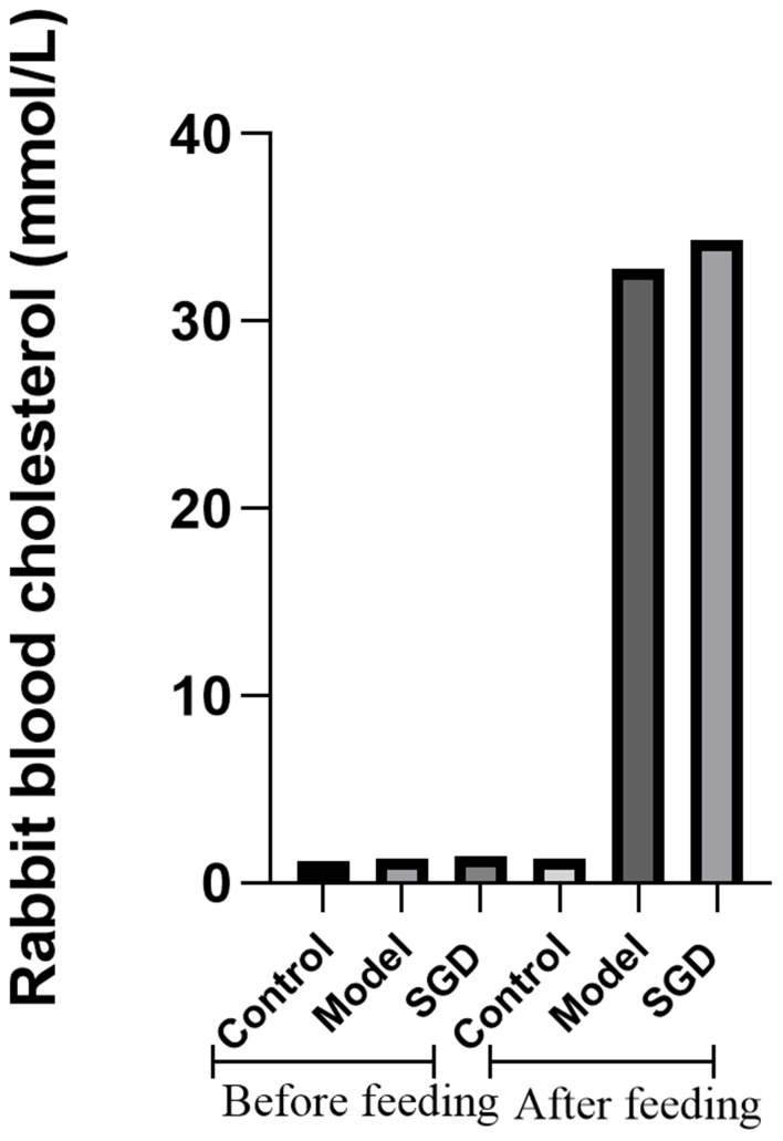
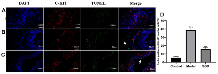
Objective: This study observes the morphological changes in the enteric nervous system (ENS) - interstitial cells of Cajal (ICC) - smooth muscle cells (SMC) network in sphincter of Oddi dysfunction (SOD) in hypercholesterolemic rabbits following treatment with Shaoyao Gancao decoction (SGD), as well as the apoptosis of the ICC.

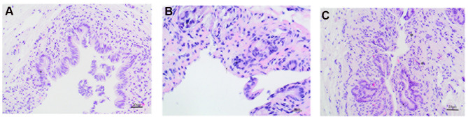
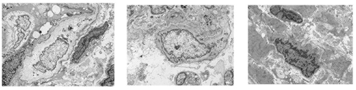
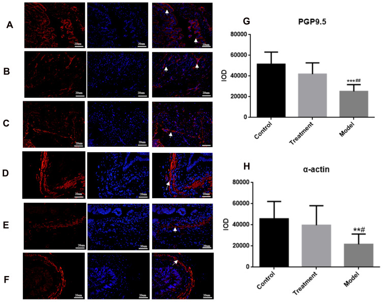
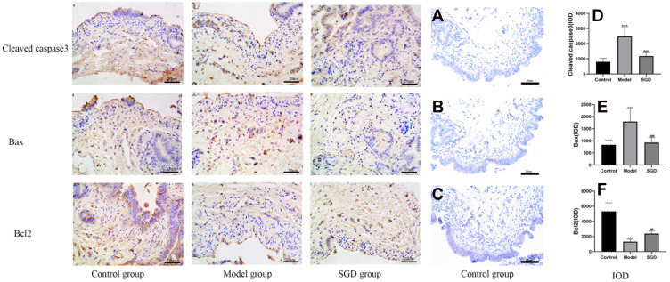
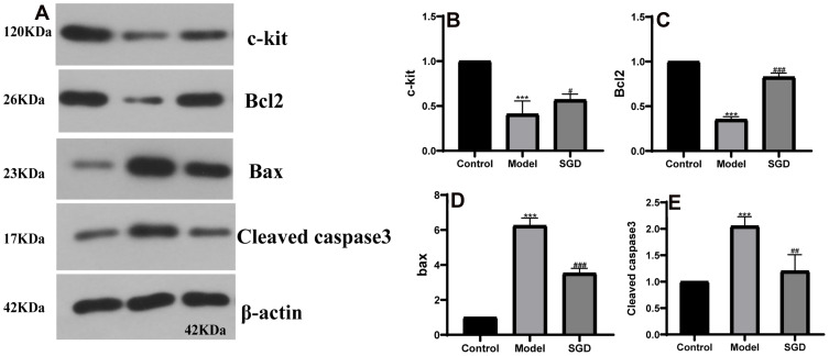
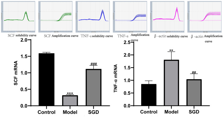
Methods: In this study, 48 healthy adult New Zealand rabbits are randomly divided into three groups (n = 16 in each group): the control, the model, and the SGD treatment groups. The hypercholesterolemic rabbit model is established. Hematoxylin and eosin staining, transmission electron microscopy, immunofluorescence, terminal deoxynucleotidyl transferase dUTP nick end labeling staining, immunohistochemistry, Western blot analysis, and reverse transcription-polymerase chain reaction are used to detect the morphological changes in the ENS-ICC-SMC network, the expression of apoptosis-related proteins in the ICC, and to observe the curative effect of SGD after treatment.

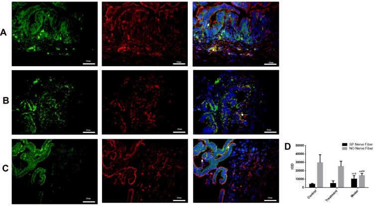
Results: Compared with the control group, the morphology and the ultrastructure of the SO are destroyed in the model group. In addition, the protein gene product 9.5 (PGP9.5), nitric oxide (NO), the SMCs, and the ICC all significantly decreased while substance P (SP) significantly increased. Compared with the model group, the SO morphology and ultrastructure are repaired in the SGD group. In addition, the PGP9.5, NO, the SMCs, and the ICC significantly increased while SP decreased. In addition, SGD may activate the stem cell factor (SCF)/c-Kit signaling pathway to treat SO dysfunction by up-regulating the expression of c-Kit and SCF. Similarly, this pathway restores SO by up-regulating the expression of Bcl2 and inhibiting cleaved caspase-3, Bax, and the tumor necrosis factor.

Conclusion: Shaoyao Gancao decoction can promote the recovery of sphincter of Oddi dysfunction in hypercholesterolemic rabbits by protecting the ENS-ICC-SMC network.

Keywords: ENS–ICC–SMC; Shaoyao Gancao Decoction; apoptosis; sphincter of Oddi dysfunction.


Conflict of interest statement

The authors report no conflicts of interest in this work.

Figures

Figure 1
Figure 1
Serum total cholesterol concentration of the rabbits before and after modeling.
Figure 2
Figure 2
Hematoxylin and eosin staining of rabbit SO tissue (400×, 20 um), (A) Normal group, (B) Model group, (C) Treatment group. The arrow (←) refers to mucosal epithelial cells, the (*) refers to inflammatory cells infiltration.
Figure 3
Figure 3
Rabbit SO tissue electron microscopy (1.0 um), (A) Normal group, (B) Model group, (C) Treatment group. The definitions of the abbreviations in the picture are as follows: interstitial cells of Cajal (ICC), smooth muscle cells (SMC), nucleus (N), nucleolus (Nu), mitochondria (M), rough endoplasmic reticulum (RER), collagen fibers (CF), dense spots (DPs), and dense bodies (DBs).
Figure 4
Figure 4
(A) Normal group, (B) Model group, (C) Treatment group: Rabbit SO tissue, nerve fiber PGP9.5 in the inner muscular layer detected by laser confocal microscopy, red light is PGP9.5, blue light is DAPI. (D) Normal group, (E) Model group, (F) SGD treatment group: α-actin labeled rabbit SO tissues were examined by laser confocal microscopy (400×, 20 um), with α-actin in red light and DAPI in blue light. (G) fluorescence intensity of PGP9.5 (IOD) in the SO tissue of rabbits in each group; (H) The fluorescence intensity (IOD) of α-actin labeled SMC in the SO tissue of rabbits in each group. Arrows indicate positive expression.*** indicates the comparison between the model group and the control group, P<0.05, ##indicates the comparison between the model group and the treatment group, P<0.05. In α-action, ** indicates the comparison between the model group and the control group, P<0.05, # indicates the comparison between the model group and the treatment group, P<0.05.
Figure 5
Figure 5
(A) Normal group, (B) Model group, (C) Treatment group: Rabbit SO tissue, nerve fiber in the inner muscular layer detected by laser confocal microscopy, SP and n-NOS respectively labeled SP nerve fibers (green light) and NO nerve fibers (red light) in rabbit SO tissue. The blue light is DAPI. Arrows indicate positive expression. ** indicates that the model group is compared with the control group, p<0.05, # indicates that the model group is compared with SGD group,p<0.05.In NO, ** indicates that the model group is compared with the control group, p<0.05, ## indicates that the model group is compared with SGD group, p<0.05.
Figure 6
Figure 6
Results of ICC and TUNEL fluorescence staining in the SO tissue of rabbits (400×, 20 um); Blue is DAPI staining, red is c-kit positive ICC cell staining, green is TUNEL apoptotic cell staining. (A) Normal group, (B) Model group, (C) Treatment group, (D) ICC double positive expression rate. The model group vs the control group, ***P ≤ 0.001. The SGD group vs the model group, ###P ≤ 0.001. Arrows indicate positive expression.
Figure 7
Figure 7
Immunohistochemical results of cleaved caspase-3, Bax, and Bcl2 in SO tissue (400×); a, b, and c: cleaved caspase-3 immunohistochemical results in the normal group, model group, and SGD treatment group, respectively; d, e, and f: Bax immunohistochemical results in the normal group, model group, and treatment group, respectively; g, h, and i: Bcl2 immunohistochemical results of the normal group, the model group, and the treatment group, respectively. The brown or brown cytoplasm is the expression of the corresponding positive protein (*). (AC) The negative controls for cleaved caspase-3, Bax, and Bcl2 using control samples, respectively. (DF) Respectively fluoresced caspase-3, Bax, and Bcl2 (IOD) in the SO tissue of rabbits in each group. The model group vs the control group, ***P ≤ 0.001. The SGD group vs the model group,##P≤0.01, ###P ≤ 0.001.
Figure 8
Figure 8
(A) the protein band; (BE) the protein results of c-kit, Bcl2, Bax, and cleaved caspase-3 in rabbit SO tissue, respectively. The model group vs the control group, ***P ≤ 0.001, the SGD group vs the model group, #P ≤ 0.05, ##P ≤ 0.01, ###P ≤ 0.001.
Figure 9
Figure 9
(A and B) the results of SCF and TNF-α mRNA in the SO tissue of rabbits in each group. The control group vs the model group, **P ≤ 0.01, ***P ≤ 0.001. The SGD group vs the model group ##P ≤ 0.01, ###P = 0.001 or less.

References

    1. Leung WD, Sherman S. Endoscopic approach to the patient with motility disorders of the bile duct and sphincter of Oddi. Gastrointest Endosc Clin N Am. 2013;23(2):405–434. doi:10.1016/j.giec.2012.12.006 - DOI - PubMed
    1. Crittenden JP, Dattilo JB. StatPearls (StatPearls Publishing Copyright © 2021. StatPearls Publishing LLC.; 2021.
    1. Behar J, Corazziari E, Guelrud M, Hogan W, Sherman S, Toouli J. Functional gallbladder and sphincter of oddi disorders. Gastroenterology. 2006;130(5):1498–1509. doi:10.1053/j.gastro.2005.11.063 - DOI - PubMed
    1. Takayama I, Daigo Y, Ward SM, Sanders KM, Yamanaka T, Fujino MA. Differential gene expression in the small intestines of wildtype and W/W(V) mice. Neurogastroenterol Motil. 2001;13(2):163–168. doi:10.1046/j.1365-2982.2001.00256.x - DOI - PubMed
    1. Müller M, Colcuc S, Drescher DG, et al. Murine genetic deficiency of neuronal nitric oxide synthase (nNOS(-/-)) and interstitial cells of Cajal (W/W(v)): implications for achalasia? J Gastroenterol Hepatol. 2014;29(10):1800–1807. doi:10.1111/jgh.12600 - DOI - PubMed