. 2021 Sep 3:12:736627.
doi: 10.3389/fphar.2021.736627. eCollection 2021.

Albuca Bracteate Polysaccharides Synergistically Enhance the Anti-Tumor Efficacy of 5-Fluorouracil Against Colorectal Cancer by Modulating β-Catenin Signaling and Intestinal Flora

Affiliations

Albuca Bracteate Polysaccharides Synergistically Enhance the Anti-Tumor Efficacy of 5-Fluorouracil Against Colorectal Cancer by Modulating β-Catenin Signaling and Intestinal Flora

Xinyu Yuan et al. Front Pharmacol. .

Abstract

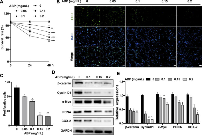
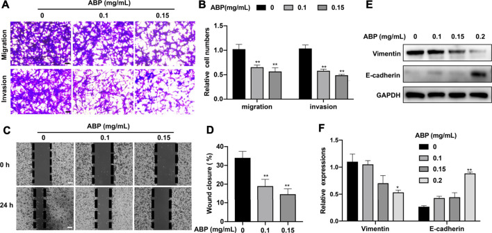
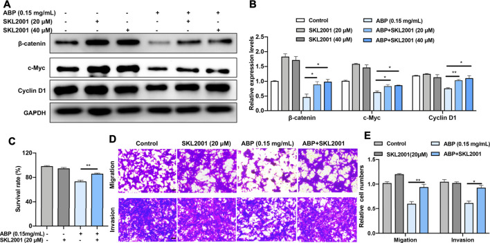
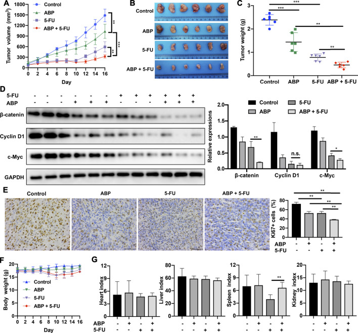
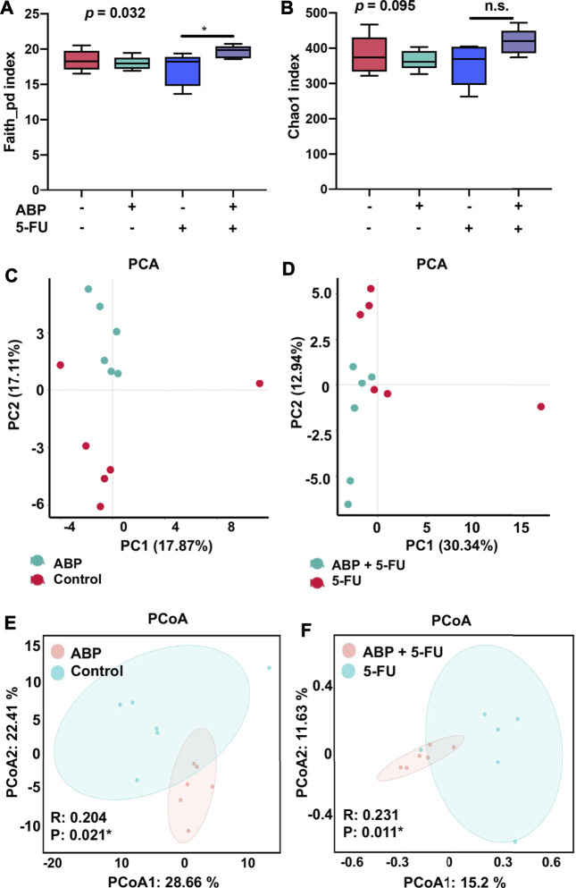
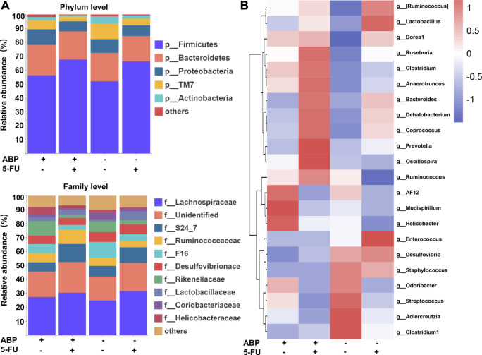
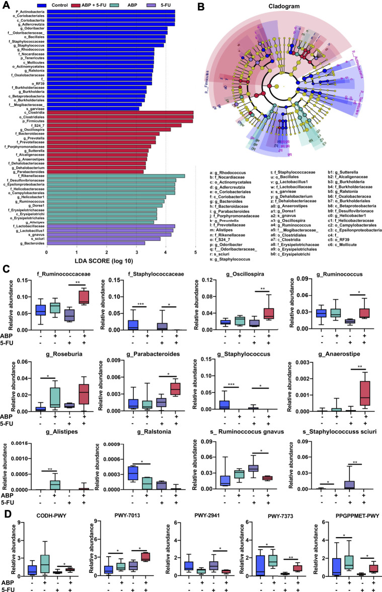
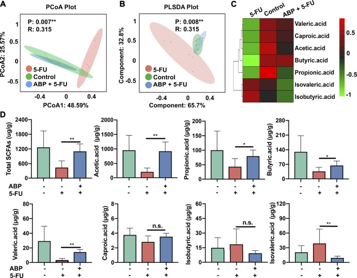
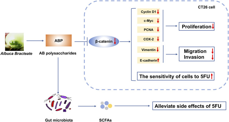
The first-line treatment for colorectal cancer (CRC) is 5-fluorouracil (5-FU). However, the efficacy of this treatment is sometimes limited owing to chemoresistance as well as treatment-associated intestinal mucositis and other adverse events. Growing evidence suggests that certain phytochemicals have therapeutic and cancer-preventing properties. Further, the synergistic interactions between many such plant-derived products and chemotherapeutic drugs have been linked to improved therapeutic efficacy. Polysaccharides extracted from Albuca bracteata (Thunb.) J.C.Manning and Goldblatt (ABP) have been reported to exhibit anti-oxidant, anti-inflammatory, and anti-tumor properties. In this study, murine CRC cells (CT26) and a murine model of CRC were used to examine the anti-tumor properties of ABP and explore the mechanism underlying the synergistic interactions between ABP and 5-FU. Our results revealed that ABP could inhibit tumor cell proliferation, invasion, and migratory activity in vitro and inhibited tumor progression in vivo by suppressing β-catenin signaling. Additionally, treatment with a combination of ABP and 5-FU resulted in better outcomes than treatment with either agent alone. Moreover, this combination therapy resulted in the specific enrichment of Ruminococcus, Anaerostipes, and Oscillospira in the intestinal microbiota and increased fecal short-chain fatty acid (SCFA) levels (acetic acid, propionic acid, and butyric acid). The improvement in the intestinal microbiota and the increase in beneficial SCFAs contributed to enhanced therapeutic outcomes and reduced the adverse effects of 5-FU. Together, these data suggest that ABP exhibits anti-neoplastic activity and can effectively enhance the efficacy of 5-FU in CRC treatment. Therefore, further research on the application of ABP in the development of novel anti-tumor drugs and adjuvant compounds is warranted and could improve the outcomes of CRC patients.

Keywords: 5-fluorouracil; colorectal cancer; gut microbiota; polysaccharide; short-chain fatty acids; β-catenin.


Conflict of interest statement

The authors declare that the research was conducted in the absence of any commercial or financial relationships that could be construed as a potential conflict of interest.

Figures

FIGURE 1
FIGURE 1
ABP treatment suppresses the proliferation of CRC cells. (A) CCK-8 assay examining the proliferation-suppressing effect of ABP on CT26 cells. (B–C) EdU uptake assay examining the proliferation-suppressing effect of ABP on CT26 cells. Scale bar = 50 μm (×200 magnification). (D–E) Western blot examining the effect of ABP on the protein levels of β-catenin, Cyclin D1, c-Myc, PCNA, COX-2, and GAPDH in CT26 cells. Compared with the untreated group, *p < 0.05, **p < 0.01.
FIGURE 2
FIGURE 2
ABP treatment suppresses the migratory and invasive activity of CRC cells. (A-B) Transwell assay examining the negative effect of ABP on the migration and invasion of CT26 cells. Scale bar = 50 μm. (C-D) Wound healing assay examining the negative effect of ABP on the migration and invasion of CT26 cells. Scale bar = 250 μm. (E-F) Western blot examining the effect of ABP on the protein levels of Vimentin, E-cadherin, and GAPDH in CT26 cells. Compared with the untreated group, *p < 0.05, **p < 0.01.
FIGURE 3
FIGURE 3
ABP treatment suppresses the growth and metastasis of CRC cells by downregulating β-catenin. (A–B) Western blot examining the effects of SKL 2001 (β-catenin activator) on β-catenin, c-Myc, and Cyclin D1 levels in CT26 cells treated with ABP. (C) CCK-8 assay examining the rescue effects of SKL2001 treatment on CT26 cells treated with ABP. (D–E) Transwell assay examining the rescue effects of SKL2001 treatment on CT26 cells treated with ABP. Scale bar = 50 μm. Compared with the ABP group, *p < 0.05, **p < 0.01.
FIGURE 4
FIGURE 4
ABP enhances the anti-tumor efficacy of 5-FU against CT26 cells. (A-B) CCK-8 assay examining the effects of XAV939 (β-catenin inhibitor) and ABP on the anti-tumor efficacy of 5-FU against CT26 cells. (C) Cellular morphology of CT26 cells treated with ABP, 5-FU, and ABP + 5-FU for 24 h. Scale bar = 50 μm. (D) Transwell assays examining the enhancing effect of ABP on the anti-tumor efficacy of 5-FU against CT26 cells. Scale bar = 50 μm. (E) Wound healing assay examining the enhancing effect of ABP on the anti-tumor efficacy of 5-FU against CT26 cells. Scale bar = 250 μm. (F) Western blot examining the protein levels of β-catenin, Cyclin D1, c-Myc, COX-2, PCNA, Vimentin, E-cadherin, an GAPDH in CT26 cells treated with ABP and 5-FU. Compared with the 5-FU group, *p < 0.05, **p < 0.01, n. s., no significance.
FIGURE 5
FIGURE 5
ABP exhibits synergistic anti-tumor effects when administered in combination with 5-FU in mice bearing CT26 tumors. (A) Tumor volumes measured at the indicated time points. Data are shown as mean ± SD (n = 6 mice at each time point). (B) Image of tumors resected from sacrificed mice at the end of the treatment period. (C) Tumor weights measured after surgical tumor removal, represented as mean ± SD. (D) Western blot examining the levels of β-catenin, Cyclin D1, and c-Myc in subcutaneous tumors, with GAPDH used as the internal control. (E) Images of Ki67 immunohistochemical staining (× 400) in tumor cells and quantification of Ki67-positive tumor cells. Scale bar = 50 μm. (F) Body weights measured every 2 days. (G) Organ indexes calculated as the ratio of organ weight (mg) to body weight (g) at the end of the treatment period. Data are means ± SD (n = 6). *p < 0.05, **p < 0.01, ***p < 0.001.
FIGURE 6
FIGURE 6
ABP treatment alters the abundance and diversity of gut microbes in mice bearing CT26 tumors. (A-B) Analysis of alpha diversity; (A) Faith_pd index. (B) chao1 index. (C-F) Analysis of beta diversity. (C-D) Principal components analysis (PCA). (E-F) principal coordination analysis (PCoA) (n = 6 mice per group).
FIGURE 7
FIGURE 7
ABP treatment improves the composition of the intestinal microflora. ( A ) Bar plots of bacterial taxa present in the feces at the phylum and family levels based on relative abundance. ( B ) Heatmap of bacterial taxa present in the feces at the genus level based on relative abundance.
FIGURE 8
FIGURE 8
ABP treatment leads to the enrichment of beneficial bacteria and influences metabolic pathways. ( A-B ) Bacterial taxa differences among the four groups, observed using LEfSe analysis. (C) Box plots demonstrating the characteristic bacteria at the family, genus, and species levels. (D) Difference in functional metabolic pathways among the groups. *p < 0.05, **p < 0.01, ***p < 0.001.
FIGURE 9
FIGURE 9
ABP treatment improves the concentrations of fecal short-chain fatty acids in CRC model mice. (A-B) PCoA and PLS-DA analysis. (C-D) Heatmap and Bar plots for fecal SCFA content. *p < 0.05, ** represents p < 0.01.
FIGURE 10
FIGURE 10
Schematic diagram depicting the mechanism of action underlying the effect of ABP as a regulator of CRC progression. ABP suppresses CRC cell proliferation, migration, and invasion by influencing the Wnt/β-catenin signaling pathway. ABP treatment further increases the sensitivity of CRC cells to 5-FU and thereby decreases the incidence of 5-FU-related adverse effects. ABP, Albuca Bracteate polysaccharides; CRC, colorectal cancer.

References

    1. Bäckhed F., Ley R. E., Sonnenburg J. L., Peterson D. A., Gordon J. I. (2005). Host-bacterial Mutualism in the Human Intestine. Science 307 (5717), 1915–1920. 10.1126/science.1104816 - DOI - PubMed
    1. Cai B., Pan J., Chen H., Chen X., Ye Z., Yuan H., et al. (2021). Oyster Polysaccharides Ameliorate Intestinal Mucositis and Improve Metabolism in 5-Fluorouracil-Treated S180 Tumour-Bearing Mice. Carbohydr. Polym. 256, 117545. 10.1016/j.carbpol.2020.117545 - DOI - PubMed
    1. Calvert P. M., Frucht H. (2002). The Genetics of Colorectal Cancer. Ann. Intern. Med. 137 (7), 603–612. 10.7326/0003-4819-137-7-200210010-00012 - DOI - PubMed
    1. Chen R., Li Y., Dong H., Liu Z., Li S., Yang S., et al. (2012). Optimization of Ultrasonic Extraction Process of Polysaccharides from Ornithogalum Caudatum Ait and Evaluation of its Biological Activities. Ultrason. Sonochem. 19 (6), 1160–1168. 10.1016/j.ultsonch.2012.03.008 - DOI - PubMed
    1. Cho Y. H., Ro E. J., Yoon J. S., Mizutani T., Kang D. W., Park J. C., et al. (2020). 5-FU Promotes Stemness of Colorectal Cancer via P53-Mediated WNT/β-catenin Pathway Activation. Nat. Commun. 11 (1), 5321. 10.1038/s41467-020-19173-2 - DOI - PMC - PubMed

LinkOut - more resources