. 2021 Sep 6:12:735643.
doi: 10.3389/fimmu.2021.735643. eCollection 2021.

Tissue-Resident-Memory CD8+ T Cells Bridge Innate Immune Responses in Neighboring Epithelial Cells to Control Human Genital Herpes

Affiliations

Tissue-Resident-Memory CD8+ T Cells Bridge Innate Immune Responses in Neighboring Epithelial Cells to Control Human Genital Herpes

Tao Peng et al. Front Immunol. .

Abstract

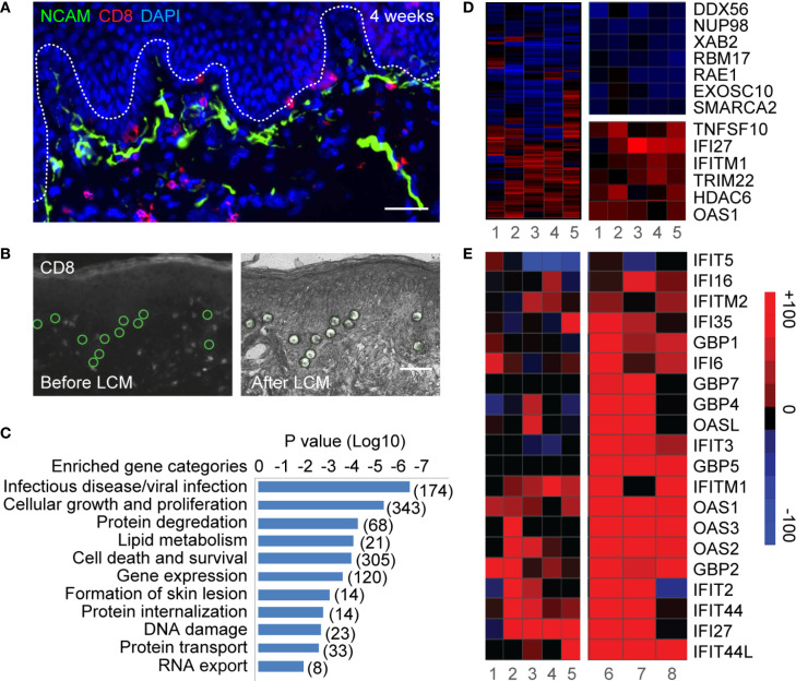
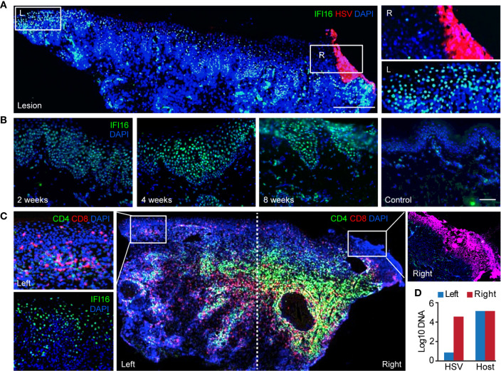
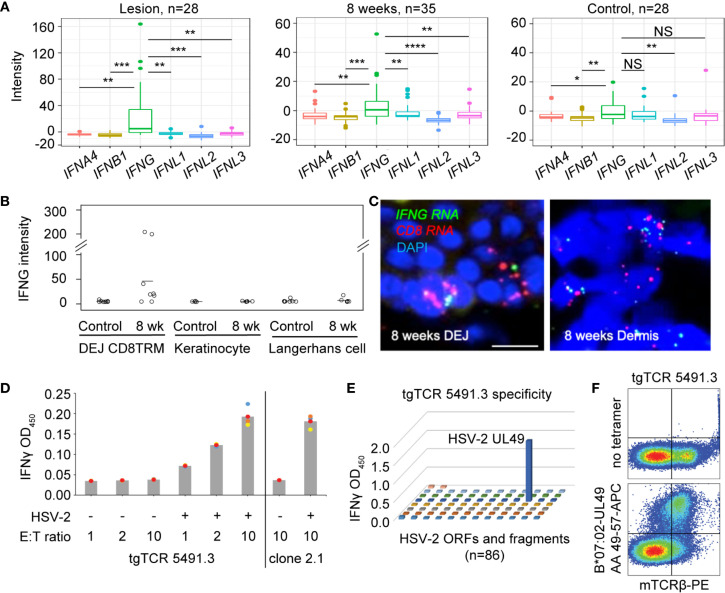
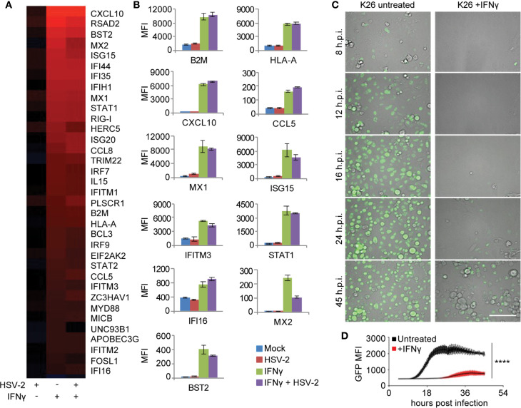
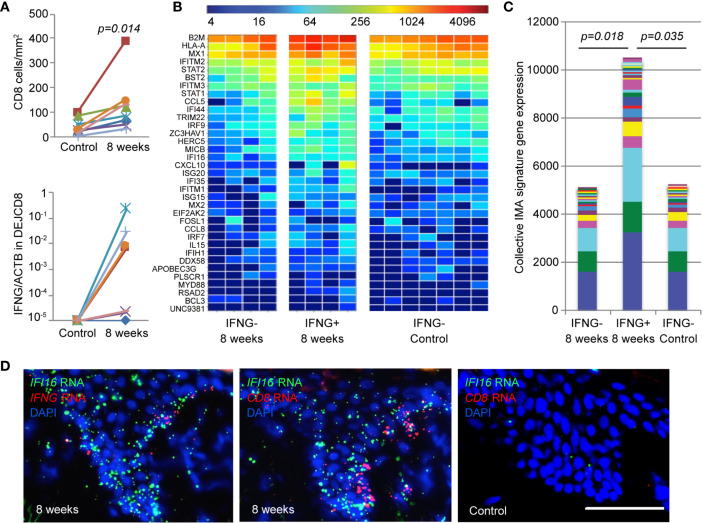
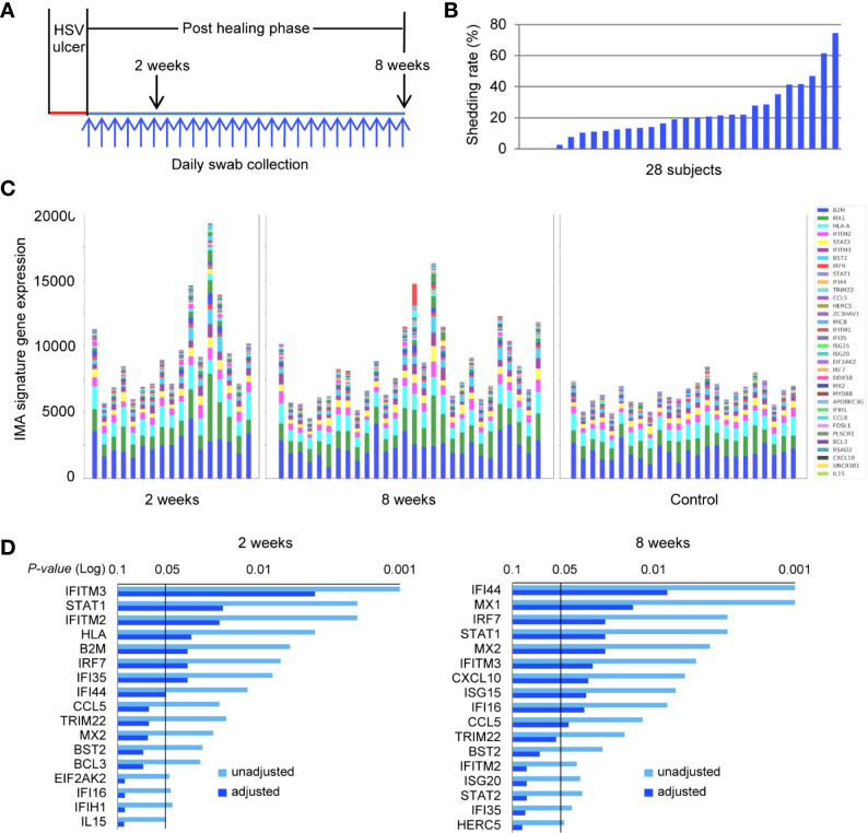
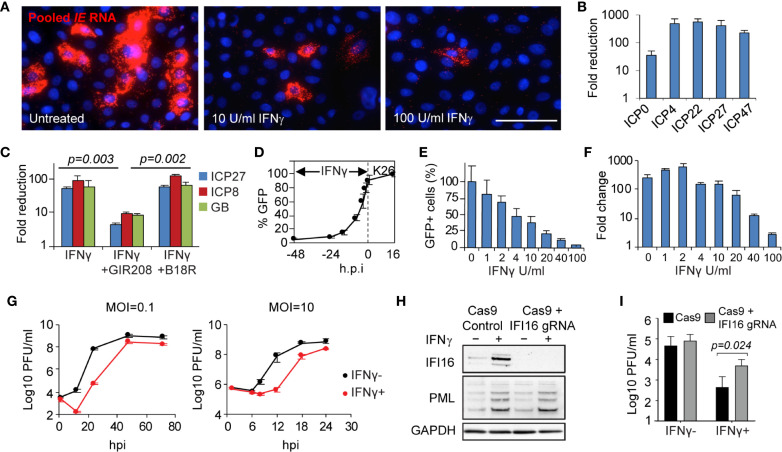
Tissue-resident-memory T cells (TRM) populate the body's barrier surfaces, functioning as frontline responders against reencountered pathogens. Understanding of the mechanisms by which CD8TRM achieve effective immune protection remains incomplete in a naturally recurring human disease. Using laser capture microdissection and transcriptional profiling, we investigate the impact of CD8TRM on the tissue microenvironment in skin biopsies sequentially obtained from a clinical cohort of diverse disease expression during herpes simplex virus 2 (HSV-2) reactivation. Epithelial cells neighboring CD8TRM display elevated and widespread innate and cell-intrinsic antiviral signature expression, largely related to IFNG expression. Detailed evaluation via T-cell receptor reconstruction confirms that CD8TRM recognize viral-infected cells at the specific HSV-2 peptide/HLA level. The hierarchical pattern of core IFN-γ signature expression is well-conserved in normal human skin across various anatomic sites, while elevation of IFI16, TRIM 22, IFITM2, IFITM3, MX1, MX2, STAT1, IRF7, ISG15, IFI44, CXCL10 and CCL5 expression is associated with HSV-2-affected asymptomatic tissue. In primary human cells, IFN-γ pretreatment reduces gene transcription at the immediate-early stage of virus lifecycle, enhances IFI16 restriction of wild-type HSV-2 replication and renders favorable kinetics for host protection. Thus, the adaptive immune response through antigen-specific recognition instructs innate and cell-intrinsic antiviral machinery to control herpes reactivation, a reversal of the canonical thinking of innate activating adaptive immunity in primary infection. Communication from CD8TRM to surrounding epithelial cells to activate broad innate resistance might be critical in restraining various viral diseases.

Keywords: IFI16 restriction factor; cell-intrinsic immunity; human genital herpes; innate antiviral response; tissue microenvironment; tissue-resident-memory T cells (TRM).


Conflict of interest statement

The authors declare that the research was conducted in the absence of any commercial or financial relationships that could be construed as a potential conflict of interest. The reviewer has declared a shared affiliation with some of the authors CS, MM, DK to the handling Editor at the time of review.

Figures

Figure 1
Figure 1
Laser capture microdissection and transcriptional profiling of keratinocytes neighboring CD8TRM in human genital HSV-2 reactivation. (A) Innervating nerve endings (green) and CD8+ T cells (red) are interconnected with basal keratinocytes at dermal-epidermal junction (DEJ) in a human genital skin biopsy obtained 4 weeks post-healing. Scale bar, 50 µm. (B) Micrographs depicting selection (left) and isolation (right) of individual basal keratinocytes in the vicinity of DEJ CD8TRM cells using laser capture microdissection. Scale bar, 50 µm. (C) Significantly enriched functional categories of differentially expressed genes in keratinocytes isolated from n = 5 post-healing biopsies compared to matched controls. (D) Hierarchical clustering of a set of 174 genes annotated to infectious disease/viral infection (left). Down-regulated genes involved in RNA transcription, processing and transportation (right top) and up-regulated genes annotated to interferon and antiviral responses (right bottom). (E) Up-regulation of a subset of ISGs in keratinocytes isolated from post healing skin (n = 5, samples 1-5) and matched HSV-2 lesion (n = 3, samples 6-8).
Figure 2
Figure 2
Widespread expression of IFI16 in genital skin during and after HSV-2 recurrence. (A) IFI16 expression in a representative HSV-2 ulcer lesion. Right box depicting actively infected epidermis with HSV-2 antigen expression. Left box showing uninfected epidermis distal to HSV lesion. Scale bars, 500 µm. (B) IFI16 expression in a representative serial skin biopsies sequentially obtained at 2-, 4-, and 8-week post-healing and in matched arm control skin. Scale bars, 100 µm. (C) Distribution of CD4+ and CD8+ T cells and IFI16 expression in an ulcerative HSV-2 lesion. IFI16 and HSV-2 antigen expression staining were performed on adjacent tissue sections. Inserts are higher magnification of the indicated boxed areas. (D) Quantitative PCR measurement of HSV-2 DNA and host genome in the right and left portion of the lesion tissue as designated in (C).
Figure 3
Figure 3
Expression of IFNG predominates in HSV-affected skin during and after viral recurrence. (A) Transcriptional analysis showing expression levels of IFN4A, IFNB1 (type I IFN), IFNG (type II IFN), IFNL1, IFNL2 and IFNL3 (type III IFN) gene in skin biopsies of n = 28 at the time of HSV-2 lesion, n = 35 at the time of 8-week post-healing, and n = 28 matched contralateral normal controls. In box plots, the center line is the median, box edges show upper and lower quartiles, whiskers represent the range and dots indicate outliers. *p < 0.05, **p < 0.01, ***p < 0.001, ****p < 0.0001, NS, not significant; student t test. (B) IFNG gene expression in DEJ CD8TRM (n = 8), keratinocytes (n = 5) and Langerhans cells (n = 7) laser microdissected from skin biopsies obtained at 8-week post-healing and their matched normal control skin, profiled by whole genome array. (C) RNA FISH images depicting co-expression of IFNG and CD8 mRNA transcript in cells (nuclei stained with DAPI) at the DEJ and in the dermis of an 8 weeks post-healing skin biopsy. Scale bars, 20 µm. (D) Recognition of autologous HSV-2-infected EBV-LCL by reconstructed transgenic TCR (tgTCR) 5491.3 reporter cells. Effectors and APC were co-incubated at indicated ratios for 24 hours. T cell activation as detected by IFN-γ release. HSV-2-specific CD8 T cell clone 2.1 as positive control. All combinations of effector cells and APC were run in quadruplicate. Each dot an individual value and bars as mean values. (E) Representative raw data from HSV-2 proteome-wide screen. Cos-7 cells were co-transfected with HLA B*07:02 cDNA and 86 individual HSV-2 genes or fragments. tgTCR5491.3 reporter cells were added for 24 h and IFN-γ release measured by ELISA. (F) tgTCR5491.3 reporter cells stained with anti-mouse TCR beta and tetrameric HLA B*07:02-HSV-2 UL49 AA 49-57 or no tetramer.
Figure 4
Figure 4
IFN-γ mediated antiviral (IMA) gene signature expression in primary human keratinocytes. (A) Signature genes differentially expressed by IFN-γ treatment annotated to the GO term “response to virus” in primary cultured human keratinocytes. Column 1: Mock treated, HSV-2 infected, HG52 MOI=1, 4 h.p.i. Column 2: IFN-γ treated, 100 U/ml for 48 hours and then mock infected. Column 3: IFN-γ treated, 100 U/ml for 48 hours and then infected with HG52, MOI=1, 4 h.p.i. Analyses were done by comparing to mock-treated and mock-infected keratinocytes. All genes listed are significantly induced by IFN-γ (FDR = 0.05). False discovery rates were derived from analysis of duplicate samples using Limma package in R. (B) Representative host gene expression in the functional category of antigen presentation, chemotaxis, and innate/cell-intrinsic antiviral defense. (C) Real-time monitoring of GFP expression in K26-infected primary human keratinocytes (MOI = 1, 45 h.p.i.), mock-treated or treated with IFN-γ 100 U/ml, 48 hours. Scale bars, 100 µm. (D) Comparison of GFP signal intensity in K26-infected primary human keratinocytes with or without IFN-γ treatment. ****p < 0.0001.
Figure 5
Figure 5
Elevated level of IMA gene signature expression correlates with IFNG expression by DEJ CD8TRM cells in HSV-2 post-healing human skin. (A) Cell density (upper) and IFNG gene expression (lower) of DEJ CD8TRM in skin biopsies taken at the sites of 8-week post-healing and their matched contralateral controls (n = 8). DEJ CD8TRM cells were laser microdissected and coupled with quantitative RT-PCR. IFNG gene expression relative to ACTB. Individual (B) and collective levels (C) of IMA gene signature expression in the 8-week and control set of skin biopsies with or without IFNG detection. (D) Micrographic images depicting spatial expression of IFI16, CD8 and IFNG mRNA transcripts in skin epidermis using RNA FISH dual probe sets of IFI16 + IFNG or IFI16 + CD8 in adjacent tissue sections of an 8-week skin biopsy, and dual probe set for IFI16 + CD8 on matched normal control skin. Scale bar, 100 µm.
Figure 6
Figure 6
IMA signature expression pattern is distinct between HSV-2-affected and normal skin in a patient cohort across a wide spectrum of genital herpes outcomes. (A) Study design. Genitalia HSV-2 reactivation was monitored by daily swab collection and PCR detection for an 8-week study period after a clinical symptomatic recurrence. Skin biopsies were obtained sequentially at the original lesion site 2- and 8-week post-healing. A control biopsy was taken at the contralateral normal site at the 8-week timepoint. Daily swabs were collected starting at day 7 after the onset of a lesion-forming HSV-2 reactivation. (B) HSV-2 shedding rates were calculated as days of positive HSV DNA detection within the total numbers of days of that swabs were collected during the study period for each participant, n = 28 subjects. (C) Skin biopsies were processed for RNA extraction and whole genome transcriptional profiling. The collective levels of IMA gene signature expression in post healing skin taken at week 2 (n = 17) and week 8 (n = 28) of prior HSV-2 recurrence and in normal contralateral control skin (n = 24). (D) IMA signature genes significantly associated with sites of prior HSV infection at 2- or 8-week post healing time. Multiplicity adjusted and unadjusted paired t-test.
Figure 7
Figure 7
Mechanism, specificity and effectiveness of IFN-γ-mediated antiviral action in primary human skin cells. (A) Evaluation of IFN-γ effect on HSV immediate early (IE) gene transcription via RNA FISH. In situ detection and visualization of IE gene expression using pooled probes specific to each of the five IE genes. Primary human keratinocytes 6 h.p.i. with K26 (MOI=1) either mock-treated or pretreated with IFN-γ (10 U/ml or 100 U/ml). Scale bar, 50 µm. (B) Quantitative assessment of IFN-γ-mediated reduction of IE gene expression via qRT-PCR. Primary human fibroblasts infected with wildtype strain KOS, MOI=1, 4 h.p.i. Mock treated compared to IFN-γ treated, 100 U/ml. (C) IFN-γ effect on HSV gene transcription (ICP27, ICP8, and glycoprotein B) in the presence or absence of interferon blockers, GIR208 and B18R. Primary human keratinocytes infected with KOS, MOI=1, 4 h.p.i. (D) Time-dependent inhibition by IFN-γ. Percentage of GFP-expressing cells in K26-infected primary human keratinocytes 16 h.p.i. (MOI=1) with IFN-γ treatment initiated at 2, 4, 8, 16, 24, or 48 hours prior to infection. (E, F) Dose-dependent inhibition by IFN-γ. GFP expression (E) and viral DNA replication (F) under increasing dosage of IFN-γ in K26-infected keratinocytes. MOI=1, 16 h.p.i. HSV genome DNA were quantified by qPCR and compared between 2 and 16 h.p.i. (G) Growth kinetics of strain KOS under low and high MOI in primary human fibroblasts with (100 U/ml) or without IFN-γ. (H) Responsiveness to IFN-γ in IFI16 knockout and Cas9 control human fibroblasts. Immunoblot of whole-cell lysate from IFI16 knockout and Cas9 control cells mock-treated or pretreated with IFN-γ (500U/mL) for 20 hours. One of three biological replicates is shown. (I) Viral yield 12 h.p.i. following wildtype HSV-2 strain 186 infection (MOI=0.1) of IFI16 knockout and Cas9 control fibroblasts mock-treated or pretreated with IFN-γ (500U/mL) for 20 hours. Two-way ANOVA with Holm-Sidak’s multiple comparisons test. Values are mean ± S.D. (error bars), from three independent experiments.

References

    1. Iwasaki A, Ruslan M. Chapter 8, Innate Responses to Viral Infections. In: Fields Virology. Philadelphia: Lippincott Williams & Wilkins; (2013).
    1. Mueller SN, Zaid A, Carbone FR. Tissue-Resident T Cells: Dynamic Players in Skin Immunity. Front Immunol (2014) 5:332. 10.3389/fimmu.2014.00332 - DOI - PMC - PubMed
    1. Park CO, Kupper TS. The Emerging Role of Resident Memory T Cells in Protective Immunity and Inflammatory Disease. Nat Med (2015) 21(7):688–97. 10.1038/nm.3883 - DOI - PMC - PubMed
    1. Schenkel JM, Masopust D. Tissue-Resident Memory T Cells. Immunity (2014) 41(6):886–97. 10.1016/j.immuni.2014.12.007 - DOI - PMC - PubMed
    1. Ariotti S, Hogenbirk MA, Dijkgraaf FE, Visser LL, Hoekstra ME, Song JY, et al. . T Cell Memory. Skin-Resident Memory CD8(+) T Cells Trigger a State of Tissue-Wide Pathogen Alert. Science (2014) 346(6205):101–5. 10.1126/science.1254803 - DOI - PubMed

Publication types

MeSH terms